Summary
Revista Brasileira de Ginecologia e Obstetrícia. 2003;25(10):725-730
DOI 10.1590/S0100-72032003001000005
PURPOSE: to evaluate the perinatal outcome of fetuses with congenital anomalies of the urinary tract. METHODS: we reviewed the perinatal outcome of 35 fetuses with congenital anomalies of the urinary tract. The following characteristics related to the uropathy were analyzed: type (hydronephrosis, dysplasia and renal agenesis), side of lesion (bilateral or unilateral), and level of the obstruction (high or low, in hydronephrosis). The perinatal outcome was evaluated according to these characteristics. The data were analyzed by the c² test and by the exact Fisher test. The level of significance was 0.05. RESULTS: the incidence of hydronephrosis was 68.6%. Half of the fetuses had unilateral hydronephrosis. Renal dysplasia occurred in 17.1% of the cases; 83.3% of these were bilateral and 16.7%, unilateral. The incidence of renal agenesis was 14.3%, all bilateral. The fetuses with dysplasia/agenesis had a 91% incidence of oligohydramnios, preterm birth, low birth weight, and death. In the group with bilateral disease the presence of oligohydramnios, preterm birth, low birth weight, death, urinary tract infections, and the need of hospitalization for a period greater than 7 days was significant when compared to the group with unilateral disease. The need of hospitalization for a period greater than 7 days in patients with low obstruction was significantly higher when compared to the patients with high obstruction. CONCLUSIONS: hydronephrosis, bilateral disease, and lower obstruction were the most frequent uropathies. The dysplasia/agenesis group had a worse prognosis when compared with the hydronephrosis group. Bilateral disease had a worse prognosis when compared with the unilateral disease group. In the low obstruction group, the need for a period of hospitalization greater than seven days was higher than in the high obstruction group.
Summary
Revista Brasileira de Ginecologia e Obstetrícia. 2000;22(6):365-371
DOI 10.1590/S0100-72032000000600007
Purpose: to evaluate the accuracy of prenatal ultrasound in the diagnosis of nephrouropathies. Methods: the authors followed-up 127 pregnancies referred to the Fetal Medicine Center of UFMG with suspicion of these anomalies. Fetal biometry, growth, vitality, and associated malformations were evaluated. Finally, a detailed description of the renal system was made to define the prenatal morphologic diagnosis of the malformations to be compared with the postnatal diagnosis. Results: based on the kappa index (statistical method that measures the concordance between different measurements, methods or measurement instruments: below 0.40, poor agreement; between 0.40 and 0.75, good agreement; above 0.75, excellent ageement), the authors found an excellent concordance (kappa index 0.95). Among the 127 cases, there were only 9 misdiagnoses, all of them of obstructive uropathies: 6 cases showed different obstruction levels after delivery and in three cases there were confounding diagnosis with multicystic kidney. Conclusions: the detailed ultrasonographic description of the renal system is a good method for prenatal diagnosis of the fetal nephropathies, allowing some options to modify the outcome of these fetuses, like to send them to specialized centers, to anticipate delivery and even to apply intrauterine therapy, in order to preserve the renal function. Serial echography and amnioinfusion can be used to improve the precision of prenatal diagnosis.
Summary
Revista Brasileira de Ginecologia e Obstetrícia. 2003;25(5):345-351
DOI 10.1590/S0100-72032003000500007
PURPOSE: to estimate the sensitivity, specificity and accuracy of patient age, ultrasound result and CA-125 marker variables for the differential diagnosis between malignant and benign ovarian tumors. In addition, to establish a risk of malignancy index (RMI) incorporating these three variables and to estimate its sensitivity, specificity and accuracy for the differential diagnosis. METHODS: one hundred patients with ovarian tumors with surgical indication were included. The age, ultrasonographic findings and CA-125 level variables were evaluated separately and later on together as the RMI. The study was performed based on the evaluation of the sensitivity, specificity and diagnostic accuracy and the use of the measurements: likelihood ratio, odds ratio, and the Student's t test, chi², and logistic regression with univariate and multivariate analysis. RESULTS: for the age variable, sensitivity, specificity and diagnostic accuracy were 58.8, 68.2 and 65.0%, respectively. For ultrasound, 88.2, 77.3 and 81.0%. For CA-125 dosage, the values were 64.7, 74.2 and 71.0%. When the three variables were put together, as the RMI, a sensitivity of 76.5%, a specificity of 87.9% and a diagnostic accuracy of 84.0% were observed. CONCLUSIONS: RMI, made up of the association of patient age, ultrasound results and CA-125 dosage variables is a valuable indicator to distinguish between malignant and benign ovarian tumor, especially in regard to its specificity.
Summary
Revista Brasileira de Ginecologia e Obstetrícia. 2003;25(4):261-268
DOI 10.1590/S0100-72032003000400007
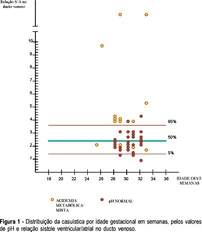
PURPOSE: to evaluate Doppler velocimetry of the ductus venosus as a noninvasive test of abnormal pH and gas analysis in preterm fetuses with "brain sparing reflex". METHODS: a cross-sectional study was performed. The studied population consisted of 48 pregnant women between the 25th and the 33rd week of gestation, whose fetuses presented brain sparing reflex (umbilical/cerebral ratio >1). The time elapsed between Doppler velocimetry and the birth (cesarean section under peridural anesthesia) was of up to 5 h. The following parameters were studied: S/A ratio of the ductus venosus, pH and base excess (BE) of fetal blood sample (collected from the umbilical vein immediately after birth). The S/A ratio of the ductus venosus was considered abnormal when superior to 3.6. The fetuses were classified according to the gas analysis result. They were considered abnormal when pH <7.26 and BE £ 6 mMol/L. Fisher's test was used for statistical analysis and considered significant when p £ 0.05. RESULTS: there was a significant correlation between umbilical blood gas analysis in preterm fetuses with brain sparing reflex and ductus venosus S/A ratio (p = 0.0000082; Fisher test). Ductus venosus Doppler velocimetry identified 10 of 14 fetuses with abnormal gas analysis. On the other hand, 32 of 34 fetuses with normal gas analysis were correctly identified. The sensitivity of the ductus venosus S/A ratio for the diagnosis of abnormal blood gas analysis was 71%, specificity 94%, false-negative rate 8%, false-positive rate 4%, positive predictive value 83% and negative predictive value 89%. Pretest likelihood, post-test posterior probability following a positive test result (post-test likelihood) and post-test posterior probability following a negative test result (post-test likelihood) were 31, 84 and 10%, respectively. CONCLUSION: the analysis of the ductus venosus S/A ratio is adequate for the diagnosis of abnormal blood gas analysis in preterm fetuses presenting brain sparing reflex.

Summary
Revista Brasileira de Ginecologia e Obstetrícia. 2000;22(8):511-517
DOI 10.1590/S0100-72032000000800007
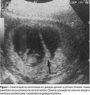
Purpose: to demonstrate the types of fetal malformations in multiple pregnancy and their relation to chorionicity. Methods: one hundred and sixty-nine multiple pregnancies were evaluated. In all cases prenatal ultrasound examination was performed during antenatal care. Chorionicity was defined by: first trimester ultrasound evaluation (absence of lambda sign); presence of two separate placentas; different fetal sex; pathological placental examination. Results: twenty-four (14.2%) fetal malformations were observed, 22 in twin and 2 in triplet pregnancy. In the group with fetal malformations 13 were monochorionic, 4 dichorionic and in 5 the chorionicity was unknown. Some malformations were unique to twins (conjoined twins n = 5, acardiac twin n = 3) and others were nonunique to twins. The gestational age at delivery was lower in the group with fetal malformations compared to the group without fetal malformations. Conclusion: the majority of malformations occurred in the monochorionic pregnancies. In multiple pregnancies early determination of chorionicity is helpful to establish the prognosis and to plan the management of pregnancy.

Summary
Revista Brasileira de Ginecologia e Obstetrícia. 2003;25(1):35-40
DOI 10.1590/S0100-72032003000100006
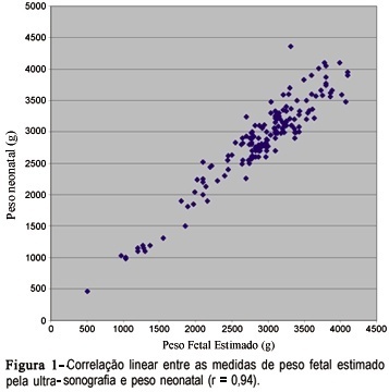
PURPOSE: tocompare the ultrasound estimation of fetal weight (EFW) with neonatal weight and to evaluate the performance of the normal EFW curve according to gestational age for the diagnosis of fetal/neonatal weight deviation and associated factors. METHODS: one hundred and eighty-six pregnant women who delivered at the institution from November 1998 to January 2000 and who had one ultra-sonographic evaluation performed until three days prior to delivery with estimation of the amniotic fluid index were included. EFW was calculated and classified in to small for gestational age (SGA), adequate for gestational age (AGA) and large for gestational age (LGA) through the normal EFW curve for this population. Neonatal weight was similarly classified. The variability of the measures and the degree of linear correlation between EFW and neonatal weight, as well as sensitivity, specificity and predictive values for the use of the normal EFW curve in the diagnosis of neonatal weight deviations were calculated. RESULTS: the difference between EFW and neonatal weight ranged from -540 to +594 g, with a mean of +46.9 g, and the two measures presented a linear correlation coefficient of 0.94. The normal EFW curve had a sensitivity of 100% and specificity of 90.5% in detecting SGA neonates and of 94.4 and 92.8%, respectively, in detecting LGA; however, the predictive positive values were low for both conditions. CONCLUSIONS:ultrasound EFW was in agreement with the neonatal weight, with a mean overweight of approximately 47 g, and its normal curve showed a good performance in the screening of SGA and LGA neonates.

Summary
Revista Brasileira de Ginecologia e Obstetrícia. 2002;24(3):195-199
DOI 10.1590/S0100-72032002000300008
Purpose: to evaluate, in a prospective way, the importance of ultrasound features of solid breast lesions in the differentiation between benign and malignant lumps. Methods: one hundred and forty-two patients with solid breast lesions, from the Department of Gynecology and Obstetrics of the Federal University of Goias (Brazil), were included in the trial. All ultrasound examinations were performed by a training doctor, always supervised by an experienced professional. The characteristics of the lesions studied were: shape, retrotumoral echoes, internal echoes, oriented diameter, halo of bright echoes and Cooper ligaments. Each of the ultrasound features was compared to the results of the histological examination. Results: among the 142 patients included in the trial, 90 (63%) had their lesions excised, and 77 (86%) had pathologic diagnoses of benign tumors and 13 (14%) of malignant tumors. The following characteristics were statistically significant in the diagnosis of the breast cancer (c²): masses with retrotumoral shadowing (p=0.0001), irregular shape (p=0.0007), heterogeneous internal echoes (p=0.0015) and vertically oriented - taller than wide (p<0.0001). The presence of halo of bright echoes anterior to the lump and the presence of wider Cooper ligaments were not related to the correct diagnosis of malignancy in this trial. Conclusion: ultrasound is a diagnostic method that can help physicians between the differentiation of benign and malignant breast lumps. The presence of retrotumoral shadowing, irregular shape, heterogeneous internal echoes and vertical orientation - lesions taller than wide - were related to the pathologic diagnosis of breast malignancies.
Summary
Revista Brasileira de Ginecologia e Obstetrícia. 2002;24(2):121-127
DOI 10.1590/S0100-72032002000200008
Purpose: to estimate the performance of ultrasound to detect gestations at risk for fetal chromosomal abnormalities. Methods: four hundred and thirty-six patients selected for the study had undergone ultrasound examination and fetal karyotyping, between March 1993 and March 1998. Two hundred and seventy-seven patients had fetal karyotype for fetal malformation detected on ultrasound and 158 for parental anxiety with normal ultrasound examination. Ultrasound sensitivity and specificity were calculated using fetal karyotype as gold standard. The relative risk for each chromosomal abnormality was calculated according to the altered system on ultrasound examination and the risks of the presence of one or more abnormalities on ultrasound, using the Epi-Info 6.0 software package for statistical analysis. Results: the relative risks for chromosomal abnormalities were 89 for face malformations, 53 for abdominal wall and cardiovascular, 49.6 for neck, 44.6 for extremities, 42.4 for lung, 32.7 for gastrointestinal tract, 27.4 for central nervous system and 23.0 for urinary tract malformations. The relative risk for fetal chromosomal anomalies for genital, thorax, spine and muscle and/or skeletal malformations was not appropriate for calculation because they occurred at very low frequencies. An isolated malformation detected by ultrasound is associated with a 7.8 times higher relative risk for chromosomal anomalies than none, and associated morphologic malformations have a 33.8 times higher relative risk for chromosomal abnormalities. Conclusion: ultrasound has good performance to detect gestations at risk for chromosomal abnormalities.