-
Original Articles
Comparison of Two- and Three-dimensional Ultrasonography in the Evaluation of Lesion Level in Fetuses with Spina Bifida
Revista Brasileira de Ginecologia e Obstetrícia. 2016;38(3):120-126
03-01-2016
Summary
Original ArticlesComparison of Two- and Three-dimensional Ultrasonography in the Evaluation of Lesion Level in Fetuses with Spina Bifida
Revista Brasileira de Ginecologia e Obstetrícia. 2016;38(3):120-126
03-01-2016Views142See morePurpose
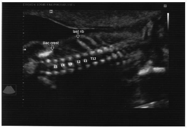
To evaluate the precision of both two- and three-dimensional ultrasonography in determining vertebral lesion level (the first open vertebra) in patients with spina bifida.
Methods
This was a prospective longitudinal study comprising of fetuses with open spina bifida who were treated in the fetal medicine division of the department of obstetrics of Hospital das Clínicas of the Universidade de São Paulo between 2004 and 2013. Vertebral lesion level was established by using both two- and three-dimensional ultrasonography in 50 fetuses (two examiners in each method). The lesion level in the neonatal period was established by radiological assessment of the spine. All pregnancies were followed in our hospital prenatally, and delivery was scheduled to allow immediate postnatal surgical correction.
Results
Two-dimensional sonography precisely estimated the spina bifida level in 53% of the cases. The estimate error was within one vertebra in 80% of the cases, in up to two vertebrae in 89%, and in up to three vertebrae in 100%, showing a good interobserver agreement. Three-dimensional ultrasonography precisely estimated the lesion level in 50% of the cases. The estimate error was within one vertebra in 82% of the cases, in up to two vertebrae in 90%, and in up to three vertebrae in 100%, also showing good interobserver agreement. Whenever an estimate error was observed, both two- and three-dimensional ultrasonography scans tended to underestimate the true lesion level (55.3% and 62% of the cases, respectively).
Conclusions
No relevant difference in diagnostic performance was observed between the two- and three-dimensional ultrasonography. The use of three-dimensional ultrasonography showed no additional benefit in diagnosing the lesion level in the fetuses with spina bifida. Errors in both methods showed a tendency to underestimate lesion level.
Views142

This is an Open Access article distributed under the terms of the Creative Commons Attribution License, which permits unrestricted use, distribution, and reproduction in any medium, provided the original work is properly cited. Summary
Original ArticlesComparison of Two- and Three-dimensional Ultrasonography in the Evaluation of Lesion Level in Fetuses with Spina Bifida
Revista Brasileira de Ginecologia e Obstetrícia. 2016;38(3):120-126
03-01-2016Views142See morePurpose
To evaluate the precision of both two- and three-dimensional ultrasonography in determining vertebral lesion level (the first open vertebra) in patients with spina bifida.
Methods
This was a prospective longitudinal study comprising of fetuses with open spina bifida who were treated in the fetal medicine division of the department of obstetrics of Hospital das Clínicas of the Universidade de São Paulo between 2004 and 2013. Vertebral lesion level was established by using both two- and three-dimensional ultrasonography in 50 fetuses (two examiners in each method). The lesion level in the neonatal period was established by radiological assessment of the spine. All pregnancies were followed in our hospital prenatally, and delivery was scheduled to allow immediate postnatal surgical correction.
Results
Two-dimensional sonography precisely estimated the spina bifida level in 53% of the cases. The estimate error was within one vertebra in 80% of the cases, in up to two vertebrae in 89%, and in up to three vertebrae in 100%, showing a good interobserver agreement. Three-dimensional ultrasonography precisely estimated the lesion level in 50% of the cases. The estimate error was within one vertebra in 82% of the cases, in up to two vertebrae in 90%, and in up to three vertebrae in 100%, also showing good interobserver agreement. Whenever an estimate error was observed, both two- and three-dimensional ultrasonography scans tended to underestimate the true lesion level (55.3% and 62% of the cases, respectively).
Conclusions
No relevant difference in diagnostic performance was observed between the two- and three-dimensional ultrasonography. The use of three-dimensional ultrasonography showed no additional benefit in diagnosing the lesion level in the fetuses with spina bifida. Errors in both methods showed a tendency to underestimate lesion level.
This is an Open Access article distributed under the terms of the Creative Commons Attribution License, which permits unrestricted use, distribution, and reproduction in any medium, provided the original work is properly cited.