-
Original Articles
Increased Expression Levels of Metalloprotease, Tissue Inhibitor of Metalloprotease, Metallothionein, and p63 in Ectopic Endometrium: An Animal Experimental Study
Revista Brasileira de Ginecologia e Obstetrícia. 2018;40(11):705-712
11-01-2018
Summary
Original ArticlesIncreased Expression Levels of Metalloprotease, Tissue Inhibitor of Metalloprotease, Metallothionein, and p63 in Ectopic Endometrium: An Animal Experimental Study
Revista Brasileira de Ginecologia e Obstetrícia. 2018;40(11):705-712
11-01-2018Views141See moreAbstract
Objective
To characterize the patterns of cell differentiation, proliferation, and tissue invasion in eutopic and ectopic endometrium of rabbits with induced endometriotic lesions via a well- known experimental model, 4 and 8 weeks after the endometrial implantation procedure.
Methods
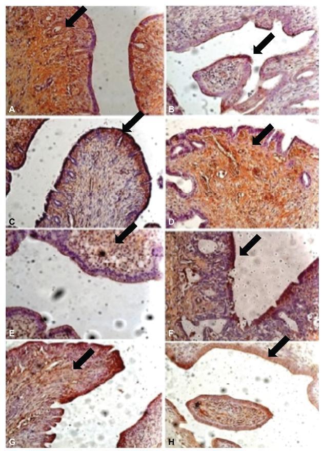
Twenty-nine female New Zealand rabbits underwent laparotomy for endometriosis induction through the resection of one uterine horn, isolation of the endometrium, and fixation of tissue segment to the pelvic peritoneum. Two groups of animals (one with 14 animals, and the other with15) were sacrificed 4 and 8 weeks after endometriosis induction. The lesion was excised along with the opposite uterine horn for endometrial gland and stroma determination. Immunohistochemical reactions were performed in eutopic and ectopic endometrial tissues for analysis of the following markers: metalloprotease (MMP-9) and tissue inhibitor of metalloprotease (TIMP-2), which are involved in the invasive capacity of the endometrial tissue; and metallothionein (MT) and p63, which are involved in cell differentiation and proliferation.
Results
The intensity of the immunostaining for MMP9, TIMP-2, MT, and p63 was higher in ectopic endometria than in eutopic endometria. However, when the ectopic lesions were compared at 4 and 8 weeks, no significant difference was observed, with the exception of the marker p63, which was more evident after 8 weeks of evolution of the ectopic endometrial tissue.
Conclusion
Ectopic endometrial lesions seem to express greater power for cell differentiation and tissue invasion, compared with eutopic endometria, demonstrating a potentially invasive, progressive, and heterogeneous presentation of endometriosis.
Views141

This is an Open Access article distributed under the terms of the Creative Commons Attribution License, which permits unrestricted use, distribution, and reproduction in any medium, provided the original work is properly cited. Summary
Original ArticlesIncreased Expression Levels of Metalloprotease, Tissue Inhibitor of Metalloprotease, Metallothionein, and p63 in Ectopic Endometrium: An Animal Experimental Study
Revista Brasileira de Ginecologia e Obstetrícia. 2018;40(11):705-712
11-01-2018Views141See moreAbstract
Objective
To characterize the patterns of cell differentiation, proliferation, and tissue invasion in eutopic and ectopic endometrium of rabbits with induced endometriotic lesions via a well- known experimental model, 4 and 8 weeks after the endometrial implantation procedure.
Methods
Twenty-nine female New Zealand rabbits underwent laparotomy for endometriosis induction through the resection of one uterine horn, isolation of the endometrium, and fixation of tissue segment to the pelvic peritoneum. Two groups of animals (one with 14 animals, and the other with15) were sacrificed 4 and 8 weeks after endometriosis induction. The lesion was excised along with the opposite uterine horn for endometrial gland and stroma determination. Immunohistochemical reactions were performed in eutopic and ectopic endometrial tissues for analysis of the following markers: metalloprotease (MMP-9) and tissue inhibitor of metalloprotease (TIMP-2), which are involved in the invasive capacity of the endometrial tissue; and metallothionein (MT) and p63, which are involved in cell differentiation and proliferation.
Results
The intensity of the immunostaining for MMP9, TIMP-2, MT, and p63 was higher in ectopic endometria than in eutopic endometria. However, when the ectopic lesions were compared at 4 and 8 weeks, no significant difference was observed, with the exception of the marker p63, which was more evident after 8 weeks of evolution of the ectopic endometrial tissue.
Conclusion
Ectopic endometrial lesions seem to express greater power for cell differentiation and tissue invasion, compared with eutopic endometria, demonstrating a potentially invasive, progressive, and heterogeneous presentation of endometriosis.
This is an Open Access article distributed under the terms of the Creative Commons Attribution License, which permits unrestricted use, distribution, and reproduction in any medium, provided the original work is properly cited.