Summary
Revista Brasileira de Ginecologia e Obstetrícia. 2015;37(7):302-307
07-01-2015
DOI 10.1590/S0100-720320150005352
To evaluate the follicular development of female Wistar rats with obesity induced by the cafeteria diet, submitted to the administration of losartan (LOS), an antagonist of the AT1 receptor of Angiotensin II.
At weaning (21 days of age), female Wistar rats were randomly divided, into two groups: control (CTL) that received standard chow and cafeteria (CAF) that received a cafeteria diet, a highly palatable and highly caloric diet. At 70 days of age, at the beginning of the reproductive age, animals of the CAF group were subdivided into two groups (n=15/group): CAF, that received water, and CAF+LOS, that received LOS for 30 days. The CTL group also received water by gavage. At 100 days of age, the animals were euthanized and body weight (BW) as well as the retroperitoneal, perigonadal and subcutaneous fat weights were analyzed. The right ovaries were isolated for counting the number of primary, secondary, antral and mature follicles. Plasma levels of FSH, LH, prolactin and progesterone hormones were analyzed. The results were expressed as mean±standard error of the mean. Data were analyzed statistically by one-way ANOVA followed by the Newman-Keuls post-test (p<0.05).
BW and fat weight, as well as the number of antral follicles, were higher in the CAF group compared to the CTL group. However, FSH and LH levels were lower in CAF animals compared to CTL animals. LOS administration attenuated the reduction of FSH and LH levels. Progesterone and PRL levels were similar among groups.
LOS could improve follicular development in obese females and could be used as an adjunctive drug in the treatment of infertility associated with obesity.
Summary
Revista Brasileira de Ginecologia e Obstetrícia. 2014;36(10):436-441
10-03-2014
DOI 10.1590/SO100-720320140005105
Pregnant women have a 2-3 fold higher probability of developing restless legs syndrome (RLS – sleep-related movement disorders) than general population. This study aims to evaluate the behavior and locomotion of rats during pregnancy in order to verify if part of these animals exhibit some RLS-like features.
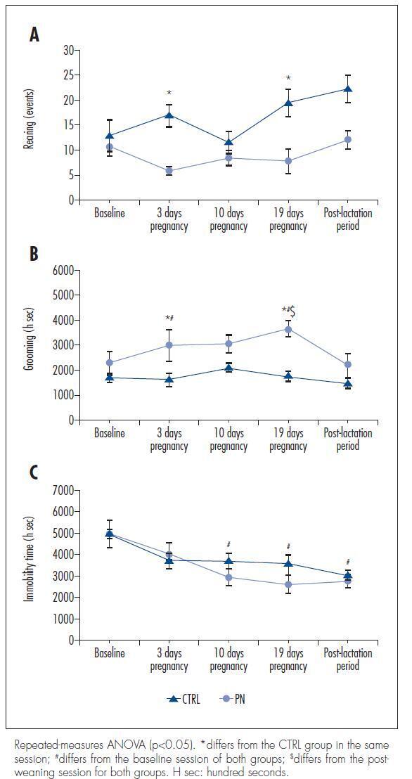
We used 14 female 80-day-old Wistar rats that weighed between 200 and 250 g. The rats were distributed into control (CTRL) and pregnant (PN) groups. After a baseline evaluation of their behavior and locomotor activity in an open-field environment, the PN group was inducted into pregnancy, and their behavior and locomotor activity were evaluated on days 3, 10 and 19 of pregnancy and in the post-lactation period in parallel with the CTRL group. The serum iron and transferrin levels in the CTRL and PN groups were analyzed in blood collected after euthanasia by decapitation.
There were no significant differences in the total ambulation, grooming events, fecal boli or urine pools between the CTRL and PN groups. However, the PN group exhibited fewer rearing events, increased grooming time and reduced immobilization time than the CTRL group (ANOVA, p<0.05).
These results suggest that pregnant rats show behavioral and locomotor alterations similar to those observed in animal models of RLS, demonstrating to be a possible animal model of this sleep disorder.

Summary
Revista Brasileira de Ginecologia e Obstetrícia. 2010;32(2):88-93
03-15-2010
DOI 10.1590/S0100-72032010000200007
PURPOSE: to evaluate the effect of the prolonged use of a high dose of tibolone on the body weight variation and lipid profile of oophorectomized female rats. METHODS: 15 Wistar rats weighing 250 g were randomly divided into two groups. The Experimental Group (n=9) received 1 mg/day of oral tibolone. The Control Group (n=6) received daily 0.5 mL of 0.5% carboxymethylcellulose by gavage. Bilateral oophorectomy was performed 30 days before the beginning of the experiment. On day 0 of the experiment, the animals began to receive the respective treatment for 20 weeks. Body weight was controlled every seven days and food consumption was measured every three to four days along the experiment, in order to establish the daily mean consumption per animal. The results were compared by the Student's t-test, with the significance level set at p<0.05. RESULTS: the daily food consumption of the Tibolone Group was significantly lower (12.7±1.2 g, p<0.001) compared to the Control Group (14.5±1.4 g). This difference was also significant when the body weight was compared between the Tibolone and Control Groups (p<0.001), with the Tibolone Group having lower weight along the experiment. At the end of the experiment, the mean body weight was 215.6±9.3 g in the Tibolone Group and 243.6±6.4 g in the Control Group. Regarding the lipid profile, the Tibolone Group had significantly (p<0.001) lower total cholesterol compared to the Control Group (30.3 versus 78.6 mg/dL). The level of HDL-c was also significantly different (p<0.001), with the Tibolone Group showing lower levels than the Control Group (9.0 versus 52.0 mg/dL). No significant difference between the groups was registered in the other biochemical parameters examined (LDL-c, VLDL-c and triglycerides). CONCLUSIONS: tibolone causes a significant reduction of HDL-c and total cholesterol and has a deleterious effect on the body weight of oophorectomized rats, which may be related to the lower food ingestion by these animals.
Summary
Revista Brasileira de Ginecologia e Obstetrícia. 2008;30(7):335-340
10-07-2008
DOI 10.1590/S0100-72032008000700003
PURPOSE: to evaluate the effect of exposure of female rats to therapeutic ultrasound in the pre-implantation phase. METHODS: pregnant Wistar female rats have been exposed to 3 MHz, 0.6 W/cm² ultrasound, pulsatile ultrasound (PUS) or continuous ultrasound (CUS), and controls, unplugged ultrasound (UUS), for five minutes. The rats were sacrificed at the 20th day post-insemination. Biochemical and hematological analyses have been done. Animals have been submitted to necropsy in order to identify lesions of internal organs, and to remove and weight the liver, kidneys and ovaries. Alive, malformed, dead and reabsorbed fetuses have been counted. RESULTS: the rats have not presented changes in their body and organs weight, and neither in their reproductive capacity, but there has been an increase in triglycerides in the PUS and CUS groups, when compared to the UUS group. The fetuses' relative weights of the heart (0.7 ± 0.9), liver (9.8 ± 0.8), kidneys (6.2 ± 0.8) and lungs (3.8 ± 0.4) increased in the CUS, when compared to the heart (0.7 ± 0.9), liver (9.8 ± 0.8), kidneys (6.2 ± 0.8) e lungs (3.8 ± 0.4) of the UUS. CONCLUSIONS: in the experimental model, the therapeutic ultrasound used has not caused meaningful maternal toxicity. Pulsatile waves have not changed fetal morphology, but continuous waves have caused increase in the relative weight of the fetuses' heart, liver, lungs and kidneys.
Summary
Revista Brasileira de Ginecologia e Obstetrícia. 2008;30(5):219-223
08-15-2008
DOI 10.1590/S0100-72032008000500003
PURPOSE: to evaluate the toxicity of tacrolimus on embryonic development in rats treated during the tubal transit period. METHODS: sixty Wistar rats were distributed into four groups (15 animals each), which received different doses of tacrolimus through intragastric administration: (T1) 1.0 mg/kg/day, (T2) 2.0 mg/kg/day and (T3) 4.0 mg/kg/day. The control group (C) received distilled water. The rats were observed daily to detect clinical signs of toxicity. The treatments were performed from the first to the fifth day of pregnancy. The following maternal variables were analyzed: body, ovary, liver, and kidney weights, food intake, number of corpora lutea, implants, alive and dead fetuses, and implantation rates. The fetuses and placentae were weighed and the former were observed in order to detect external malformation. Statistical analysis was performed by one way: analysis of variance (ANOVA), folowed by the Dunnet test (alpha=0.05). RESULTS: there were no signs of maternal toxicity, such as body weight loss, decrease in food intake or in organ weights (p>0.05). There was also no significant difference among weights of fetuses (C: 1.8±0.6; T1: 2.2±0.5; T2: 1.9±0.5 and T3: 2.0±0.5 g) and placentae (C: 1,6±0.4; T1: 1.5±0.4; T2: 1.8±0.4 e T3: 1.6±0.4 g), with p>0.05; no external malformation was detected in the fetuses. CONCLUSIONS: the administration of tacrolimus to pregnant rats during the tubal transit period does not seem to generate any toxic effect to mother or embryo.
Summary
Revista Brasileira de Ginecologia e Obstetrícia. 2007;29(8):408-414
11-01-2007
DOI 10.1590/S0100-72032007000800005
PURPOSE: to analyze the macroscopic and histological changes that occur with the use of sinvastatin in experimental endometriosis in female rats. METHODS: forty Wistar female rats were submitted to the technique of uterine self-transplant in mesenterium. After three weeks, 24 of them developed experimental endometriosis grade III, and were divided in two groups: one group received sinvastatin orally (20 mg/kg/day) and the other (control group) received 0.9% of sodium chloride orally (1 mL/100 g of body weight/day). Both groups received gavage for 14 days, followed by death. The implant volume was calculated [4pi (lenght/2) x (width/2) x (height/2)/3] at the surgical intervention and after the animal’s death. The self-transplants were removed, dyed with hematoxylin-eosin and analyzed by light microscopy. The Mann-Whitney’s test was used in the independent samples and the Wilcoxon’s test for the related samples. The Fisher’s exact test was used for the histological evaluation, with a significance level of 5%. RESULTS: the difference between groups of the initial average volumes of the self-transplants was not significant (p=1.00), but became significant for the final average volumes (p=0.04). There was a significant increase (p=0.01) between the initial and final average volumes in the control group, and a no significant decrease in the sinvastatin group (p=0.95). Histologically, the sinvastatin group (n=9) presented seven cases (77.8%) of moderately preserved and two cases (22.2%) of well preserved epithelial wall, while the control group (n=12) presented seven cases (58.3%) of moderately preserved and five cases (41.7%) of well preserved epithelial wall. CONCLUSIONS: sinvastatin prevented the growth of experimental endometriosis. Studies with sinvastatin for longer periods are promising.