-
Original Article05-01-2017
The Effect of Mesenchymal Stem Cells on Fertility in Experimental Retrocervical Endometriosis
Revista Brasileira de Ginecologia e Obstetrícia. 2017;39(5):217-223
Abstract
Original ArticleThe Effect of Mesenchymal Stem Cells on Fertility in Experimental Retrocervical Endometriosis
Revista Brasileira de Ginecologia e Obstetrícia. 2017;39(5):217-223
Views160See moreAbstract
Purpose
To evaluate the effect of mesenchymal stem cells (MSCs) on fertility in experimental retrocervical endometriosis.
Methods
A total of 27 New Zealand rabbits were divided into three groups: endometriosis, in which endometrial implants were created; mesenchymal, in which MSCs were applied in addition to the creation of endometrial implants; and control, the group without endometriosis. Fisher’s exact test was performed to compare the dichotomous qualitative variables among the groups. The quantitative variables were compared by the nonparametric Mann-Whitney and Kruskal-Wallis tests. The MannWhitney test was used for post-hoc multiple comparison with Boniferroni correction.
Results
Regarding the beginning of the fertile period, the three groups had medians of 14±12.7, 40±5, and 33±8.9 days respectively (p = 0.005). With regard to fertility (number of pregnancies), the endometriosis and control groups showed a rate of 77.78%, whereas the mesenchymal group showed a rate of 11.20% (p = 0.015). No differences in Keenan’s histological classification were observed among the groups (p = 0.730). With regard to the macroscopic appearance of the lesions, the mesenchymal group showed the most pelvic adhesions.
Conclusion
The use of MSCs in endometriosis negatively contributed to fertility, suggesting the role of these cells in the development of this disease.
Views160

This is an Open Access article distributed under the terms of the Creative Commons Attribution License, which permits unrestricted use, distribution, and reproduction in any medium, provided the original work is properly cited. Abstract
Original ArticleThe Effect of Mesenchymal Stem Cells on Fertility in Experimental Retrocervical Endometriosis
Revista Brasileira de Ginecologia e Obstetrícia. 2017;39(5):217-223
Views160See moreAbstract
Purpose
To evaluate the effect of mesenchymal stem cells (MSCs) on fertility in experimental retrocervical endometriosis.
Methods
A total of 27 New Zealand rabbits were divided into three groups: endometriosis, in which endometrial implants were created; mesenchymal, in which MSCs were applied in addition to the creation of endometrial implants; and control, the group without endometriosis. Fisher’s exact test was performed to compare the dichotomous qualitative variables among the groups. The quantitative variables were compared by the nonparametric Mann-Whitney and Kruskal-Wallis tests. The MannWhitney test was used for post-hoc multiple comparison with Boniferroni correction.
Results
Regarding the beginning of the fertile period, the three groups had medians of 14±12.7, 40±5, and 33±8.9 days respectively (p = 0.005). With regard to fertility (number of pregnancies), the endometriosis and control groups showed a rate of 77.78%, whereas the mesenchymal group showed a rate of 11.20% (p = 0.015). No differences in Keenan’s histological classification were observed among the groups (p = 0.730). With regard to the macroscopic appearance of the lesions, the mesenchymal group showed the most pelvic adhesions.
Conclusion
The use of MSCs in endometriosis negatively contributed to fertility, suggesting the role of these cells in the development of this disease.
This is an Open Access article distributed under the terms of the Creative Commons Attribution License, which permits unrestricted use, distribution, and reproduction in any medium, provided the original work is properly cited. 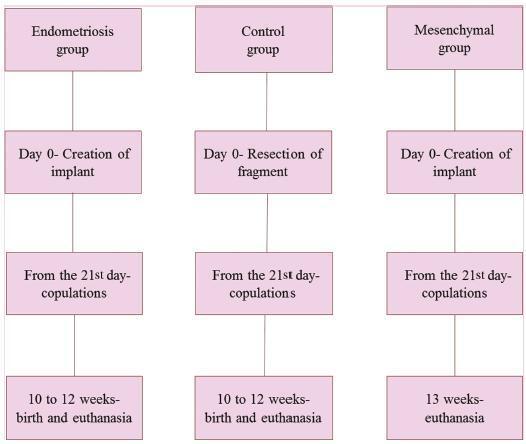
-
Original Article04-10-2005
Histological aspects of rabbit ovarian tissue after cryopreservation
Revista Brasileira de Ginecologia e Obstetrícia. 2005;27(11):642-649
Abstract
Original ArticleHistological aspects of rabbit ovarian tissue after cryopreservation
Revista Brasileira de Ginecologia e Obstetrícia. 2005;27(11):642-649
DOI 10.1590/S0100-72032005001100002
Views147See morePURPOSE: to evaluate follicular preservation and histologic characteristics of the cryopreserved ovarian tissue and to compare with the fresh one, in rabbits. METHODS: ten adult female white rabbits were submitted to right oophorectomy. The dried ovary was dissected and the cortex was maintained with approximately 1.5 millimeter thickness. The tissue was fractionated into small sections, some reserved for control histologic study and others destined for cryopreservation. Six weeks later the ovarian tissue was thawed and evaluated histologically. After histologic processing, the control and the experimental samples were stained with hematoxylin and eosin and treated immunohistochemically by the PCNA technique for evaluation of DNA preservation. Histologic alterations present in the fresh and in the cryopreserved tissues were identified, and cryopreserved tissue viability was evaluated. RESULTS: in the cryopreserved tissue only primordial follicles persisted. Reversible alterations were identified: cytoplasmatic vacuolation (p=0,039), stromal lysis (p=0.648) and oocytes with irregular contours (p=0.007). Irreversible alterations: (hyalin degeneration and pyknosis) were found, but not at significant levels (p=0.210). The immunohistochemical analysis showed PCNA staining of follicles at different stages of development in the fresh tissue and primordial follicles in the cryopreserved tissue, indicating the presence of active DNA in both tissues. CONCLUSION: in the cryopreserved ovarian tissue the following were observed: survival of only primordial follicles; significant reversible histologic alterations (cytoplasmic vacuolation, stromal lysis and oocytes with irregular contours); irreversible alterations (hyalin degeneration and pyknosis), and PCNA staining of all follicles.
Views147This is an Open Access article distributed under the terms of the Creative Commons Attribution License, which permits unrestricted use, distribution, and reproduction in any medium, provided the original work is properly cited. Abstract
Original ArticleHistological aspects of rabbit ovarian tissue after cryopreservation
Revista Brasileira de Ginecologia e Obstetrícia. 2005;27(11):642-649
DOI 10.1590/S0100-72032005001100002
Views147See morePURPOSE: to evaluate follicular preservation and histologic characteristics of the cryopreserved ovarian tissue and to compare with the fresh one, in rabbits. METHODS: ten adult female white rabbits were submitted to right oophorectomy. The dried ovary was dissected and the cortex was maintained with approximately 1.5 millimeter thickness. The tissue was fractionated into small sections, some reserved for control histologic study and others destined for cryopreservation. Six weeks later the ovarian tissue was thawed and evaluated histologically. After histologic processing, the control and the experimental samples were stained with hematoxylin and eosin and treated immunohistochemically by the PCNA technique for evaluation of DNA preservation. Histologic alterations present in the fresh and in the cryopreserved tissues were identified, and cryopreserved tissue viability was evaluated. RESULTS: in the cryopreserved tissue only primordial follicles persisted. Reversible alterations were identified: cytoplasmatic vacuolation (p=0,039), stromal lysis (p=0.648) and oocytes with irregular contours (p=0.007). Irreversible alterations: (hyalin degeneration and pyknosis) were found, but not at significant levels (p=0.210). The immunohistochemical analysis showed PCNA staining of follicles at different stages of development in the fresh tissue and primordial follicles in the cryopreserved tissue, indicating the presence of active DNA in both tissues. CONCLUSION: in the cryopreserved ovarian tissue the following were observed: survival of only primordial follicles; significant reversible histologic alterations (cytoplasmic vacuolation, stromal lysis and oocytes with irregular contours); irreversible alterations (hyalin degeneration and pyknosis), and PCNA staining of all follicles.
This is an Open Access article distributed under the terms of the Creative Commons Attribution License, which permits unrestricted use, distribution, and reproduction in any medium, provided the original work is properly cited.