Summary
Revista Brasileira de Ginecologia e Obstetrícia. 2005;27(2):86-91
DOI 10.1590/S0100-72032005000200008
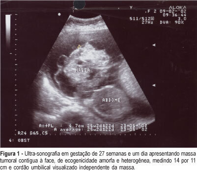
Oropharyngeal teratoma is the most rare type of teratoma, with only 2% of fetal teratomas. The diagnosis must be established as early as possible, preferably during the prenatal period. The prognosis will depend on the size and location of the lesion, growth rate of the lesion, degree of intracranial spread, its resectability, and immediate care at birth by a multisciplinary team. We report aparticular case of congenital oropharyngeal teratoma (epignathus). The diagnosis was made during the prenatal period by ultrasound, and the fetus evolved to intrauterine death at the 29th week. The anatomopathological examination revealed a female fetus, compatible with 27-28 weeks, oropharyngeal teratoma and congenital malformations.
Summary
Revista Brasileira de Ginecologia e Obstetrícia. 2005;27(2):86-91
DOI 10.1590/S0100-72032005000200008
Oropharyngeal teratoma is the most rare type of teratoma, with only 2% of fetal teratomas. The diagnosis must be established as early as possible, preferably during the prenatal period. The prognosis will depend on the size and location of the lesion, growth rate of the lesion, degree of intracranial spread, its resectability, and immediate care at birth by a multisciplinary team. We report aparticular case of congenital oropharyngeal teratoma (epignathus). The diagnosis was made during the prenatal period by ultrasound, and the fetus evolved to intrauterine death at the 29th week. The anatomopathological examination revealed a female fetus, compatible with 27-28 weeks, oropharyngeal teratoma and congenital malformations.
Summary
Revista Brasileira de Ginecologia e Obstetrícia. 2005;27(2):80-85
DOI 10.1590/S0100-72032005000200007
PURPOSE: to evaluate the evolution of pregnancy and the maternofetal prognosis in women with uterine leiomyomas. METHODS: a descriptive retrospective analysis of the medical records of 75 pregnant women with leiomyomas attended at the University Hospital, Faculty of Medicine of Ribeirão Preto, University of São Paulo, from January 1992 to January 2002. RESULTS: seventy-five pregnant women with leiomyomas were identified in a population of 34,467 pregnant women attended during this period (incidence of 0.2%). The diagnosis was made before pregnancy in 18 patients (24%), during the current pregnancy in 41 (54.6%), and during cesarean section in 16 (21.3%), of whom only six were not submitted to ultrasound scan during the prenatal period. Ten deliveries with preterm fetuses and five cases of premature rupture of the amniotic membranes were observed. Forty-seven patients (75.8%) were submitted to cesarean section, with the indication being directly related to the leiomyomas in 38.3% of them (anomalous presentation, obstruction of the birth canal, or uterine scar due to a previous myomectomy). Four cases of central necrosis, two cases of hyaline degeneration and one case of malignant potential of the leiomyoma were identified in patients submitted to postpartum myomectomy or hysterectomy. Sixty-one newborns (98.4%) had an Apgar score above 7 at the fifth minute of life, and surgery did not lead to a worse maternofetal prognosis when performed during pregnancy. CONCLUSIONS: the incidence of leiomyomas during pregnancy was 0.2% during the study period, with ultrasonography failing to diagnose 10 patients. Cesarean section was frequently indicated for this group of patients, but the presence of leiomyomas during pregnancy did not compromise the Apgar score of the newborns.
Summary
Revista Brasileira de Ginecologia e Obstetrícia. 2004;26(10):799-805
DOI 10.1590/S0100-72032004001000007
PURPOSE: to assess risk factors for low Apgar score. METHODS: this was a cross-sectional study preformed in a random sample of patients admitted to a level III maternity hospital in 2001. The outcome was low Apgar score defined as an Apgar score 1-6 (study group) versus Apgar score 7-10 (control group) in the first minute of life. The first step was the evaluation of the association of each possible risk factor with low Apgar score. The second step was multivariate analysis with the backward stepwise logistic regression model. RESULTS: there were 39 (14%) depressed newborns which were compared to 238 (86%) not depressed babies. The final analysis (multivariate) showed an association between low Apgar score and previous case of stillbirth (OR=52.6), preterm labor threat (OR=33.8), low birth weight, less than 2,500 g body weight (OR=11.2) and previous cesarean section (OR=7.4). Some factors acted as a protection, including birth weight, in grams (OR=0.9), female sex of the newborn (OR=0.1), medical complications (OR=0.4) and prematurity (gestational age < 37 weeks, OR=0.1). CONCLUSION: the study may help in the identification of fetuses at great risk of asphyxia, allowing proper reference within the health system and planning of effective assistance in neonatal intensive care units.
Summary
Revista Brasileira de Ginecologia e Obstetrícia. 2004;26(9):697-701
DOI 10.1590/S0100-72032004000900004
PURPOSE: to evaluate perinatal results in pregnant women over 35 years old and to check differences between two groups: 35 to 39-year-old women and women older than 40. METHODS: a retrospective survey was made during the period between January/2000 and July/2003, through the analysis of obstetric charts of 3,093 pregnant women who delivered in the "Hospital do Servidor Público Estadual - Francisco Morato de Oliveira", excluding 933 patients. The patients were divided into 3 groups: 18 to 29 years old (control group), 35 to 39 years old, and over 40 years old. Data collection was done with standardized forms, and the data were transferred to an electronic spreadsheet (Excel - Microsoft Office 2000). Statistical analysis was performed using the chi2 test and the Fisher test. The alpha risk was less or equal to 5% and the confidence interval 95%. RESULTS: cesarean section was the most used method not only in the 35 to 39-year-old group (438/792; 55.3%) but also in the group of women over 40 (153/236; 64.8%). The rates of prematurity (39/236; 16.5%), low weight (37/236; 15.7%), and restriction of fetal growth (38/236; 16.1%) were significantly higher in the group of women over 40, when compared to the other groups. Concerning fetal death, a five times higher incidence was observed in the group over 40 years old, as compared to the other groups, a statistically significant difference. CONCLUSION: the only difference between the 35 to 39-year-old group and the control group was the cesarean section rate. This allows us to suggest a differentiated prenatal attendance for pregnant women over 40.
Summary
Revista Brasileira de Ginecologia e Obstetrícia. 2004;26(6):477-482
DOI 10.1590/S0100-72032004000600009
OBJECTIVE: to evaluate the neonatal morbidity and mortality related to mothers at the age of 35 or older than that. METHODS: in 2377 births in a year, 316 newborns (13.26%) from mothers at the age of 35 or more were selected for the study. These women were compared to pregnant controls aged 20 to 29, randomly selected among the 1170 women in the same age group (49,2%). For the inclusion criteria, pregnancies should have been over 22 weeks and the newborns should have weighted 500g or more at birth. Fourteen twin cases were excluded. To evaluate mortality and morbidity the following variables were considered: Apgar Index, birth weight, newborn health conditions, fetal malformations and neonatal mortality until hospital discharge. RESULTS: when analyzed as a whole, nulliparous and multiparous women showed significantly less favorable perinatal results for the selected group of women at 35 or more years old as compared with pregnant controls, what was not sustained when the nulliparous were excluded. Multiparous at the age of 35 or over presented a higher rate of low Apgar index in the 1st minute: 21.3 and 13.1%: (p<0,0033); small NB for the gestational age: 15.2% and 6.7% (p<0,02); big NB for the gestational age: 5.7 and 0.0% (p<0,02); low weight at birth: 23.8 and 14,5% (p<0,01), and prematurity, 16,7 and 6,7%, (p<0,005). Significant differences were not found for the Apgar index in the 5th minute, fetal malformations, newborn health conditions at hospital discharge and neonatal mortality. CONCLUSIONS: Neonatal morbidity increased among pregnant women at the age of 35 and older, but not the neonatal mortality.
Summary
Revista Brasileira de Ginecologia e Obstetrícia. 2004;26(3):177-184
DOI 10.1590/S0100-72032004000300002
PURPOSE: to assess the use of antenatal corticosteroid (AC) by mothers and its repercussion on the birth conditions of preterm babies at the eight university neonatal units belonging to the Brazilian Network of Neonatal Research. METHODS: an observational prospective cohort study. All 463 pregnant women with a gestational age (GA) of 23 to 34 weeks and their 514 newborn babies were evaluated during the period from August 1 to December 31, 2001. The data were obtained by maternal interview, by the analysis of the medical records and by the follow-up of the newborn infants, and analyzed statistically using chi2, Mann-Whitney and ANOVA tests and multiple logistic regression, with the level of significance set at 0.05. RESULTS: 60.1% (282/463) of the pregnant women (a variation from 12.5 to 87.3% among units) received at least one AC dose. The AC use was directly associated with the number of prenatal visits, with maternal hypertension and with the antenatal use of tocolytic agents. Babies from treated pregnant women presented higher birth weight (1,379±421 vs 1,244±543 g), longer gestational age (30.9±2.0 vs 29.5±3.5 weeks), better Apgar scores at the 1st and 5th minute, and a reduced need for intervention in the delivery room. The use of AC, the GA and a baby small for GA independently improved the birth conditions. CONCLUSIONS: at most centers, AC was administered at frequencies below the desired ones, and in 50% of cases in an inadequate manner. Treatment was applied more to mothers who received appropriate prenatal care and was associated with better birth conditions.
Summary
Revista Brasileira de Ginecologia e Obstetrícia. 2004;26(2):147-151
DOI 10.1590/S0100-72032004000200010
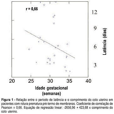
PURPOSE: to assess the length of the uterine cervix by transvaginal ultrasonography in pregnant women with preterm premature rupture of membranes. METHODS: the study group (Ge) consisted of 26 pregnant women with gestational age between 24 and 36 weeks and the control group (Gc) of 49 clinically normal patients at the same gestational age. The patients were evaluated between the 24th to 28th, 28th to 32th and 32th to 36th weeks. The groups were divided into subgroups Ge24-28, Ge28-32, Ge32-36 and Gc24-28, Gc28-32, Gc32-36, according to the study or control group. The cervix length was measured by transvaginal ultrasonography as the linear distance between the internal and external cervical os. RESULTS: we observed significant differences in cervix length between Ge24-28 and Gc24-28 groups whose values were, respectively, 24.3 and 33.0 mm (p=0.04), and between Ge32-36 and Gc32-36, 20.1 and 28.0 mm, respectively (p=0.005). The latency periods of Ge24-28, Ge28-32 and Ge32-36 were, respectively, seven, five and three days, showing a positive correlation with cervix length (r=0.66) and a negative correlation with gestational age (r=-0.27). CONCLUSIONS: the length of the uterine cervix varied with the gestational age when premature preterm rupture of the membranes was detected, with the length being shorter in the study group than in the control group betweeen the 24th and 28th and 32th and 36th weeks. In addition, it was demonstrated that, the shorter the cervix length, the shorter the latency time, with a reduction in the latency period with increasing gestational age at the time of rupture.
Summary
Revista Brasileira de Ginecologia e Obstetrícia. 2004;26(2):147-151
DOI 10.1590/S0100-72032004000200010
PURPOSE: to assess the length of the uterine cervix by transvaginal ultrasonography in pregnant women with preterm premature rupture of membranes. METHODS: the study group (Ge) consisted of 26 pregnant women with gestational age between 24 and 36 weeks and the control group (Gc) of 49 clinically normal patients at the same gestational age. The patients were evaluated between the 24th to 28th, 28th to 32th and 32th to 36th weeks. The groups were divided into subgroups Ge24-28, Ge28-32, Ge32-36 and Gc24-28, Gc28-32, Gc32-36, according to the study or control group. The cervix length was measured by transvaginal ultrasonography as the linear distance between the internal and external cervical os. RESULTS: we observed significant differences in cervix length between Ge24-28 and Gc24-28 groups whose values were, respectively, 24.3 and 33.0 mm (p=0.04), and between Ge32-36 and Gc32-36, 20.1 and 28.0 mm, respectively (p=0.005). The latency periods of Ge24-28, Ge28-32 and Ge32-36 were, respectively, seven, five and three days, showing a positive correlation with cervix length (r=0.66) and a negative correlation with gestational age (r=-0.27). CONCLUSIONS: the length of the uterine cervix varied with the gestational age when premature preterm rupture of the membranes was detected, with the length being shorter in the study group than in the control group betweeen the 24th and 28th and 32th and 36th weeks. In addition, it was demonstrated that, the shorter the cervix length, the shorter the latency time, with a reduction in the latency period with increasing gestational age at the time of rupture.
Summary
Revista Brasileira de Ginecologia e Obstetrícia. 2003;25(10):745-748
DOI 10.1590/S0100-72032003001000008
Ehlers-Danlos syndrome is a connective tissue disease that is rarely associated with pregnancy, but with potentially fatal complications during pregnancy and puerperium, such as vascular and intestinal ruptures. It can also be associated with joint laxity and pain in women; during pregnancy there is a greater risk of prematurity, because of premature rupture of membranes and/or cervical insufficiency. Uterine rupture and inversion can also be associated with this syndrome. In the present study, we describe the case of a pregnant woman with Ehlers-Danlos syndrome, with a favorable evolution, without fetal complications and a good perinatal outcome.