Summary
Revista Brasileira de Ginecologia e Obstetrícia. 2014;36(11):489-496
DOI 10.1590/S0100-720320140005090
To evaluate the predictive clinical factors for the development of endometrial polyps in postmenopausal women.
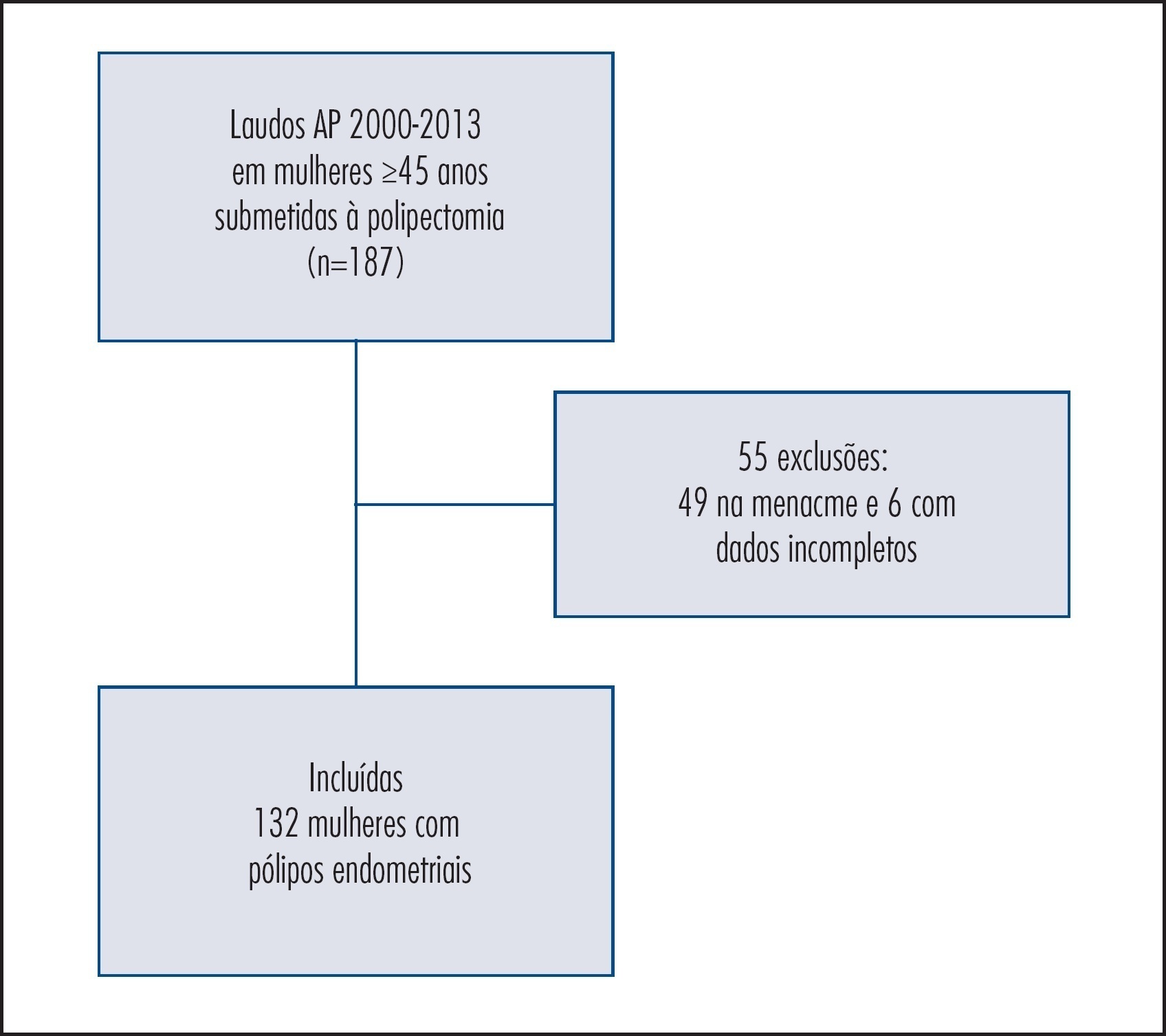
Observational cohort study with postmenopausal women who had been at a public university hospital. Clinical, anthropometrical, laboratorial, and ultrasonographic data of 132 patients with a histopathological diagnosis of endometrial polyps and 264 women without endometrial alterations (control) were compared in order to evaluate the predictive factors of endometrial polyps. Women with amenorrhea ≥12 months and ≥45 years of age were included in the study at a proportion of 1 case for 2 controls. The Student's t, χ2, and logistic regression tests were used for statistical analysis – odds ratio (OR).
Patients with endometrial polyps were older and had been in menopause for a longer time compared to control (p<0.0001). The percentage of obese women with polyps (72.0%) was higher compared to the Control Group (39%; p<0.0001). The measurement of waist circumference was superior among patients with polyps (p=0.0001). We observed a higher incidence of diabetes, hypertension and dyslipidemia in patients with endometrial polyps (p<0.0001). According to the US National Cholesterol Education Program/Adult Treatment Panel III (NCEP/ATP III) criteria, 48.5% of women with polyps and 33.3% of the Control Group were classified as having metabolic syndrome (p=0.004). Analysis of risk for endometrial polyps formation showed higher chances of occurrence of the disorder in patients with: BMI≥25 kg/m2 (OR=4.6; 95%CI 2.1–10.0); glucose ≥100 mg/dL (OR=2.8; 95%CI 1.3–5.9); dyslipidemia (OR=7.0; 95%CI 3.7–13.3); diabetes (OR=2.5; 95%CI 1.0–6.3), and metabolic syndrome (OR=2.7; 95%CI 1.1–6.4) compared to the Control Group.
In postmenopausal women, obesity, dyslipidemia, hyperglycemia and presence of metabolic syndrome were predictive factors for the development of endometrial polyps.
Summary
Revista Brasileira de Ginecologia e Obstetrícia. 2014;36(11):489-496
DOI 10.1590/S0100-720320140005090
To evaluate the predictive clinical factors for the development of endometrial polyps in postmenopausal women.
Observational cohort study with postmenopausal women who had been at a public university hospital. Clinical, anthropometrical, laboratorial, and ultrasonographic data of 132 patients with a histopathological diagnosis of endometrial polyps and 264 women without endometrial alterations (control) were compared in order to evaluate the predictive factors of endometrial polyps. Women with amenorrhea ≥12 months and ≥45 years of age were included in the study at a proportion of 1 case for 2 controls. The Student's t, χ2, and logistic regression tests were used for statistical analysis – odds ratio (OR).
Patients with endometrial polyps were older and had been in menopause for a longer time compared to control (p<0.0001). The percentage of obese women with polyps (72.0%) was higher compared to the Control Group (39%; p<0.0001). The measurement of waist circumference was superior among patients with polyps (p=0.0001). We observed a higher incidence of diabetes, hypertension and dyslipidemia in patients with endometrial polyps (p<0.0001). According to the US National Cholesterol Education Program/Adult Treatment Panel III (NCEP/ATP III) criteria, 48.5% of women with polyps and 33.3% of the Control Group were classified as having metabolic syndrome (p=0.004). Analysis of risk for endometrial polyps formation showed higher chances of occurrence of the disorder in patients with: BMI≥25 kg/m2 (OR=4.6; 95%CI 2.1–10.0); glucose ≥100 mg/dL (OR=2.8; 95%CI 1.3–5.9); dyslipidemia (OR=7.0; 95%CI 3.7–13.3); diabetes (OR=2.5; 95%CI 1.0–6.3), and metabolic syndrome (OR=2.7; 95%CI 1.1–6.4) compared to the Control Group.
In postmenopausal women, obesity, dyslipidemia, hyperglycemia and presence of metabolic syndrome were predictive factors for the development of endometrial polyps.
Summary
Revista Brasileira de Ginecologia e Obstetrícia. 2013;35(6):243-248
DOI 10.1590/S0100-72032013000600002
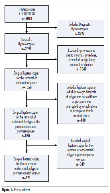
PURPOSE: To evaluate the accuracy of sonographic endometrial thickness and hysteroscopic characteristics in predicting malignancy in postmenopausal women undergoing surgical resection of endometrial polyps. METHODS: Five hundred twenty-one (521) postmenopausal women undergoing hysteroscopic resection of endometrial polyps between January 1998 and December 2008 were studied. For each value of sonographic endometrial thickness and polyp size on hysteroscopy, the sensitivity, specificity, positive predictive value (PPV) and negative predictive value (NPV) were calculated in relation to the histologic diagnosis of malignancy. The best values of sensitivity and specificity for the diagnosis of malignancy were determined by the Receiver Operating Characteristic (ROC) curve. RESULTS: Histologic diagnosis identified the presence of premalignancy or malignancy in 4.1% of cases. Sonographic measurement revealed a greater endometrial thickness in cases of malignant polyps when compared to benign and premalignant polyps. On surgical hysteroscopy, malignant endometrial polyps were also larger. An endometrial thickness of 13 mm showed a sensitivity of 69.6%, specificity of 68.5%, PPV of 9.3%, and NPV of 98% in predicting malignancy in endometrial polyps. Polyp measurement by hysteroscopy showed that for polyps 30 mm in size, the sensitivity was 47.8%, specificity was 66.1%, PPV was 6.1%, and NPV was 96.5% for predicting cancer. CONCLUSIONS: Sonographic endometrial thickness showed a higher level of accuracy than hysteroscopic measurement in predicting malignancy in endometrial polyps. Despite this, both techniques showed low accuracy for predicting malignancy in endometrial polyps in postmenopausal women. In suspected cases, histologic evaluation is necessary to exclude malignancy.
Summary
Revista Brasileira de Ginecologia e Obstetrícia. 2013;35(6):243-248
DOI 10.1590/S0100-72032013000600002
PURPOSE: To evaluate the accuracy of sonographic endometrial thickness and hysteroscopic characteristics in predicting malignancy in postmenopausal women undergoing surgical resection of endometrial polyps. METHODS: Five hundred twenty-one (521) postmenopausal women undergoing hysteroscopic resection of endometrial polyps between January 1998 and December 2008 were studied. For each value of sonographic endometrial thickness and polyp size on hysteroscopy, the sensitivity, specificity, positive predictive value (PPV) and negative predictive value (NPV) were calculated in relation to the histologic diagnosis of malignancy. The best values of sensitivity and specificity for the diagnosis of malignancy were determined by the Receiver Operating Characteristic (ROC) curve. RESULTS: Histologic diagnosis identified the presence of premalignancy or malignancy in 4.1% of cases. Sonographic measurement revealed a greater endometrial thickness in cases of malignant polyps when compared to benign and premalignant polyps. On surgical hysteroscopy, malignant endometrial polyps were also larger. An endometrial thickness of 13 mm showed a sensitivity of 69.6%, specificity of 68.5%, PPV of 9.3%, and NPV of 98% in predicting malignancy in endometrial polyps. Polyp measurement by hysteroscopy showed that for polyps 30 mm in size, the sensitivity was 47.8%, specificity was 66.1%, PPV was 6.1%, and NPV was 96.5% for predicting cancer. CONCLUSIONS: Sonographic endometrial thickness showed a higher level of accuracy than hysteroscopic measurement in predicting malignancy in endometrial polyps. Despite this, both techniques showed low accuracy for predicting malignancy in endometrial polyps in postmenopausal women. In suspected cases, histologic evaluation is necessary to exclude malignancy.
Summary
Revista Brasileira de Ginecologia e Obstetrícia. 2013;35(1):05-09
DOI 10.1590/S0100-72032013000100002
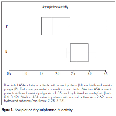
PURPOSE: To assess if arylsulfatase A activity (ASA) and sulfatide (SL) concentration in the human endometrium can be predictive of the development of endometrial polyps over the years, since ASA activity reflects the endometrial sensitivity to hormones. METHODS: ASA activity and SL concentration were determined by biochemical procedures on endometrial samples collected between 1990 and 1994 in non-menopausal women. These women underwent a new endometrial sampling following the clinical indication some years after the first endometrial sampling. The histological assessment of the second endometrial specimens found four patients with normal endometrial pattern and 10 patients with one or more endometrial polyps. ASA activity/years elapsed and SL concentration/years elapsed were compared using two tailed Mann-Whitney test for unpaired data between patients with normal pattern and patients with endometrial polyps. RESULTS: Median ASA activities were 2.62 (normal pattern) versus 1.85 (endometrial polyps) nmol hydrolized substrate/min. Median activity/years elapsed is higher in patients with second endometrial sample presenting normal pattern (p=0.006) and median SL concentration/years elapsed does not differ significantly among groups, even if median SL concentration seems to be higher in patients who subsequently developed polyps (1031 µg/g of fresh tissue versus 341,5 µg/g of fresh tissue). CONCLUSIONS: ASA activity can predict the onset of endometrial polyps over the years.
Summary
Revista Brasileira de Ginecologia e Obstetrícia. 2013;35(1):05-09
DOI 10.1590/S0100-72032013000100002
PURPOSE: To assess if arylsulfatase A activity (ASA) and sulfatide (SL) concentration in the human endometrium can be predictive of the development of endometrial polyps over the years, since ASA activity reflects the endometrial sensitivity to hormones. METHODS: ASA activity and SL concentration were determined by biochemical procedures on endometrial samples collected between 1990 and 1994 in non-menopausal women. These women underwent a new endometrial sampling following the clinical indication some years after the first endometrial sampling. The histological assessment of the second endometrial specimens found four patients with normal endometrial pattern and 10 patients with one or more endometrial polyps. ASA activity/years elapsed and SL concentration/years elapsed were compared using two tailed Mann-Whitney test for unpaired data between patients with normal pattern and patients with endometrial polyps. RESULTS: Median ASA activities were 2.62 (normal pattern) versus 1.85 (endometrial polyps) nmol hydrolized substrate/min. Median activity/years elapsed is higher in patients with second endometrial sample presenting normal pattern (p=0.006) and median SL concentration/years elapsed does not differ significantly among groups, even if median SL concentration seems to be higher in patients who subsequently developed polyps (1031 µg/g of fresh tissue versus 341,5 µg/g of fresh tissue). CONCLUSIONS: ASA activity can predict the onset of endometrial polyps over the years.
Summary
Revista Brasileira de Ginecologia e Obstetrícia. 2012;34(3):122-127
DOI 10.1590/S0100-72032012000300006
PURPOSE: To compare the diagnostic accuracy of sonohysterography (HSN) and conventional transvaginal ultrasound (USG) in assessing the uterine cavity of infertile women candidate to assisted reproduction techniques (ART). METHODS: Comparative cross-sectional study with 120 infertile women candidate to ART, assisted at Centro de Reprodução Assistida (CRA) of Hospital Regional da Asa Sul (HRAS), Brasília - DF, from August 2009 to November 2010. Sonohysterography was performed with saline solution infusion in a close system. The sonohysterography finding was compared to previous USG results. The uterine cavity was considered abnormal when the endometrium was found to be thicker than expected during the menstrual cycle and when an endometrial polyp, a submucous myoma and an abnormal shape of the uterine cavity were observed. The statistical analysis was done using absolute frequencies, percentage values and the χ², with the level of significance set at 5%. RESULTS: HSN revealed that 92 (76.7%) infertile women candidate to ART had a normal uterine cavity, while 28 (23.3%) had the following abnormalities: 15 polyps (12.5%), 9 cases of abnormal shape of the uterine cavity (7.5%), 6 submucous myomas (5%), 4 cases of inadequate endometrial thickness for the menstrual cycle phase (3.3%), and 2 cases of uterine septum (1.7%); 5 women presented more than one abnormality (4.2%). While USG showed alteration in the cavity only in 5 (4.2%) women, the sonohysterography confirmed 4 out of the 5 abnormalities shown by USG and detected an abnormal uterine cavity in 24 other women, who had not been detected by USG. This means that sonohysterography was able to detect more abnormalities in the uterine cavity than USG, with a statistically significant difference (p=0.002). CONCLUSION: The sonohysterography was more accurate than USG in the assessment of the uterine cavity of this cohort of infertile women candidate to ART. The sonohysterography can be easily incorporated into the investigation of these women and contribute to reducing embryo implantation failures.
Summary
Revista Brasileira de Ginecologia e Obstetrícia. 2010;32(8):393-397
DOI 10.1590/S0100-72032010000800006
PURPOSE: to describe hysteroscopy findings in infertile patients. METHODS: this was a retrospective series of 953 patients with diagnosis of infertility evaluated by hysteroscopy. A total of 957 patients investigated for infertility were subjected to hysteroscopy, preferentially during the first phase of the menstrual cycle. When necessary, directed biopsies (under direct visualization during the exam) or guided biopsies were obtained using a Novak curette after defining the site to be biopsied during the hysteroscopic examination. Outcome frequencies were determined as percentages, and the χ2 test was used for the correlations. The statistical software EpiInfo 2000 (CDC) was used for data analysis. RESULTS: a normal uterine cavity was detected in 436 cases (45.8%). This was the most frequent diagnosis for women with primary infertility and for women with one or no abortion (p<0.05). Abnormal findings were obtained in 517 of 953 cases (54.2%), including intrauterine synechiae in 185 patients (19.4%), endometrial polyps in 115 (12.1%), endocervical polyps in 66 (6.0%), submucosal myomas in 47 (4.9%), endometrial hyperplasia in 39 (4.1%), adenomyosis in five (0.5%), endometritis (with histopathological confirmation) in four (0.4%), endometrial bone metaplasia in two (0.4%), and cancer of the endometrium in one case (0.1%). Morphological and functional changes of the uterus were detected in 5.6% of the cases, including uterine malformations in 32 (3.4%) and isthmus-cervical incompetence in 21 (2.2%). CONCLUSIONS: intrauterine synechiae were the most frequent abnormal findings in patients evaluated for infertility. Patients with a history of abortion and infertility should be submitted to hysteroscopy in order to rule out intrauterine synechiae as a possible cause of infertility.
Summary
Revista Brasileira de Ginecologia e Obstetrícia. 2010;32(7):327-333
DOI 10.1590/S0100-72032010000700004
PURPOSE: to evaluate the clinical and epidemiological risk factors for endometrial cancer in postmenopausal women with endometrial polyps, as well as the genetic polymorphism of the progesterone receptor (PROGINS). METHODS: a case-control study was designed with 160 postmenopausal women with endometrial polyps, compared to a normal Control Group of 400 postmenopausal women. The genotyping of PROGINS polymorphism was determined by the polymerase chain reaction. Clinical and epidemiological data were compared between benign endometrial polyps and 118 of the control subjects. Variables were also compared with regard to benign and malignant endometrial polyps. RESULTS: comparison of the epidemiological variables between groups showed a significant difference for age, ethnicity, time since menopause, parity, tamoxifen use, hypertension and breast cancer, all of them more prevalent in the polyp group. After adjustment for age, statistical significance remained only for parity (OR=1.1), hypertension (OR=2.2) and breast cancer (OR=14.4). There were six cases of malignant polyps (3.7%). The frequency of bleeding was 23.4% for benign polyps and 100% for malignant polyps, with large polyps being detected in 54.6% of the benign cases and in 100 of the malignnat ones. The frequency of arterial hypertension was 54.5% for benign polyps and 83.3% for the malignant ones. The frequency of PROGINS T1/T1, T1/T2 and T2/T2 polymorphism was 79.9%, 19.5% and 0.6%, respectively, for the polyp group, and 78.8%, 20.8% and 0.5% for the Control Group. CONCLUSIONS: elderly age, hypertension, and breast cancer were significantly associated with endometrial polyps. The presence of PROGINS polymorphism was not significantly associated with endometrial polyps. The incidence of malignant polyps was low and strongly associated with bleeding, large-sized polyp and arterial hypertension.
Summary
Revista Brasileira de Ginecologia e Obstetrícia. 2009;31(3):131-137
DOI 10.1590/S0100-72032009000300005
PURPOSE: to evaluate the effects of tamoxifen on the expression of TGF-β and p27 proteins in polyps and adjacent endometrium of women after menopause. METHODS: prospective study with 30 post-menopausal women with diagnosis of breast cancer, taking tamoxifen (20 mg/day), presenting diagnosis of suspect endometrial polyps through transvaginal ultrasonography, and submitted to diagnostic and surgical hysterectomy to withdraw the polyps and adjacent endometrium. A immunohistochemical study has been done to verify the expression of the TGF-β and p27 proteins in the polyps and adjacent endometrium. These proteins' quantification has been done by morphometry. RESULTS: the patients' average age was 61.7 years old; their average age at the menopause onset was 49.5; and the average of using tamoxifen was 25.3 months. The average concentration of positive cells for TGF-β protein in the glandular and stroma polyp epithelium was 62.6±4.5 cells/mm². For the p27, in the glandular polyp epithelium, it was 24.2±18.6 cells/mm² and for the stroma, 19.2±15.2 cells/mm². There was no significant difference between the expression of TGF-β and p27 in the glandular epithelial form the polyps and the adjacent endometrium. The expression of proteins in the polyp and adjacent endometrium with its respective glandular and stroma epithelium showed a significant difference for the p27 protein (r=0.9, p<0.05). CONCLUSIONS: we have concluded that the TGF-β expression is not related to the effect of tamoxifen on the growing of endometrial polyps, but the absence of polyps' malignization by tamoxifen may be explained by the high expression of p27 protein in its glandular epithelium.
Summary
Revista Brasileira de Ginecologia e Obstetrícia. 2006;28(1):18-23
DOI 10.1590/S0100-72032006000100004
PURPOSE: to characterize postmenopausal endometrial polyps and to determine risk for concomitant premalignant and malignant pathology. METHODS: a retrospective study including 82 postmenopausal women with a histological diagnosis of endometrial polyps who underwent hysteroscopic polypectomy, after a diagnosis of endometrial thickening made by transvaginal ultrasound, was performed. Medical reports provided clinical and gynecological history, data related to the operative hysteroscopy and definitive histological findings. RESULTS: among the 82 patients who underwent hysteroscopic polypectomy, 10.9% were receiving some type of hormonal therapy. Twenty-eight women (34.1%) reported abnormal vaginal bleeding. Single polyp was encountered in 56 women (68.3%), two polyps were found in 19 cases (23.2%) and in 7 cases (3.6%), three or more polyps were found. The definitive histopathologic analysis revealed 63 (76.8%) benign polyps, 17 (20.8%) hyperplastic polyps (10 cases 12.2% - of simple endometrial hyperplasia without cytologic atypia and 7 cases 8.6% - of complex endometrial hyperplasia without cytologic atypia). Two polyps (2.4%) were diagnosed as harboring neoplasia. For the statistical analysis we employed chi2 test improved by Yates. The authors correlated the polyps' histology with the occurrence of abnormal vaginal bleeding (p=0.0056), number of endometrial polyps (p=0.921) and time after menopause (p=0.720). CONCLUSIONS: endometrial polyps are commonly found entities in postmenopausal women, related with low frequency to endometrial hyperplasia or carcinomas and only histological evaluation seems to allow the exclusion of premalignant and malignant pathology.