Summary
Revista Brasileira de Ginecologia e Obstetrícia. 2008;30(5):241-247
DOI 10.1590/S0100-72032008000500006
PURPOSE: to evaluate the meiotic spindle and the chromosome distribution of in vitro matured oocytes obtained from stimulated cycles of infertile women with polycystic ovary syndrome (PCOS) and with male factor and/or tubal infertility (Control Group) and compare in vitro maturation (IVM) rates between the groups analyzed. METHODS: five infertile patients with PCOS and eight controls, submitted to stimulated cycles for intracytoplasmic sperm injection, were selected prospectively and consecutively, and respectively assigned to the study group and the Control Group. Immature oocytes (21 and 29, respectively, from PCOS and Control Group) were submitted to IVM. After IVM, oocytes with first polar body extruded were fixed and submitted to immunostaining and fluorescence microscopy for morphological evaluation of the spindle and of chromosome distribution. Statistical analysis was performed by the Fisher test with significance, when p<0.05. RESULTS: IVM rates were similar between groups (47.6 e 44.8%, respectively, for PCOS and Control Group). Six of the ten oocytes (60%) from the study group and four of the 12 oocytes (33.3%) from the Control Group presented meiotic anomalies of the spindle and/or anomalous chromosome distribution, without statistical difference between groups. CONCLUSIONS: data from the present study did not demonstrate significant difference neither in IVM rates nor in the proportions of meiotic anomalies between in vitro matured oocytes obtained from stimulated cycles from PCOS patients and control ones.
Summary
Revista Brasileira de Ginecologia e Obstetrícia. 2008;30(4):201-209
DOI 10.1590/S0100-72032008000400008
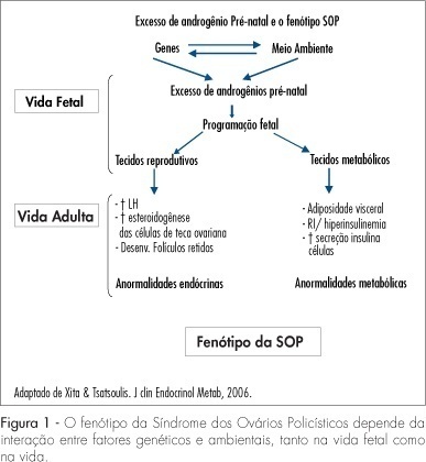
Polycystic ovary syndrome (PCOS) occurs in 6 to 10% of women during the reproductive age. Insulin resistance and compensatory hyperinsulinemia are currently two of the main factors involved in the etiopathogenesis of PCOS. The objective of the present review was to discuss the controversies related to the treatment of infertile women with PCOS and during their pregnancy, focusing on the European Society of Human Reproduction and Embryology (ESHRE) and the American Society for Reproductive Medicine (ASRM) current consensus.
Summary
Revista Brasileira de Ginecologia e Obstetrícia. 2008;30(4):201-209
DOI 10.1590/S0100-72032008000400008
Polycystic ovary syndrome (PCOS) occurs in 6 to 10% of women during the reproductive age. Insulin resistance and compensatory hyperinsulinemia are currently two of the main factors involved in the etiopathogenesis of PCOS. The objective of the present review was to discuss the controversies related to the treatment of infertile women with PCOS and during their pregnancy, focusing on the European Society of Human Reproduction and Embryology (ESHRE) and the American Society for Reproductive Medicine (ASRM) current consensus.
Summary
Revista Brasileira de Ginecologia e Obstetrícia. 2007;29(5):241-247
DOI 10.1590/S0100-72032007000500004
PURPOSE: to evaluate the ultra-sensitive C-Reactive Protein level (us-CRP) in patients with Polycystic Ovary Syndrome (PCOS), and the correlation of clinical and laboratory parameters with the us-CRP level. Methods: in this cross-sectional study, 46 women with Polycystic Ovary Syndrome, according to the Rotterdam criteria, and 44 control women have been included. Serum was analyzed for C reactive protein (CRP) levels. Body mass index (BMI), age, circumference waist, HOMA-IR, total, low and high density lipoprotein cholesterol, triglycerides, glucose, testosterone and insulin levels were correlated to CRP level through a linear regression model. RESULTS: PCOS patients not only were older and had higher BMI, but their waist circumference, fasting insulin, HOMA-IR, total and LDL cholesterol were also higher, as compared to the women from the control group. A significant difference was observed in the us-CRP level between the PCOS (2.7 mg/dL±2.17) the control (1.6 mg/dL±1.49) groups. When us-CRP levels were categorized as of low (<1.0 mg/L), moderate (1-3.0 mg/L) and high (3.0 mg/L) risk for cardiovascular episodes, only 28.3% women with PCOS had us-CRP levels defined as low, 34.8% as moderate and 37% as high risk. The prevalence of Metabolic Syndrome was higher in the women with PCOS (30.4%) than in the women from the control group (6.8%). Through a stepwise linear regression model, only waist circumference, presence of metabolic syndrome and age had a confounding effect in the relation between us-CRP and PCOS. After adjustment for confounding factors, PCOS showed an independent effect on us-CRP level. CONCLUSIONS: the us-CRP levels were higher in the PCOS women than in the healthy controls. By a regression model, PCOS showed an independent effect on us-CRP level.
Summary
Revista Brasileira de Ginecologia e Obstetrícia. 2007;29(3):141-146
DOI 10.1590/S0100-72032007000300005
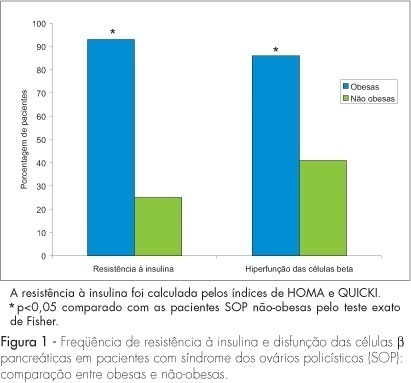
PURPOSE: to evaluate the effect of obesity on beta-cell function in patients with polycystic ovary syndrome (PCOS). METHODS: this cross-section study evaluated 82 patients with PCOS selected consecutively, at the moment of the diagnosis. We compared 31 PCOS obese women (BMI >30 kg/m²) to 51 age-matched PCOS nonobese patients (BMI <30 kg/m²). Using fasting glucose and insulin levels, homeostatic model assessment values for insulin resistance (HOMA-IR and QUICKI) and percent beta-cell function (HOMA-%beta-cell) were calculated. As secondary variables, the age at PCOS diagnosis, age of menarche, hormonal levels (testosterone, prolactin, FSH and LH), total cholesterol, triglycerides, HDL cholesterol and LDL cholesterol were also analyzed. RESULTS: menarche was significantly earlier in obese PCOS patients (11.7±1.8 years) than in nonobese patients (12.67±1.86 years) (p<0.05). Obese patients presented lower LH levels (7.9±5 mIU/mL) than did nonobese patients (10.6±6 mIU/mL) (p<0.05). Both groups presented mean HDL cholesterol levels below 50 mg/dL. Obese patients presented significantly higher baseline insulin levels (32.5±25.2 mIU/mL) and fasting blood glucose levels (115.9±40.7 mg/dL) than did nonobese patients (8.8±6.6 mIU/mL and 90.2±8.9 mg/dL, respectively) (p<0.01). Of the obese PCOS patients, 93% presented insulin resistance versus 25% of nonobese PCOS patients (p<0.01). Eighty-six perecent of the obese women had hyperfunction of beta-cell versus 41% of nonobese with PCOS (p<0.0001). CONCLUSIONS: obese PCOS patients presented higher prevalence of insulin resistance and hyperfunction of beta-cell than did nonobese PCOS patients.
Summary
Revista Brasileira de Ginecologia e Obstetrícia. 2007;29(3):141-146
DOI 10.1590/S0100-72032007000300005
PURPOSE: to evaluate the effect of obesity on beta-cell function in patients with polycystic ovary syndrome (PCOS). METHODS: this cross-section study evaluated 82 patients with PCOS selected consecutively, at the moment of the diagnosis. We compared 31 PCOS obese women (BMI >30 kg/m²) to 51 age-matched PCOS nonobese patients (BMI <30 kg/m²). Using fasting glucose and insulin levels, homeostatic model assessment values for insulin resistance (HOMA-IR and QUICKI) and percent beta-cell function (HOMA-%beta-cell) were calculated. As secondary variables, the age at PCOS diagnosis, age of menarche, hormonal levels (testosterone, prolactin, FSH and LH), total cholesterol, triglycerides, HDL cholesterol and LDL cholesterol were also analyzed. RESULTS: menarche was significantly earlier in obese PCOS patients (11.7±1.8 years) than in nonobese patients (12.67±1.86 years) (p<0.05). Obese patients presented lower LH levels (7.9±5 mIU/mL) than did nonobese patients (10.6±6 mIU/mL) (p<0.05). Both groups presented mean HDL cholesterol levels below 50 mg/dL. Obese patients presented significantly higher baseline insulin levels (32.5±25.2 mIU/mL) and fasting blood glucose levels (115.9±40.7 mg/dL) than did nonobese patients (8.8±6.6 mIU/mL and 90.2±8.9 mg/dL, respectively) (p<0.01). Of the obese PCOS patients, 93% presented insulin resistance versus 25% of nonobese PCOS patients (p<0.01). Eighty-six perecent of the obese women had hyperfunction of beta-cell versus 41% of nonobese with PCOS (p<0.0001). CONCLUSIONS: obese PCOS patients presented higher prevalence of insulin resistance and hyperfunction of beta-cell than did nonobese PCOS patients.
Summary
Revista Brasileira de Ginecologia e Obstetrícia. 2007;29(1):10-17
DOI 10.1590/S0100-72032007000100003
PURPOSE: to evaluate the prevalence of metabolic syndrome in women with polycystic ovary syndrome (PCOS). METHODS: forty six women with PCOS, in accord with Rotterdam criteria (2003), and 44 women with regular menses, without any clinical or laboratorial hyperandrogenism features, and no ultrasonographic ovarian microcysts (control group) were evaluated. For metabolic syndrome, the National Cholesterol Education Program (NCEP, 2002) and the International Diabetes Federation (IDF, 2005) guidelines were considered. RESULTS: the prevalence of metabolic syndrome were 30.4% (NCEP) and 32.6% (IDF) for the women with PCOS, nearly 4-fold higher than that reported for the control group (p<0.004), which were 6.8% (NCEP) and 9.1% (IDF). Women with PCOS had persistently higher prevalence rates of the metabolic syndrome, regardless of matched age and body mass index. The most prevalent factor of the metabolic syndrome among the PCOS subjects was low serum HDL cholesterol which was below 50 mg/dl (52.2%). Waist circumference above 88 cm (47.8%), blood pressure above 130/85 mmHg and fasting glycemia above 110 mg/dl (4.3%) were significantly more frequent among women with PCOS than among control women. CONCLUSIONS: the metabolic syndrome is significantly more frequent in women with PCOS, placing them at higher risk for cardiovascular disease.
Summary
Revista Brasileira de Ginecologia e Obstetrícia. 2000;22(2):89-94
DOI 10.1590/S0100-72032000000200005
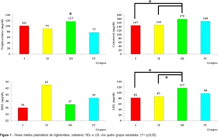
Purpose: to evaluate the lipid profile (cholesterol, triglycerides, HDL and LDL) of women with polycystic ovary syndrome (PCO) and compare it to that of women with ovulatory menstrual cycles. Methods: the patients were divided into two groups, obese and nonobese, based on body mass index, so that it would be possible to determine the joint effect of PCO and obesity on the lipid metabolism of the studied women. We studied 117 women divided into 4 groups: group I (PCO--obese), n = 33; group II (PCO--nonobese), n = 27; group III (control--obese), n = 28; group IV (control--nonobese), n = 29. Results: cholesterol levels were elevated (179 mg/dl) in obese patients with ovulatory cycles (group III) compared to group I (147 mg/dl) and group II (149 mg/dl), as also were triglyceride levels (117 mg/dl) compared to group IV (77 mg/dl) and LDL levels (117 mg/dl) compared to group I (82 mg/dl). Conclusion: these data suggest that alterations in lipid profile are related to obesity only.
Summary
Revista Brasileira de Ginecologia e Obstetrícia. 2000;22(2):89-94
DOI 10.1590/S0100-72032000000200005
Purpose: to evaluate the lipid profile (cholesterol, triglycerides, HDL and LDL) of women with polycystic ovary syndrome (PCO) and compare it to that of women with ovulatory menstrual cycles. Methods: the patients were divided into two groups, obese and nonobese, based on body mass index, so that it would be possible to determine the joint effect of PCO and obesity on the lipid metabolism of the studied women. We studied 117 women divided into 4 groups: group I (PCO--obese), n = 33; group II (PCO--nonobese), n = 27; group III (control--obese), n = 28; group IV (control--nonobese), n = 29. Results: cholesterol levels were elevated (179 mg/dl) in obese patients with ovulatory cycles (group III) compared to group I (147 mg/dl) and group II (149 mg/dl), as also were triglyceride levels (117 mg/dl) compared to group IV (77 mg/dl) and LDL levels (117 mg/dl) compared to group I (82 mg/dl). Conclusion: these data suggest that alterations in lipid profile are related to obesity only.
Summary
Revista Brasileira de Ginecologia e Obstetrícia. 2004;26(9):727-733
DOI 10.1590/S0100-72032004000900009
PURPOSE: to evaluate the results of ovulation hyperinduction followed by in vitro fertilization (IVF) in women with polycystic ovary syndrome (POS), as compared to normal cycle women. METHODS: a controlled retrospective study conducted on 36 women with POS (POS group) and on 44 women with infertility due to mild male factor (control group), submitted to IVF from 1997 to 2003. Subject ages ranged from 18 to 36 years. Ovulation hyperinduction was obtained with recombinant follicle-stimulating hormone and a gonadotrophin-releasing hormone agonist. The analyzed variables were the follicles with a mean diameter of 14 to 17 mm and the follicles with diameters of 18 mm or above on the day of human chorionic gonadotrophin administration, percentage of follicles >18 mm, the number of retrieved oocytes, fertilization rate, cleavage rate, incidence of ovarian hyperstimulation syndrome (OHS), clinical pregnancy rate, and abortion rate. The variables were analyzed by the unpaired t test, Fisher exact test and Mann-Whitney test, with level of significance set at p<0.05. RESULTS: the POS group presented a larger number of retrieved follicles, most of them measuring 14 to 17 mm in diameter, compared to the control group (64.8 vs 53.9%), a lower fertilization rate (59.43 vs 79.57%) and a higher incidence of OHS (38.9 vs 9.1%). The number of retrieved oocytes, cleavage rates, pregnancy rates per embryo transfer, abortion rates and live born rates did not differ between groups. CONCLUSION: the success of IVF is impaired in women with POS due to their larger number of retrieved follicles of reduced diameter, reduced fertilization rate and high OHS rates.
Summary
Revista Brasileira de Ginecologia e Obstetrícia. 2001;23(5):307-312
DOI 10.1590/S0100-72032001000500006
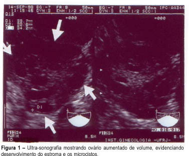
Purpose: to evaluate the effectiveness of color Doppler as a diagnosis method for polycystic ovary syndrome (PCOS) through blood flow variations in the ovarian stroma, in the uterine arteries and in the subendometrial tissue. Methods: thirty patients divided into two groups were selected: fifteen patients with amenorrhea or oligomenorrhea, hirsutism (Ferriman and Gallwey score >8), body mass index >25 kg/m² and echographic examination identifying increased hyperechogenic stromal and ovarian polycystosis (study group), and an identical number of patients presenting normal menstrual cycles, with no signs of hirsutism and with normal ultrasonography (control group). Transvaginal Doppler flowmetry measured systolic peak velocity or maximal velocity (Vmax) pulsatility index (PI) and resistance of ovarian stromal vessels, uterine arteries and subendometrial layer. Results: Doppler velocimetry showed significantly higher Vmax layer (p<=0,0004) in the ovarian stromal of patients with PCOS (12.2 cm/s) when compared to the control group (8.05 cm/s); the uterine artery PI was also higher in the PCOS group (3.3 cm/s) versus the control group (2.7 cm/s); other Doppler velocimetry parameters did not show significant differences. As we established a cutoff = 9 cm/s for the sample for Vmax, we obtained the percentages of 95.2 for sensitivity, 80.0 for specificity, 83.3 for positive predictive value and 94.1 for negative predictive value. Conclusion: Doppler velocimetry might constitute an additional tool to be incorporated in clinical and ultrasonographic investigation concerning the PCOS diagnosis.
Summary
Revista Brasileira de Ginecologia e Obstetrícia. 2001;23(5):307-312
DOI 10.1590/S0100-72032001000500006
Purpose: to evaluate the effectiveness of color Doppler as a diagnosis method for polycystic ovary syndrome (PCOS) through blood flow variations in the ovarian stroma, in the uterine arteries and in the subendometrial tissue. Methods: thirty patients divided into two groups were selected: fifteen patients with amenorrhea or oligomenorrhea, hirsutism (Ferriman and Gallwey score >8), body mass index >25 kg/m² and echographic examination identifying increased hyperechogenic stromal and ovarian polycystosis (study group), and an identical number of patients presenting normal menstrual cycles, with no signs of hirsutism and with normal ultrasonography (control group). Transvaginal Doppler flowmetry measured systolic peak velocity or maximal velocity (Vmax) pulsatility index (PI) and resistance of ovarian stromal vessels, uterine arteries and subendometrial layer. Results: Doppler velocimetry showed significantly higher Vmax layer (p<=0,0004) in the ovarian stromal of patients with PCOS (12.2 cm/s) when compared to the control group (8.05 cm/s); the uterine artery PI was also higher in the PCOS group (3.3 cm/s) versus the control group (2.7 cm/s); other Doppler velocimetry parameters did not show significant differences. As we established a cutoff = 9 cm/s for the sample for Vmax, we obtained the percentages of 95.2 for sensitivity, 80.0 for specificity, 83.3 for positive predictive value and 94.1 for negative predictive value. Conclusion: Doppler velocimetry might constitute an additional tool to be incorporated in clinical and ultrasonographic investigation concerning the PCOS diagnosis.