Summary
. ;:307-312
DOI 10.1590/S0100-72032001000500006
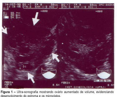
Purpose: to evaluate the effectiveness of color Doppler as a diagnosis method for polycystic ovary syndrome (PCOS) through blood flow variations in the ovarian stroma, in the uterine arteries and in the subendometrial tissue. Methods: thirty patients divided into two groups were selected: fifteen patients with amenorrhea or oligomenorrhea, hirsutism (Ferriman and Gallwey score >8), body mass index >25 kg/m² and echographic examination identifying increased hyperechogenic stromal and ovarian polycystosis (study group), and an identical number of patients presenting normal menstrual cycles, with no signs of hirsutism and with normal ultrasonography (control group). Transvaginal Doppler flowmetry measured systolic peak velocity or maximal velocity (Vmax) pulsatility index (PI) and resistance of ovarian stromal vessels, uterine arteries and subendometrial layer. Results: Doppler velocimetry showed significantly higher Vmax layer (p<=0,0004) in the ovarian stromal of patients with PCOS (12.2 cm/s) when compared to the control group (8.05 cm/s); the uterine artery PI was also higher in the PCOS group (3.3 cm/s) versus the control group (2.7 cm/s); other Doppler velocimetry parameters did not show significant differences. As we established a cutoff = 9 cm/s for the sample for Vmax, we obtained the percentages of 95.2 for sensitivity, 80.0 for specificity, 83.3 for positive predictive value and 94.1 for negative predictive value. Conclusion: Doppler velocimetry might constitute an additional tool to be incorporated in clinical and ultrasonographic investigation concerning the PCOS diagnosis.

Summary
. ;:481-488
DOI 10.1590/S0100-72032001000800002
Purpose: to investigate leptin levels in patients with polycystic ovary syndrome (PCOS), and relationships with testosterone, estradiol, follicle-stimulating hormone (FSH) and insulin levels. Methods: transversal study on 40 patients with PCOS divided into two groups: Group I (n = 20)- obese women (body mass index - BMI > or = 28 kg/m²), and Group II (n = 20) - non obese women (BMI <28 kg/m²). Results: BMI was different between the two groups (p=0.04). We observed that leptin concentrations were significantly correlated with BMI (p<0.001). After adjusting for BMI, no correlation between leptin, insulin (p=0.194), FSH (p=0.793), and total (p=0.441) and free (p=0.422) testosterone was found. However, we only observed positive correlations between leptin and estradiol (p=0.043). Conclusions: there is a strong correlation between leptin levels, BMI and estradiol levels in women with PCOS.