-
Original Article
Effect of Testosterone on Proliferation Markers and Apoptosis in Breasts of Ovariectomized Rats
- Jussara Celi Conceição Oliveira,
- Marcelo Luis Steiner,
- Thérèse Rachell Theodoro,
- Ana Maria Amaral Antonio Mader,
- Giuliana Petri, [ ... ],
- Luciano Melo Pompei

02-03-2019
Summary
Original ArticleEffect of Testosterone on Proliferation Markers and Apoptosis in Breasts of Ovariectomized Rats
Revista Brasileira de Ginecologia e Obstetrícia. 2019;41(12):703-709
02-03-2019- Jussara Celi Conceição Oliveira,
- Marcelo Luis Steiner,
- Thérèse Rachell Theodoro,
- Ana Maria Amaral Antonio Mader,
- Giuliana Petri,
- Luiz Carlos Abreu,
- Maria Aparecida da Silva Pinhal,
- César Eduardo Fernandes,
- Luciano Melo Pompei
Views167See moreAbstract
Objective
To investigate the action of testosterone (T), isolated or associated with estradiol benzoate (EB), on the proliferation markers and apoptosis of breasts of ovariectomized rats.
Methods
A total of 48 castrated female Wistar rats were divided into 6 groups, and each of them were submitted to one of the following treatments for 5 weeks: 1) control; 2) EB 50 mcg/day + T 50 mcg/day; 3) T 50mcg/day; 4) EB 50 mcg +T 300 mcg/day; 5) T 300 mcg/day; and 6) EB 50 mcg/day. After the treatment, the mammary tissue was submitted to a histological analysis and immunoexpression evaluation of proliferation markers (proliferating cell nuclear antigen, PCNA) and apoptosis (caspase-3).
Results
There was a statistically significant difference among the groups regarding microcalcifications and secretory activity, with higher prevalence in the groups treated with EB. There was no difference among the groups regarding atrophy, but a higher prevalence of atrophy was found in the groups that received T versus those that received EB +T. There was a difference among the groups regarding the PCNA (p = 0.028), with higher expression in the group submitted to EB +T 300 mcg/day. Regarding caspase-3, there was no difference among the groups; however, in the group submitted to EB +T 300 mcg/day, the expression was higher than in the isolated T group.
Conclusion
Isolated T did not have a proliferative effect on the mammary tissue, contrary to EB. Testosterone in combination with EB may or may not decrease the proliferation, depending on the dose of T.
Views167

This is an Open Access article distributed under the terms of the Creative Commons Attribution License, which permits unrestricted use, distribution, and reproduction in any medium, provided the original work is properly cited. Summary
Original ArticleEffect of Testosterone on Proliferation Markers and Apoptosis in Breasts of Ovariectomized Rats
Revista Brasileira de Ginecologia e Obstetrícia. 2019;41(12):703-709
02-03-2019- Jussara Celi Conceição Oliveira,
- Marcelo Luis Steiner,
- Thérèse Rachell Theodoro,
- Ana Maria Amaral Antonio Mader,
- Giuliana Petri,
- Luiz Carlos Abreu,
- Maria Aparecida da Silva Pinhal,
- César Eduardo Fernandes,
- Luciano Melo Pompei
Views167See moreAbstract
Objective
To investigate the action of testosterone (T), isolated or associated with estradiol benzoate (EB), on the proliferation markers and apoptosis of breasts of ovariectomized rats.
Methods
A total of 48 castrated female Wistar rats were divided into 6 groups, and each of them were submitted to one of the following treatments for 5 weeks: 1) control; 2) EB 50 mcg/day + T 50 mcg/day; 3) T 50mcg/day; 4) EB 50 mcg +T 300 mcg/day; 5) T 300 mcg/day; and 6) EB 50 mcg/day. After the treatment, the mammary tissue was submitted to a histological analysis and immunoexpression evaluation of proliferation markers (proliferating cell nuclear antigen, PCNA) and apoptosis (caspase-3).
Results
There was a statistically significant difference among the groups regarding microcalcifications and secretory activity, with higher prevalence in the groups treated with EB. There was no difference among the groups regarding atrophy, but a higher prevalence of atrophy was found in the groups that received T versus those that received EB +T. There was a difference among the groups regarding the PCNA (p = 0.028), with higher expression in the group submitted to EB +T 300 mcg/day. Regarding caspase-3, there was no difference among the groups; however, in the group submitted to EB +T 300 mcg/day, the expression was higher than in the isolated T group.
Conclusion
Isolated T did not have a proliferative effect on the mammary tissue, contrary to EB. Testosterone in combination with EB may or may not decrease the proliferation, depending on the dose of T.
This is an Open Access article distributed under the terms of the Creative Commons Attribution License, which permits unrestricted use, distribution, and reproduction in any medium, provided the original work is properly cited. -
Original Article
A Long-term Estrogen Deficiency in Ovariectomized Mice is Associated with Disturbances in Fatty Acid Oxidation and Oxidative Stress
Revista Brasileira de Ginecologia e Obstetrícia. 2018;40(5):251-259
05-01-2018
Summary
Original ArticleA Long-term Estrogen Deficiency in Ovariectomized Mice is Associated with Disturbances in Fatty Acid Oxidation and Oxidative Stress
Revista Brasileira de Ginecologia e Obstetrícia. 2018;40(5):251-259
05-01-2018Views175See moreAbstract
Objective
The aim of this work was to evaluate the changes caused by estrogen deficiency in lipid metabolism.
Methods
This study encompassed direct measurements of plasma biochemical analyses, liver lipid contents, and assessments of the mitochondrial β-oxidation capacity as well as an evaluation of the liver redox status in an animal model of estrogen deficiency.
Results
When compared with control mice, the livers of ovariectomized (OVX) mice presented considerable accretions in their lipid contents, which were accompanied by increased levels of lipid peroxidation in liver homogenates andmitochondria from OVX groups and decreased reduced glutathione (GSH) contents. In isolated mitochondria, estrogen deficiency inhibited mitochondrial β-oxidation of fatty acids irrespective of their chain length. The liver mitochondrial and peroxisomal H2O2 generations in OVX mice were increased. Additionally, the activities of all antioxidant enzymes assessed were decreased.
Conclusion
These data provide one potential explanation for the increased susceptibility to metabolic diseases observed after menopause.
Views175This is an Open Access article distributed under the terms of the Creative Commons Attribution License, which permits unrestricted use, distribution, and reproduction in any medium, provided the original work is properly cited. Summary
Original ArticleA Long-term Estrogen Deficiency in Ovariectomized Mice is Associated with Disturbances in Fatty Acid Oxidation and Oxidative Stress
Revista Brasileira de Ginecologia e Obstetrícia. 2018;40(5):251-259
05-01-2018Views175See moreAbstract
Objective
The aim of this work was to evaluate the changes caused by estrogen deficiency in lipid metabolism.
Methods
This study encompassed direct measurements of plasma biochemical analyses, liver lipid contents, and assessments of the mitochondrial β-oxidation capacity as well as an evaluation of the liver redox status in an animal model of estrogen deficiency.
Results
When compared with control mice, the livers of ovariectomized (OVX) mice presented considerable accretions in their lipid contents, which were accompanied by increased levels of lipid peroxidation in liver homogenates andmitochondria from OVX groups and decreased reduced glutathione (GSH) contents. In isolated mitochondria, estrogen deficiency inhibited mitochondrial β-oxidation of fatty acids irrespective of their chain length. The liver mitochondrial and peroxisomal H2O2 generations in OVX mice were increased. Additionally, the activities of all antioxidant enzymes assessed were decreased.
Conclusion
These data provide one potential explanation for the increased susceptibility to metabolic diseases observed after menopause.
This is an Open Access article distributed under the terms of the Creative Commons Attribution License, which permits unrestricted use, distribution, and reproduction in any medium, provided the original work is properly cited. -
Original Articles
Loss of Ovarian Function Results in Increased Loss of Skeletal Muscle in Arthritic Rats
Revista Brasileira de Ginecologia e Obstetrícia. 2016;38(2):56-64
02-01-2016
Summary
Original ArticlesLoss of Ovarian Function Results in Increased Loss of Skeletal Muscle in Arthritic Rats
Revista Brasileira de Ginecologia e Obstetrícia. 2016;38(2):56-64
02-01-2016Views144See moreObjective
We studied the effects of loss of ovarian function (ovariectomy) onmuscle mass of gastrocnemius and themRNA levels of IGF-1, atrogin-1, MuRF-1, andmyostatin in an experimental model of rheumatoid arthritis in rats.
Methods
We randomly allocated 24 female Wistar rats (9 weeks, 195.3±17.4 grams) into four groups: control (CT-Sham; n = 6); rheumatoid arthritis (RA; n = 6); ovariectomy without rheumatoid arthritis (OV; n = 6); ovariectomy with rheumatoid arthritis (RAOV; n = 6). We performed the ovariectomy (OV and RAOV) or Sham (CTSham or RA) procedures at the same time, fifteen days before the rheumatoid arthritis induction. The RA and RAOV groups were immunized and then were injected with Met- BSA in the tibiotarsal joint. After 15 days of intra-articular injections the animals were euthanized. We evaluated the external manifestations of rheumatoid arthritis (perimeter joint) as well as animal weight, and food intake throughout the study. We also analyzed the cross-sectional areas (CSA) of gastrocnemius muscle fibers in 200 fibers (H&E method). In the gastrocnemius muscle, we analyzed mRNA expression by quantitative real time PCR followed by the Livak method (ΔΔCT).
Results
The rheumatoid arthritis induced reduction in CSA of gastrocnemius muscle fibers. The RAOV group showed a lower CSA of gastrocnemius muscle fibers compared to RA and CT-Sham groups. Skeletal muscle IGF-1 mRNA increased in arthritics and ovariectomized rats. The increased IGF-1 mRNA was higher in OV groups than in the RA and RAOV groups. Antrogin-1 mRNA also increased in the gastrocnemius muscle of arthritic and ovariectomized rats. However, the increased atrogin-1 mRNA was higher in RAOV groups than in the RA and OV groups. Gastrocnemius muscle MuRF-1 mRNA increased in the OVand RAOVgroups, but not in the RA and Shamgroups. However, the RAOV group showed higher MuRF-1 mRNA than the OV group. The myostatin gene expression was similar in all groups.
Conclusion
Loss of ovarian function results in increased loss of skeletal musclerelated ubiquitin ligases atrogin-1 and MuRF-1 in arthritic rats.
Views144This is an Open Access article distributed under the terms of the Creative Commons Attribution License, which permits unrestricted use, distribution, and reproduction in any medium, provided the original work is properly cited. Summary
Original ArticlesLoss of Ovarian Function Results in Increased Loss of Skeletal Muscle in Arthritic Rats
Revista Brasileira de Ginecologia e Obstetrícia. 2016;38(2):56-64
02-01-2016Views144See moreObjective
We studied the effects of loss of ovarian function (ovariectomy) onmuscle mass of gastrocnemius and themRNA levels of IGF-1, atrogin-1, MuRF-1, andmyostatin in an experimental model of rheumatoid arthritis in rats.
Methods
We randomly allocated 24 female Wistar rats (9 weeks, 195.3±17.4 grams) into four groups: control (CT-Sham; n = 6); rheumatoid arthritis (RA; n = 6); ovariectomy without rheumatoid arthritis (OV; n = 6); ovariectomy with rheumatoid arthritis (RAOV; n = 6). We performed the ovariectomy (OV and RAOV) or Sham (CTSham or RA) procedures at the same time, fifteen days before the rheumatoid arthritis induction. The RA and RAOV groups were immunized and then were injected with Met- BSA in the tibiotarsal joint. After 15 days of intra-articular injections the animals were euthanized. We evaluated the external manifestations of rheumatoid arthritis (perimeter joint) as well as animal weight, and food intake throughout the study. We also analyzed the cross-sectional areas (CSA) of gastrocnemius muscle fibers in 200 fibers (H&E method). In the gastrocnemius muscle, we analyzed mRNA expression by quantitative real time PCR followed by the Livak method (ΔΔCT).
Results
The rheumatoid arthritis induced reduction in CSA of gastrocnemius muscle fibers. The RAOV group showed a lower CSA of gastrocnemius muscle fibers compared to RA and CT-Sham groups. Skeletal muscle IGF-1 mRNA increased in arthritics and ovariectomized rats. The increased IGF-1 mRNA was higher in OV groups than in the RA and RAOV groups. Antrogin-1 mRNA also increased in the gastrocnemius muscle of arthritic and ovariectomized rats. However, the increased atrogin-1 mRNA was higher in RAOV groups than in the RA and OV groups. Gastrocnemius muscle MuRF-1 mRNA increased in the OVand RAOVgroups, but not in the RA and Shamgroups. However, the RAOV group showed higher MuRF-1 mRNA than the OV group. The myostatin gene expression was similar in all groups.
Conclusion
Loss of ovarian function results in increased loss of skeletal musclerelated ubiquitin ligases atrogin-1 and MuRF-1 in arthritic rats.
This is an Open Access article distributed under the terms of the Creative Commons Attribution License, which permits unrestricted use, distribution, and reproduction in any medium, provided the original work is properly cited. 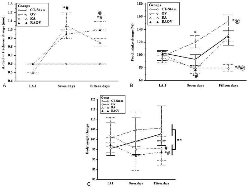
-
Artigos Originais
Effects of hyperprolactinemia and ovariectomy on the tibial epiphyseal growth plate and bone formation in mice
Revista Brasileira de Ginecologia e Obstetrícia. 2014;36(8):359-366
08-01-2014
Summary
Artigos OriginaisEffects of hyperprolactinemia and ovariectomy on the tibial epiphyseal growth plate and bone formation in mice
Revista Brasileira de Ginecologia e Obstetrícia. 2014;36(8):359-366
08-01-2014DOI 10.1590/SO100-720320140005065
Views65See morePURPOSE:
To evaluate the effects of ovariectomy and the hyperprolactinemia procedure in the tibial epiphyseal growth plate of female mice.
METHODS:
In this study, the epiphyseal growth plate of ovariectomized (OVX) and/or rendered hyperprolactinemic female mice by 50 days of treatment with 200 μg metoclopramide (M) was evaluated morphologically, morphometrically and immuno-histochemically. Forty female and adult mice were divided into four groups according to treatment: V group - animals treated with saline solution; H group - hyperprolactinemic animals; Ovx/V group - ovariectomized animals and treated with saline solution; Ovx/H group - hyperprolactinemic and ovariectomized animals. After the treatment period, the animals were sacrificed, tibia was removed and fixed in 10% buffered formalin and decalcified in 10% formic acid. The material was immersed in paraffin and subjected to histological processing in paraffin. The sections were stained with Masson's trichrome and immunohistochemistry was carried out for the pro-apoptotic protein BCL-2. The images for the morphological and morphometric study were analyzed with the imaging program AxioVision 4.8 (Carl-Zeiss(r), Germany).
RESULTS:
The combination of hyperprolactinemia and the ovariectomy procedure decreased the number of resting chondrocytes 1.5-fold, the number of proliferative chondrocytes 1.8-fold; the percentage of resting cartilage 2.4-fold and the percentage of trabecular bone 2.1-fold, compared with respective control animals.
CONCLUSION:
The procedure of ovariectomy combined with the metoclopramide-induced hyperprolactinemia in female mice has showed marked bone degeneration due to significant decrease of cell proliferation in the epiphyseal growth plate and bone formation.
Views65This is an Open Access article distributed under the terms of the Creative Commons Attribution License, which permits unrestricted use, distribution, and reproduction in any medium, provided the original work is properly cited. Summary
Artigos OriginaisEffects of hyperprolactinemia and ovariectomy on the tibial epiphyseal growth plate and bone formation in mice
Revista Brasileira de Ginecologia e Obstetrícia. 2014;36(8):359-366
08-01-2014DOI 10.1590/SO100-720320140005065
Views65See morePURPOSE:
To evaluate the effects of ovariectomy and the hyperprolactinemia procedure in the tibial epiphyseal growth plate of female mice.
METHODS:
In this study, the epiphyseal growth plate of ovariectomized (OVX) and/or rendered hyperprolactinemic female mice by 50 days of treatment with 200 μg metoclopramide (M) was evaluated morphologically, morphometrically and immuno-histochemically. Forty female and adult mice were divided into four groups according to treatment: V group - animals treated with saline solution; H group - hyperprolactinemic animals; Ovx/V group - ovariectomized animals and treated with saline solution; Ovx/H group - hyperprolactinemic and ovariectomized animals. After the treatment period, the animals were sacrificed, tibia was removed and fixed in 10% buffered formalin and decalcified in 10% formic acid. The material was immersed in paraffin and subjected to histological processing in paraffin. The sections were stained with Masson's trichrome and immunohistochemistry was carried out for the pro-apoptotic protein BCL-2. The images for the morphological and morphometric study were analyzed with the imaging program AxioVision 4.8 (Carl-Zeiss(r), Germany).
RESULTS:
The combination of hyperprolactinemia and the ovariectomy procedure decreased the number of resting chondrocytes 1.5-fold, the number of proliferative chondrocytes 1.8-fold; the percentage of resting cartilage 2.4-fold and the percentage of trabecular bone 2.1-fold, compared with respective control animals.
CONCLUSION:
The procedure of ovariectomy combined with the metoclopramide-induced hyperprolactinemia in female mice has showed marked bone degeneration due to significant decrease of cell proliferation in the epiphyseal growth plate and bone formation.
This is an Open Access article distributed under the terms of the Creative Commons Attribution License, which permits unrestricted use, distribution, and reproduction in any medium, provided the original work is properly cited. 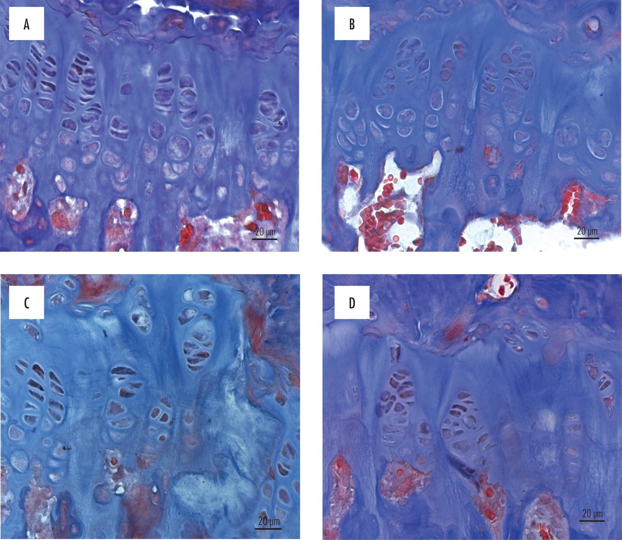
-
Artigos Originais
Effect of high doses of tibolone in body weight and lipid profile of ovariectomized rats
Revista Brasileira de Ginecologia e Obstetrícia. 2010;32(2):88-93
03-15-2010
Summary
Artigos OriginaisEffect of high doses of tibolone in body weight and lipid profile of ovariectomized rats
Revista Brasileira de Ginecologia e Obstetrícia. 2010;32(2):88-93
03-15-2010DOI 10.1590/S0100-72032010000200007
Views95See morePURPOSE: to evaluate the effect of the prolonged use of a high dose of tibolone on the body weight variation and lipid profile of oophorectomized female rats. METHODS: 15 Wistar rats weighing 250 g were randomly divided into two groups. The Experimental Group (n=9) received 1 mg/day of oral tibolone. The Control Group (n=6) received daily 0.5 mL of 0.5% carboxymethylcellulose by gavage. Bilateral oophorectomy was performed 30 days before the beginning of the experiment. On day 0 of the experiment, the animals began to receive the respective treatment for 20 weeks. Body weight was controlled every seven days and food consumption was measured every three to four days along the experiment, in order to establish the daily mean consumption per animal. The results were compared by the Student's t-test, with the significance level set at p<0.05. RESULTS: the daily food consumption of the Tibolone Group was significantly lower (12.7±1.2 g, p<0.001) compared to the Control Group (14.5±1.4 g). This difference was also significant when the body weight was compared between the Tibolone and Control Groups (p<0.001), with the Tibolone Group having lower weight along the experiment. At the end of the experiment, the mean body weight was 215.6±9.3 g in the Tibolone Group and 243.6±6.4 g in the Control Group. Regarding the lipid profile, the Tibolone Group had significantly (p<0.001) lower total cholesterol compared to the Control Group (30.3 versus 78.6 mg/dL). The level of HDL-c was also significantly different (p<0.001), with the Tibolone Group showing lower levels than the Control Group (9.0 versus 52.0 mg/dL). No significant difference between the groups was registered in the other biochemical parameters examined (LDL-c, VLDL-c and triglycerides). CONCLUSIONS: tibolone causes a significant reduction of HDL-c and total cholesterol and has a deleterious effect on the body weight of oophorectomized rats, which may be related to the lower food ingestion by these animals.
Views95This is an Open Access article distributed under the terms of the Creative Commons Attribution License, which permits unrestricted use, distribution, and reproduction in any medium, provided the original work is properly cited. Summary
Artigos OriginaisEffect of high doses of tibolone in body weight and lipid profile of ovariectomized rats
Revista Brasileira de Ginecologia e Obstetrícia. 2010;32(2):88-93
03-15-2010DOI 10.1590/S0100-72032010000200007
Views95See morePURPOSE: to evaluate the effect of the prolonged use of a high dose of tibolone on the body weight variation and lipid profile of oophorectomized female rats. METHODS: 15 Wistar rats weighing 250 g were randomly divided into two groups. The Experimental Group (n=9) received 1 mg/day of oral tibolone. The Control Group (n=6) received daily 0.5 mL of 0.5% carboxymethylcellulose by gavage. Bilateral oophorectomy was performed 30 days before the beginning of the experiment. On day 0 of the experiment, the animals began to receive the respective treatment for 20 weeks. Body weight was controlled every seven days and food consumption was measured every three to four days along the experiment, in order to establish the daily mean consumption per animal. The results were compared by the Student's t-test, with the significance level set at p<0.05. RESULTS: the daily food consumption of the Tibolone Group was significantly lower (12.7±1.2 g, p<0.001) compared to the Control Group (14.5±1.4 g). This difference was also significant when the body weight was compared between the Tibolone and Control Groups (p<0.001), with the Tibolone Group having lower weight along the experiment. At the end of the experiment, the mean body weight was 215.6±9.3 g in the Tibolone Group and 243.6±6.4 g in the Control Group. Regarding the lipid profile, the Tibolone Group had significantly (p<0.001) lower total cholesterol compared to the Control Group (30.3 versus 78.6 mg/dL). The level of HDL-c was also significantly different (p<0.001), with the Tibolone Group showing lower levels than the Control Group (9.0 versus 52.0 mg/dL). No significant difference between the groups was registered in the other biochemical parameters examined (LDL-c, VLDL-c and triglycerides). CONCLUSIONS: tibolone causes a significant reduction of HDL-c and total cholesterol and has a deleterious effect on the body weight of oophorectomized rats, which may be related to the lower food ingestion by these animals.
This is an Open Access article distributed under the terms of the Creative Commons Attribution License, which permits unrestricted use, distribution, and reproduction in any medium, provided the original work is properly cited.