Summary
Revista Brasileira de Ginecologia e Obstetrícia. 2024;46:e-rbgo63
Management of suspect adnexal masses involves surgery to define the best treatment. Diagnostic choices include a two-stage procedure for histopathology examination (HPE) or intraoperative histological analysis – intraoperative frozen section (IFS) and formalin-fixed and paraffin-soaked tissues (FFPE). Preoperative assessment with ultrasound may also be useful to predict malignancy. We aimed at determining the accuracy of IFS to evaluate adnexal masses stratified by size and morphology having HPE as the diagnostic gold standard.
A retrospective chart review of 302 patients undergoing IFS of adnexal masses at Hospital de Clínicas de Porto Alegre, between January2005 and September2011 was performed. Data were collected regarding sonographic size (≤10cm or >10cm), characteristics of the lesion, and diagnosis established in IFS and HPE. Eight groups were studied: unilocular lesions; septated/cystic lesions; heterogeneous (solid/cystic) lesions; and solid lesions, divided in two main groups according to the size of lesion, ≤10cm or >10cm. Kappa agreement between IFS and HPE was calculated for each group.
Overall agreement between IFS and HPE was 96.1% for benign tumors, 96.1% for malignant tumors, and 73.3% for borderline tumors. Considering the combination of tumor size and morphology, 100% agreement between IFS and HPE was recorded for unilocular and septated tumors ≤10cm and for solid tumors.
Stratification of adnexal masses according to size and morphology is a good method for preoperative assessment. We should wait for final HPE for staging decision, regardless of IFS results, in heterogeneous adnexal tumors of any size, solid tumors ≤10cm, and all non-solid tumors >10cm.
Summary
Revista Brasileira de Ginecologia e Obstetrícia. 2019;41(7):440-448
To describe a series of cases of ovarian Sertoli-Leydig cell tumors (SLCTs).
Retrospective review of 12 cases of SLCT treated at the Hospital do Câncer de Barretos, Barretos, state of São Paulo, Brazil, between October 2009 and August 2017.
The median age of the patients was 31 years old (15-71 years old). A total of 9 patients (75.0%) presented symptoms: 8 (66.7%) presented with abdominal pain, 5 (41.7%) presented with abdominal enlargement, 2 (16.7%) presentedwith virilizing signs, 2 (16.7%) presented with abnormal uterine bleeding, 1 (8.3%) presented with dyspareunia, and 1 (8.3%) presented with weight loss. The median preoperative lactate dehydrogenase (LDH) was 504.5 U/L (138-569 U/L), alpha-fetoprotein (AFP) was 2.0 ng/ml (1.1-11.3 ng/ml), human chorionic gonadotropin (β-hCG) was 0.6 mUI/ml (0.0-2.3 mUI/ml), carcinoembryonic antigen (CEA) was 0.9 ng/ml (0.7-3.4 ng/ml), and cancer antigen 125 (CA-125) was 26.0 U/ml (19.1-147.0 U/ml). All of the tumors were unilateral and surgically treated. Lymphadenectomy was performed in 3 (25.0%) patients, but none of the three patients submitted to lymphadenectomy presented lymph node involvement. In the anatomopathological exam, 1 (8.3%) tumor was well-differentiated, 8 (66.7%) were moderately differentiated, and 3 (25.0%) were poorly differentiated. A total of 5 (55.6%) tumors were solid-cystic, 2 (22.2%) were purely cystic, 1 (11.1%) was cystic with vegetations, and 1 (11.1%) was purely solid, but for 3 patients this information was not available. The median lesion size was 14.2 cm (3.2-23.5 cm). All of the tumors were at stage IA of the 2014 classification of the International Federation ofGynecology andObstetrics (FIGO). A total of 2 (16.7%) patients received adjuvant treatment; 1 of themunderwent 3 cycles of paclitaxel and carboplatin every 21days, and the other underwent 4 cycles of ifosfamide, cisplatin and etoposide every 21 days. None of all of the patients had recurrence, and one death related to complications after surgical staging occurred.
Abdominal pain was the most frequent presentation. There was no ultrasonographic pattern. All of the SLCTs were at stage IA, and most of them were moderately differentiated. Relapses did not occur, but one death related to the surgical staging occurred.
Summary
Revista Brasileira de Ginecologia e Obstetrícia. 2017;39(12):676-685
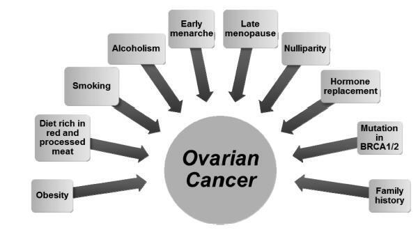
Ovarian cancer is the leading cause of death among gynecologic tumors because in most of the cases (75%), the disease is diagnosed in advanced stages. Screening methods are not available since the disease is rare, and the tested methods, such as ultrasound and CA125, were not able to decrease the mortality rate for this type of cancer. This article discusses the main risk factors for ovarian cancer, and the potential clinical and surgical strategies for the prevention of this disease.
Summary
Revista Brasileira de Ginecologia e Obstetrícia. 2017;39(12):676-685
Ovarian cancer is the leading cause of death among gynecologic tumors because in most of the cases (75%), the disease is diagnosed in advanced stages. Screening methods are not available since the disease is rare, and the tested methods, such as ultrasound and CA125, were not able to decrease the mortality rate for this type of cancer. This article discusses the main risk factors for ovarian cancer, and the potential clinical and surgical strategies for the prevention of this disease.
Summary
Revista Brasileira de Ginecologia e Obstetrícia. 2015;37(6):283-290
DOI 10.1590/SO100-720320150005292
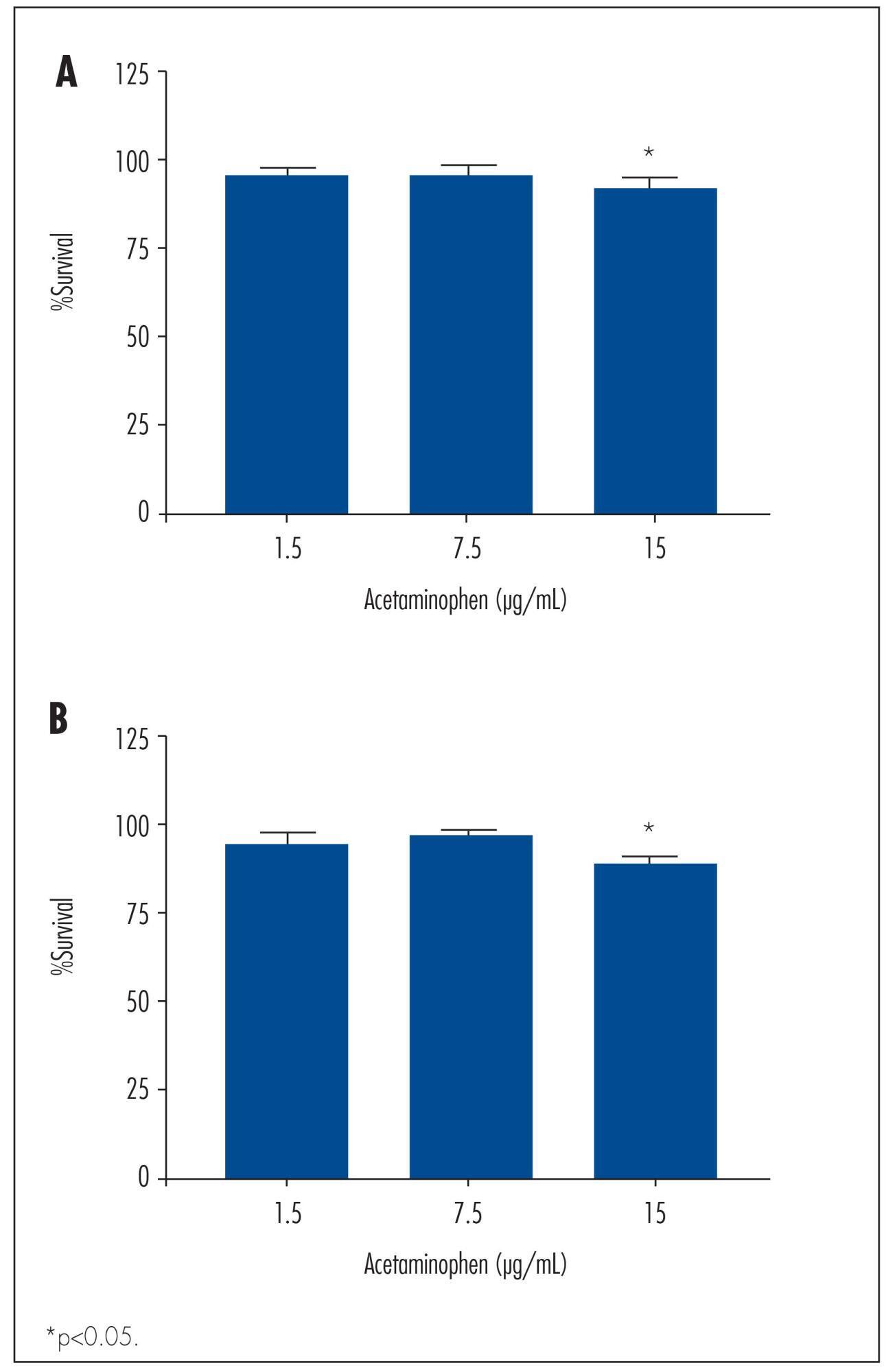
To determine the basic expression of ABC transporters in an epithelial ovarian cancer cell line, and to investigate whether low concentrations of acetaminophen and ibuprofen inhibited the growth of this cell line in vitro.
TOV-21 G cells were exposed to different concentrations of acetaminophen (1.5 to 15 μg/mL) and ibuprofen (2.0 to 20 μg/mL) for 24 to 48 hours. The cellular growth was assessed using a cell viability assay. Cellular morphology was determined by fluorescence microscopy. The gene expression profile of ABC transporters was determined by assessing a panel including 42 genes of the ABC transporter superfamily.
We observed a significant decrease in TOV-21 G cell growth after exposure to 15 μg/mL of acetaminophen for 24 (p=0.02) and 48 hours (p=0.01), or to 20 μg/mL of ibuprofen for 48 hours (p=0.04). Assessing the morphology of TOV-21 G cells did not reveal evidence of extensive apoptosis. TOV-21 G cells had a reduced expression of the genes ABCA1, ABCC3, ABCC4, ABCD3, ABCD4 and ABCE1 within the ABC transporter superfamily.
This study provides in vitro evidence of inhibitory effects of growth in therapeutic concentrations of acetaminophen and ibuprofen on TOV-21 G cells. Additionally, TOV-21 G cells presented a reduced expression of the ABCA1, ABCC3, ABCC4, ABCD3, ABCD4 and ABCE1 transporters.
Summary
Revista Brasileira de Ginecologia e Obstetrícia. 2015;37(6):283-290
DOI 10.1590/SO100-720320150005292
To determine the basic expression of ABC transporters in an epithelial ovarian cancer cell line, and to investigate whether low concentrations of acetaminophen and ibuprofen inhibited the growth of this cell line in vitro.
TOV-21 G cells were exposed to different concentrations of acetaminophen (1.5 to 15 μg/mL) and ibuprofen (2.0 to 20 μg/mL) for 24 to 48 hours. The cellular growth was assessed using a cell viability assay. Cellular morphology was determined by fluorescence microscopy. The gene expression profile of ABC transporters was determined by assessing a panel including 42 genes of the ABC transporter superfamily.
We observed a significant decrease in TOV-21 G cell growth after exposure to 15 μg/mL of acetaminophen for 24 (p=0.02) and 48 hours (p=0.01), or to 20 μg/mL of ibuprofen for 48 hours (p=0.04). Assessing the morphology of TOV-21 G cells did not reveal evidence of extensive apoptosis. TOV-21 G cells had a reduced expression of the genes ABCA1, ABCC3, ABCC4, ABCD3, ABCD4 and ABCE1 within the ABC transporter superfamily.
This study provides in vitro evidence of inhibitory effects of growth in therapeutic concentrations of acetaminophen and ibuprofen on TOV-21 G cells. Additionally, TOV-21 G cells presented a reduced expression of the ABCA1, ABCC3, ABCC4, ABCD3, ABCD4 and ABCE1 transporters.
Summary
Revista Brasileira de Ginecologia e Obstetrícia. 2014;36(3):124-130
DOI 10.1590/S0100-72032014000300006
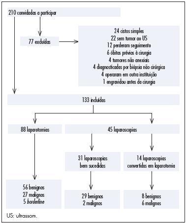
To assess clinical factors, histopathologic diagnoses, operative time and differences in complication rates between women undergoing laparoscopy or laparotomy to diagnose and treat an adnexal mass and their association with laparoscopy failure.
In this prospective study, 210 women were invited to participate and 133 of them were included. Eighty-eight women underwent laparotomy and 45 underwent laparoscopy. Fourteen of the 45 laparoscopies were converted to laparotomy intraoperatively. We assessed whether age, body mass index (BMI), previous abdominal surgeries, CA-125, Index of Risk of Malignancy (IRM), tumor diameter, histological diagnosis, operative time and surgical complication rates differed between the laparoscopy group and the group converted to laparotomy and whether those factors were associated with conversion of laparoscopy to laparotomy. We also assessed surgical logs to evaluate the reasons, as stated by the surgeons, to convert a laparoscopy to laparotomy.
In this research, 30% of the women had malignant tumors. CA-125, IRM, tumor diameter and operative times were higher for the laparotomy group than the laparoscopy group. Complication rates were similar for both groups and also for the successful laparoscopy and unsuccessful laparoscopy groups. The surgical complication rate in women with benign tumors was lower for the laparoscopy group than for the laparotomy group. The factors associated with conversion to laparotomy were tumor diameter and malignancy. During laparoscopy, adhesions a large tumor diameter were the principal causes of conversion.
This study suggests that laparoscopy for the diagnosis and treatment of adnexal masses is safe and does not increase complication rates even in patients who need conversion to laparotomy. However, when doubt about the safety of the procedure and about the presence of malignancy persists, consultation with an expert gynecology-oncologist with experience in advanced laparoscopy is recommended. A large tumor diameter was associated with the necessity of conversion to laparotomy.
Summary
Revista Brasileira de Ginecologia e Obstetrícia. 2014;36(3):124-130
DOI 10.1590/S0100-72032014000300006
To assess clinical factors, histopathologic diagnoses, operative time and differences in complication rates between women undergoing laparoscopy or laparotomy to diagnose and treat an adnexal mass and their association with laparoscopy failure.
In this prospective study, 210 women were invited to participate and 133 of them were included. Eighty-eight women underwent laparotomy and 45 underwent laparoscopy. Fourteen of the 45 laparoscopies were converted to laparotomy intraoperatively. We assessed whether age, body mass index (BMI), previous abdominal surgeries, CA-125, Index of Risk of Malignancy (IRM), tumor diameter, histological diagnosis, operative time and surgical complication rates differed between the laparoscopy group and the group converted to laparotomy and whether those factors were associated with conversion of laparoscopy to laparotomy. We also assessed surgical logs to evaluate the reasons, as stated by the surgeons, to convert a laparoscopy to laparotomy.
In this research, 30% of the women had malignant tumors. CA-125, IRM, tumor diameter and operative times were higher for the laparotomy group than the laparoscopy group. Complication rates were similar for both groups and also for the successful laparoscopy and unsuccessful laparoscopy groups. The surgical complication rate in women with benign tumors was lower for the laparoscopy group than for the laparotomy group. The factors associated with conversion to laparotomy were tumor diameter and malignancy. During laparoscopy, adhesions a large tumor diameter were the principal causes of conversion.
This study suggests that laparoscopy for the diagnosis and treatment of adnexal masses is safe and does not increase complication rates even in patients who need conversion to laparotomy. However, when doubt about the safety of the procedure and about the presence of malignancy persists, consultation with an expert gynecology-oncologist with experience in advanced laparoscopy is recommended. A large tumor diameter was associated with the necessity of conversion to laparotomy.
Summary
Revista Brasileira de Ginecologia e Obstetrícia. 2013;35(7):331-335
DOI 10.1590/S0100-72032013000700008
The sclerosing stromal tumor of the ovary is an extremely rare benign tumor more common in young women and without specific symptoms in most cases. Less than 150 cases have been described, of which 8 were diagnosed during pregnancy. In this report, we describe the association between sclerosing stromal tumor of the ovary, Meigs' syndrome and elevated levels of CA-125 in term pregnancy.
Summary
Revista Brasileira de Ginecologia e Obstetrícia. 2012;34(11):511-517
DOI 10.1590/S0100-72032012001100006
PURPOSE: To assess the association between clinical symptoms and the diagnosis of malignancy in women with adnexal tumors who underwent surgery. METHODS: Cross-sectional study, in which 105 women with adnexal tumors and indication for laparotomy/laparoscopy were included. All women were treated at a teaching hospital in the state of São Paulo between November 2009 and March 2011. All patients underwent a structured interview about the occurrence of 18 symptoms associated with ovarian cancer. The interview included the severity, frequency, and duration of these symptoms in the 12 months prior to the first medical consultation. The CA125 levels and the ultrasound classification of the tumors were also evaluated. We calculated for each symptom the prevalence ratio with 95% confidence intervals. The golden-standard was the result of the pathological examination of the surgical specimens. RESULTS: Of the 105 women included, 75 (71.4%) had benign tumors and 30 (28.6%) had malignant ones. In women with malignant tumors, the most frequent symptoms were: abdominal bloating (70%), increased abdominal size (67%), pelvic pain (60%), menstrual irregularity (60%), swelling (53%), abdominal pain (50%), backache (50%), and early repletion (50%). Women with benign tumors showed essentially pelvic pain (61%), menstrual irregularities (61%), and abdominal swelling (47%). Symptoms significantly associated with malignancy were: bloating (PR=2.0; 95%CI 1.01 - 3.94), increased abdominal size (PR=2.16; 95%CI 1.12 - 4.16), backache (RP=1.97; 95%CI 1.09 - 3.55), swelling (PR=2.25; 95%CI 1.25 - 4.07), early repletion (RP=2.06; 95%CI 1.14 - 3.70), abdominal mass (PR=1.83; 95%CI 1.01 - 3.30), eating difficulties (PR=1.98; 95%CI 1.10 - 3.56), and postmenopausal bleeding (PR=2.91; 95%CI 1.55 - 5.44). The presence of pelvic pain, constipation, dyspareunia, fatigue, abdominal pain, nausea or vomiting, menstrual irregularity, weight loss, diarrhea, and bleeding after intercourse was similar in both groups. CONCLUSIONS: In women with adnexal tumors including indication of surgical treatment, the preoperative evaluation of symptoms may help predicting malignancy.
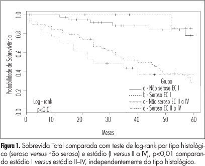
Summary
Revista Brasileira de Ginecologia e Obstetrícia. 2012;34(5):196-202
DOI 10.1590/S0100-72032012000500002
Summary
Revista Brasileira de Ginecologia e Obstetrícia. 2012;34(5):196-202
DOI 10.1590/S0100-72032012000500002