Summary
Revista Brasileira de Ginecologia e Obstetrícia. 2021;43(4):329-333
03-30-2021
Malignant mesonephric tumors are uncommon in the female genital tract, and they are usually located where embryonic remnants of Wolffian ducts are detected, such as the uterine cervix. The information about these tumors, their treatment protocol, and prognosis are scarce.
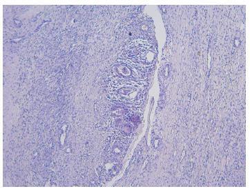
A 60-year-old woman with postmenopausal vaginal bleeding was initially diagnosed with endometrial carcinoma. After suspicion co-testing, the patient underwent a loop electrosurgical excision of the cervix and was eventually diagnosed with mesonephric adenocarcinoma. She was subjected to a radical hysterectomy, which revealed International Federation of Gynecology and Obstetrics (FIGO) IB1 stage, and adjuvant radiotherapy. The follow-up showed no evidence of recurrence after 60 months.
We present the case of a woman with cervical mesonephric adenocarcinoma. When compared with the literature, this case had the longest clinical follow-up without evidence of recurrence, which reinforces the concept that these tumors are associated with a favorable prognosis if managed according to the guidelines defined for the treatment of patients with cervical adenocarcinomas. Though a rare entity, it should be kept in mind as a differential diagnosis for other cervical cancers.
Summary
Revista Brasileira de Ginecologia e Obstetrícia. 2018;40(2):86-91
02-01-2018
To compare the quality of cervicovaginal samples obtained from basic health units (BHUs) of the Unified Health System (SUS) and those obtained fromprivate clinics to screen precursor lesions of cervical cancer.
It was an intervention study whose investigated variables were: adequacy of the samples; presence of epithelia in the samples, and cytopathological results. A total of 940 forms containing the analysis of the biological samples were examined: 470 forms of women attended at BHUs of the SUS and 470 forms of women examined in private clinics in January and February of 2016.
All the unsatisfactory samples were collected at BHUs and corresponded to 4% of the total in this sector (p < 0.0001). There was a higher percentage of samples containing only squamous cells in the SUS (43.9%). There was squamocolumnar junction (SJC) representativeness in 82.1% of the samples from the private clinics (p < 0.0001). Regarding negative results for intraepithelial lesions and/or malignancies, the percentages obtained were 95.9% and 99.1% (p < 0.0049) in the exams collected in the private system and SUS, respectively. Less serious lesions corresponded to 0.89% of the samples from the SUS and 2.56% of the tests from the private sector; more serious lesions were not represented in the samples obtained from BHUs, whereas the percentage was 1.49% in private institutions.
Unsatisfactory cervical samples were observed only in exams performed at the SUS. There is a need for guidance and training of professionals who perform this procedure to achieve higher reliability in the results and more safety for women who undergo this preventive test.
Summary
Revista Brasileira de Ginecologia e Obstetrícia. 2017;39(5):249-254
05-01-2017
The occurrence of Manson’s schistosomiasis in organs of the female reproductive tract is an uncommon event, given that the etiological agent for this disease is a blood parasite that inhabits the mesenteric veins. In this case report, a 45-year-old female patient reported that her first symptoms had been strong pain in the left iliac region around two years earlier. An endovaginal pelvic ultrasonography showed that the left ovary was enlarged, and the report suggested that this finding might be correlated with clinical data and tumor markers. After being examined at several healthcare services, the patient was referred to an oncology service due to suspected neoplasia, where she underwent a left ovariectomy. The result from the histopathological examination showed the presence of granulomatous inflammatory processes surrounding both viable and calcified eggs of Schistosoma mansoni. There was no evidence of any neoplastic tissue. The patient was medicated and followed-up as an outpatient.
