Summary
Revista Brasileira de Ginecologia e Obstetrícia. 2023;45(7):393-400
Endometriosis causes a decrease in oocyte quality. However, this mechanism is not fully understood. The present study aimed to analyze the effect of endometriosis on cumulus cell adenosine triphosphate ATP level, the number of mitochondria, and the oocyte maturity level.
A true experimental study with a post-test only control group design on experimental animals. Thirty-two mice were divided into control and endometriosis groups. Cumulus oocyte complex (COC) was obtained from all groups. Adenosine triphosphate level on cumulus cells was examined using the Elisa technique, the number of mitochondria was evaluated with a confocal laser scanning microscope and the oocyte maturity level was evaluated with an inverted microscope.
The ATP level of cumulus cells and the number of mitochondria in the endometriosis group increased significantly (p < 0.05; p < 0.05) while the oocyte maturity level was significantly lower (p < 0.05). There was a significant relationship between ATP level of cumulus cells and the number of mitochondrial oocyte (p < 0.01). There was no significant relationship between cumulus cell ATP level and the number of mitochondrial oocytes with oocyte maturity level (p > 0.01; p > 0.01). The ROC curve showed that the number of mitochondrial oocytes (AUC = 0.672) tended to be more accurate than cumulus cell ATP level (AUC = 0.656) in determining the oocyte maturity level.
In endometriosis model mice, the ATP level of cumulus cells and the number of mitochondrial oocytes increased while the oocyte maturity level decreased. There was a correlation between the increase in ATP level of cumulus cells and an increase in the number of mitochondrial oocytes.
Summary
Revista Brasileira de Ginecologia e Obstetrícia. 2018;40(5):251-259
The aim of this work was to evaluate the changes caused by estrogen deficiency in lipid metabolism.
This study encompassed direct measurements of plasma biochemical analyses, liver lipid contents, and assessments of the mitochondrial β-oxidation capacity as well as an evaluation of the liver redox status in an animal model of estrogen deficiency.
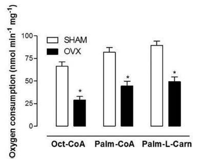
When compared with control mice, the livers of ovariectomized (OVX) mice presented considerable accretions in their lipid contents, which were accompanied by increased levels of lipid peroxidation in liver homogenates andmitochondria from OVX groups and decreased reduced glutathione (GSH) contents. In isolated mitochondria, estrogen deficiency inhibited mitochondrial β-oxidation of fatty acids irrespective of their chain length. The liver mitochondrial and peroxisomal H2O2 generations in OVX mice were increased. Additionally, the activities of all antioxidant enzymes assessed were decreased.
These data provide one potential explanation for the increased susceptibility to metabolic diseases observed after menopause.
Summary
Revista Brasileira de Ginecologia e Obstetrícia. 2018;40(5):251-259
The aim of this work was to evaluate the changes caused by estrogen deficiency in lipid metabolism.
This study encompassed direct measurements of plasma biochemical analyses, liver lipid contents, and assessments of the mitochondrial β-oxidation capacity as well as an evaluation of the liver redox status in an animal model of estrogen deficiency.
When compared with control mice, the livers of ovariectomized (OVX) mice presented considerable accretions in their lipid contents, which were accompanied by increased levels of lipid peroxidation in liver homogenates andmitochondria from OVX groups and decreased reduced glutathione (GSH) contents. In isolated mitochondria, estrogen deficiency inhibited mitochondrial β-oxidation of fatty acids irrespective of their chain length. The liver mitochondrial and peroxisomal H2O2 generations in OVX mice were increased. Additionally, the activities of all antioxidant enzymes assessed were decreased.
These data provide one potential explanation for the increased susceptibility to metabolic diseases observed after menopause.