Summary
Revista Brasileira de Ginecologia e Obstetrícia. 2015;37(4):186-191
04-01-2015
DOI 10.1590/SO100-720320150005252
To evaluate genes differentially expressed in ovaries from lean (wild type) and obese (ob/ob) female mice and cyclic AMP production in both groups.
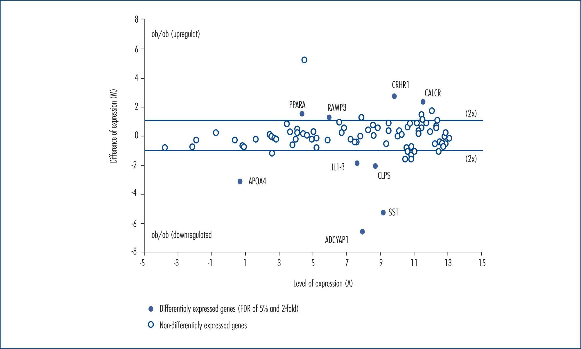
The expression on messenger RNA levels of 84 genes concerning obesity was analyzed through the PCR array, and cyclic AMP was quantified by the enzyme immunoassay method.
The most downregulated genes in the Obesity Group included adenylate cyclase-activating polypeptide type 1, somatostatin, apolipoprotein A4, pancreatic colipase, and interleukin-1 beta. The mean decrease in expression levels of these genes was around 96, 40, 9, 4.2 and 3.6-fold, respectively. On the other hand, the most upregulated genes in the Obesity Group were receptor (calcitonin) activity-modifying protein 3, peroxisome proliferator activated receptor alpha, calcitonin receptor, and corticotropin-releasing hormone receptor 1. The increase means in the expression levels of such genes were 2.3, 2.7, 4.8 and 6.3-fold, respectively. The ovarian cyclic AMP production was significantly higher in ob/ob female mice (2,229±52 fMol) compared to the Control Group (1,814±45 fMol).
Obese and anovulatory female mice have reduced reproductive hormone levels and altered ovogenesis. Several genes have their expression levels altered when leptin is absent, especially adenylate cyclase-activating polypeptide type 1.
Summary
Revista Brasileira de Ginecologia e Obstetrícia. 2015;37(4):186-191
04-01-2015
DOI 10.1590/SO100-720320150005252
To evaluate genes differentially expressed in ovaries from lean (wild type) and obese (ob/ob) female mice and cyclic AMP production in both groups.
The expression on messenger RNA levels of 84 genes concerning obesity was analyzed through the PCR array, and cyclic AMP was quantified by the enzyme immunoassay method.
The most downregulated genes in the Obesity Group included adenylate cyclase-activating polypeptide type 1, somatostatin, apolipoprotein A4, pancreatic colipase, and interleukin-1 beta. The mean decrease in expression levels of these genes was around 96, 40, 9, 4.2 and 3.6-fold, respectively. On the other hand, the most upregulated genes in the Obesity Group were receptor (calcitonin) activity-modifying protein 3, peroxisome proliferator activated receptor alpha, calcitonin receptor, and corticotropin-releasing hormone receptor 1. The increase means in the expression levels of such genes were 2.3, 2.7, 4.8 and 6.3-fold, respectively. The ovarian cyclic AMP production was significantly higher in ob/ob female mice (2,229±52 fMol) compared to the Control Group (1,814±45 fMol).
Obese and anovulatory female mice have reduced reproductive hormone levels and altered ovogenesis. Several genes have their expression levels altered when leptin is absent, especially adenylate cyclase-activating polypeptide type 1.
Summary
Revista Brasileira de Ginecologia e Obstetrícia. 2014;36(10):449-455
10-03-2014
DOI 10.1590/SO100-720320140004946
To assess cardiometabolic risk factors during normal pregnancy and the influence of maternal obesity on them.
This study included 25 healthy pregnant women with a single pregnancy and a gestational age of less than twenty weeks. Longitudinal analysis of blood pressure, body weight, body mass index (BMI), serum concentrations of leptin, adiponectin, cortisol, total cholesterol and fractions, triglycerides, uric acid, fasting glucose, oral glucose tolerance test, HOMA-IR and insulin/glucose ratio was performed each trimester during pregnancy. In order to evaluate the impact of obesity, pregnant women were divided into two groups based on BMI for the first quarter of pregnancy: Gpn for pregnant women with BMI<25 kg/m2 and Gso for BMI≥25 kg/m2. One-Way ANOVA for repeated measurements or Friedman test and Student-t or Mann-Whitney tests for statistical comparisons and Pearson correlations test were used for statistical analysis.
The mean values for the first quarter of pregnancy for the following parameters were: age: 22 years; weight: 66.3 kg and BMI 26.4 kg/m2, with 20.2 and 30.7 kg/m2 for the Gpn and Gso groups, respectively. Mean weight gain during pregnancy was ±12.7 kg with 10.3 kg for the Gso group and 15.2 kg for the Gpn group. Regarding plasma determinations, cortisol, uric acid and lipid profile increased during all trimesters of pregnancy, except for HDL-cholesterol, which did not change. Blood pressure, insulin and HOMA-IR only increased in the third quarter of pregnancy. The Gso group tended to gain more weight and to show higher concentrations of leptin, total cholesterol, LDL-cholesterol, VLDL-cholesterol, TG, glucose, insulin, HOMA-IR, besides lower HDL-cholesterol and greater diastolic blood pressure in the 3rdquarter of pregnancy. Three pregnant women developed gestational hypertension, presented prepregnancy obesity, excessive weight gain, hyperleptinemia and an insulin/glucose ratio greater than two. Weight and BMI were positively correlated with total cholesterol and its LDL fraction, TG, uric acid, fasting blood glucose, insulin and HOMA-IR; and were negatively correlated with adiponectin and HDL-cholesterol. Leptin level was positively correlated with blood pressure.
The metabolic changes in pregnancy are more significant in obese women, suggesting, as expected, an increased risk of cardiometabolic complications. During their first visit for prenatal care, obese women should be informed about these risks, have their BMI and insulin/glucose ratio calculated along with their lipid profile to identify pregnant women at higher risk for cardiovascular diseases.
Summary
Revista Brasileira de Ginecologia e Obstetrícia. 2012;34(6):268-273
07-13-2012
DOI 10.1590/S0100-72032012000600005
PURPOSE: To evaluate the correlation between maternal waist circumference measured before the 12th week of gestation and serum leptin levels during pregnancy, as well as to compare the leptin levels of women with and without abdominal obesity diagnosed in early pregnancy. METHODS: Prospective study including 40 pregnant women receiving low-risk prenatal care, older than 20 years, nonsmokers, with singleton pregnancies and without chronic disease. Waist circumference was measured before the 12th week and serum leptin levels were measured between the 9th and 12th, 25th and 28th and 34th and 37th weeks of gestation. According to waist circumference measurement, the cohort was divided into two groups: with and without abdominal obesity. The Mann-Whitney and χ² tests were used to assess the differences between groups. The Pearson correlation coeffient was used to assess the association between waist circumference and serum leptin levels during pregnancy. The level of significance was set at p<0.05. RESULTS: The mean weight and body mass index of patients with abdominal obesity (74.4±11.0 kg/28.9±4.1) was higher than that of patients without abdominal obesity (55.6±5.9 kg/21.1±2.4) (p=0.001). The mean leptin levels in pregnant patients with abdominal obesity (41.9±3.5 ng/mL) was higher than in patients without abdominal obesity (23.6±2.7 ng/mL) (p<0.0002). A positive correlation was obtained between the waist circumference measured during the same period and the mean serum leptin levels (r=0.7; p<0.0001). CONCLUSIONS: Waist circumference measured before the 12th week of pregnancy is a valid and simple method to predict the serum leptin levels throughout pregnancy. Pregnant women with abdominal obesity diagnosed before 12th week have higher mean serum leptin levels during pregnancy than those without abdominal obesity.
Summary
Revista Brasileira de Ginecologia e Obstetrícia. 2005;27(4):216-221
07-30-2005
DOI 10.1590/S0100-72032005000400009
PURPOSE: to identify the relationship between serum levels of leptin and the levels of estradiol and follicle-stimulating hormone (FSH) in women with pituitary suppression and to evaluate its possible interference on the reproductive axis. METHODS: a total of 64 patients submitted to controlled ovarian hyperstimulation with recombinant FSH for assisted reproduction, due to a male or tubal factor, and 20 patients using estradiol valerate, for endometrial preparation in order to be submitted to oocyte donation treatment were studied. All patients used GnRH analogues before starting treatment in order to avoid premature LH surge. Data were analyzed statistically by the chi2 test, Student's t-test and the Pearson correlation test, when appropriate, with the level of significance set at p<0,05. RESULTS: it was observed that leptin levels correlated with body mass index (BMI) even though they had not influenced growth rate of these hormones. A positive correlation was observed between estradiol and leptin levels in both groups, as leptin levels increased from 10.42 to 15.68 ng/mL in the FSH group and from 11.09 to 14.5 ng/mL in the estradiol group, following estradiol increase. The growth rate of leptin was higher in women with higher estradiol levels, i.e., who had induced cycles with recombinant FSH, than in those who received estradiol valerate (159.60±58.01 and 136.73±34.17, respectively). CONCLOSION: we may state that leptin correlated with BMI and that both FSH and estradiol do interfere in the regulation of leptin production in women.
Summary
Revista Brasileira de Ginecologia e Obstetrícia. 2004;26(9):691-695
01-19-2004
DOI 10.1590/S0100-72032004000900003
PURPOSE: to evaluate the importance of circulating maternal and fetal leptin in the healthy gestation, using its association with maternal, placental and fetal anthropometric variables, obtained at birth, and the relationship between the evaluated compartments. METHODS: in a transversal study a population of 33 single, healthy and term gestations was studied. The evaluated variables were maternal age, maternal weight, body mass index (BMF), weight of the newborn, placental weight, and placental index. Samples of maternal blood were immediately obtained before birth and from fetal umbilical cord blood at birth. Determination of serum leptin was performed using conventional radioimmunoassay. The relationships between serum leptin concentrations in maternal blood, umbilical artery and vein and the studied variables were assessed through linear regression. RESULTS: leptin levels were detected in the blood of all 33 pregnant women and their respective newborns, with maternal blood concentration (17.1±1.77 ng/mL) higher than that of umbilical vessels (vein: 9.0±1.16 ng/mL; artery: 8.23±1.02 ng/mL), p<0.0001. Leptin concentrations in the maternal blood were correlated with leptin concentrations in fetal blood (artery: coef. 0.63, p=0.037; vein: coef. 0.72, p=0.006). Regarding the anthropometric variables, leptin measured in the maternal blood was associated with initial and final maternal BMF (coef. 1.13; p=0.002; coef. 1,18, p=0.001) and cord leptin levels were correlated with the fetal weight at birth (vein: coef. 0.007, p=0.02; artery: coef. 0.006, p=0.02). CONCLUSION: there was a correlation between maternal and fetal leptin production and probably by the action of similar stimuli during gestation. Serum leptin was associated with the weight of the compartment where it circulates.
Summary
Revista Brasileira de Ginecologia e Obstetrícia. 2004;26(6):429-433
09-02-2004
DOI 10.1590/S0100-72032004000600002
OBJECTIVE: to correlate serum leptin concentration with bone mineral density (BMD) in postmenopausal women. METHODS: twenty-two healthy postmenopausal women were included in the present study. BMD was measured by dual energy X-ray absorptiometry at the lumbar spine and femoral neck. Serum leptin concentrations were determined using an immunoradiometric assay. Statistical analysis was performed by ANOVA and Dunn and Pearson's correlation tests. RESULTS: mean BMD values were 0.898 ± 0.140 g/cm² at the lumbar spine and 0.760 ± 0.152 g/cm² at the femoral neck. Mean serum leptin concentration was 17.2 ± 9.4 ng/ml and no significant differences were observed among women with normal BMD, osteopenia and osteoporosis (18.6 ± 7.8, 18.9 ± 9.9 and 15.6 ± 10.6, respectively; p > 0.05). No significant correlations were observed between serum leptin levels and BMD measurements at the lumbar spine and femoral neck, when the whole sample was considered and when patients were divided into groups with osteoporosis and/or osteopenia and a control group. We observed a positive significant correlation between serum leptin levels and body mass index (BMI) (r = 0.66; p = 0.0044). CONCLUSIONS: there was no direct correlation between leptin and BMD in postmenopausal women, although we observed positive significant correlation between leptin and BMI. This fact indicates a possible indirect effect of leptin on bone metabolism.
Summary
Revista Brasileira de Ginecologia e Obstetrícia. 2001;23(5):283-287
06-26-2001
DOI 10.1590/S0100-72032001000500003
Purpose: to study in primigravid adolescents the behavior of serum leptin levels during the evolution of normal pregnancy, comparing the results with those obtained from preeclamptics. Methods: prospective, longitudinal study conducted in 15 normotensive pregnant adolescents and 5 preeclamptic adolescents. Serum leptin levels (ng/mL) were determined by radioimmunoassay. Blood pressure was measured by the oscilometric method by using DINAMAP 1846. Patients were evaluated in two different gestational periods: between the 21st and 30th week and between the 31st and 40th week. The ratio leptin/body mass index (BMI) was used to correct changes observed in BMI throughout gestation. Preeclamptic pregnant patients were diagnosed when the blood pressure was > or = 140/90 mmHg, proteinuria >300 mg/24 h and when arteriolar spasm was present in the optic fundi. Results: there was a trend towards an elevation of serum leptin at the end of pregnancy in both groups although more pronounced in preeclamptic patients. In pregnant normotensive patients serum leptin increased from 11.9±1.20 (21stto 30th week) to 13.9±2.23 ng/mL (31st to 40th week), and in preeclamptic from 11.9±1.20 to 17.6±4.565 ng/mL. In preeclamptic patients the BMI increased significantly in the period from the 21st to 30th week when compared to the period between the 31st and 40thweek: 21.5±0.8 vs 27.4±1.7 kg/m², p<0.05.In normotensive these values were maintained stable: 24.9±1.5 vs 25.1±1.00 kg/m². At the end of gestation the ratio leptin/BMI was significantly higher in preeclamptics: 0.56±0.06 (21stto 30thweek) vs 0.70±0.15, p<0.05. The values of the ratio leptin/BMI in normotensive pregnants varied from 0.44±0.02 between the 21st and 30th week to 0.41±0.04 between the 31st to 40thweek. In normal pregnants there was a direct and significant correlation between the levels of leptin and BMI in both periods of pregnancy: r = 0.7, p<0.004 (31st to 40th ) vs r = 0.94, p<0.0001 (31st to 40th week). These correlations were lost in preeclamptic patients in both gestational periods. Conclusion: the higher concentrations of leptin and leptin/BMI ratio observed when preeclamptics were compared with normotensive patients, in both gestational periods, suggest a leptin resistance pattern in preeclampsia.
Summary
Revista Brasileira de Ginecologia e Obstetrícia. 2001;23(8):481-488
06-24-2001
DOI 10.1590/S0100-72032001000800002
Purpose: to investigate leptin levels in patients with polycystic ovary syndrome (PCOS), and relationships with testosterone, estradiol, follicle-stimulating hormone (FSH) and insulin levels. Methods: transversal study on 40 patients with PCOS divided into two groups: Group I (n = 20)- obese women (body mass index - BMI > or = 28 kg/m²), and Group II (n = 20) - non obese women (BMI <28 kg/m²). Results: BMI was different between the two groups (p=0.04). We observed that leptin concentrations were significantly correlated with BMI (p<0.001). After adjusting for BMI, no correlation between leptin, insulin (p=0.194), FSH (p=0.793), and total (p=0.441) and free (p=0.422) testosterone was found. However, we only observed positive correlations between leptin and estradiol (p=0.043). Conclusions: there is a strong correlation between leptin levels, BMI and estradiol levels in women with PCOS.