Summary
Revista Brasileira de Ginecologia e Obstetrícia. 2023;45(4):207-214
06-30-2023
Supplementation with folic acid (FA) during gestation has been recommended by medical society all over the world, but some studies have shown that intake of high folic acid diet may unleash damages to the descendants. Objectives: Describing the effects of maternal supplementation with FA during gestation on offspring's kidney at late life stages. Data Source: It is a systematic review by which were consulted the following databases: Medline, through Pubmed, Lilacs, and SciELO. The research was performed using the keywords “Folic acid”, “Gestation” and “Kidney”. Study Selection: Eight studies were regarded for this systematic review. Data Collection: Only studies that evaluated folic acid consumption during gestation and its effects exclusively on descendants' kidney at several phases of life were regarded. Results: Gestational FA intake did not change the renal volume, glomerular filtration rate and the expression of some essential genes in the kidney of puppies whose dams were supplemented with FA. Maternal consumption of double FA plus selenium diet was effective in preserving antioxidant enzymes activity in the kidney of descendants from mothers exposed to alcohol. FA supplementation decreased some gross anomalies in the puppies caused by teratogenic drug despite of had not been effective in preventing some renal architectural damages. Conclusion: FA supplementation did not cause renal toxicity; it exerted an antioxidant protective effect and mitigated some renal disorders caused by severe aggressions.
Summary
Revista Brasileira de Ginecologia e Obstetrícia. 2019;41(3):199-202
03-13-2019
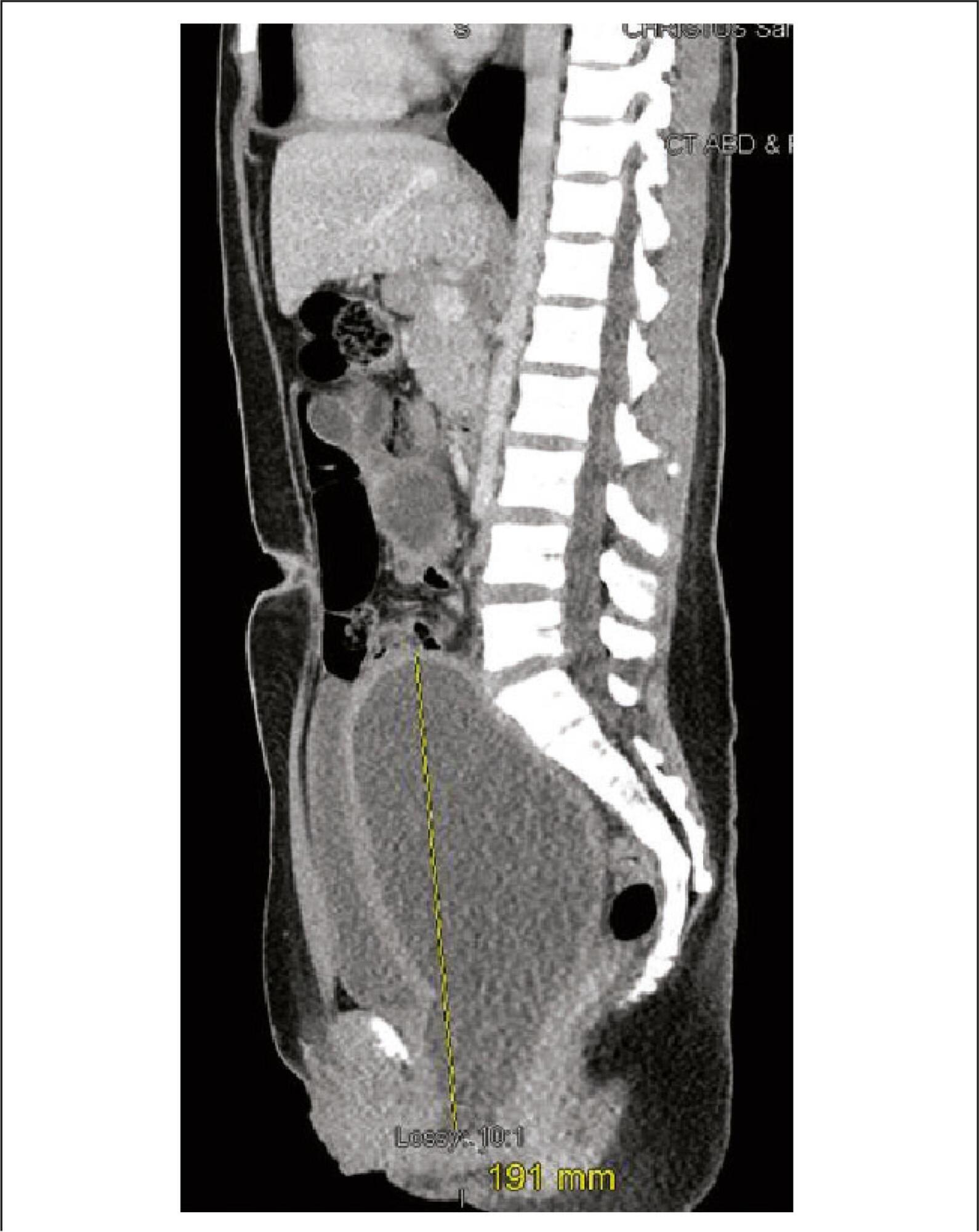
Angiomyolipomas (AMLs) are rare benign tumors derived from mesenchymal tissue and composed of varying degrees of adipose tissue, muscle and blood vessels. Renal AMLs (RAMLs) are the result of a sporadic event, and, in most of cases, the diagnosis is usually incidental, but hemorrhage and shock may be present. During pregnancy, the size of AMLs may increase and they may rupture, probably due to the high expression of hormone receptors, and the increase in maternal circulation and abdominal pressure. The authors present a case of a woman with ruptured RAML submitted to urgent endovascular treatment four days after giving birth by cesarean section.
Summary
Revista Brasileira de Ginecologia e Obstetrícia. 2019;41(3):199-202
03-13-2019
Angiomyolipomas (AMLs) are rare benign tumors derived from mesenchymal tissue and composed of varying degrees of adipose tissue, muscle and blood vessels. Renal AMLs (RAMLs) are the result of a sporadic event, and, in most of cases, the diagnosis is usually incidental, but hemorrhage and shock may be present. During pregnancy, the size of AMLs may increase and they may rupture, probably due to the high expression of hormone receptors, and the increase in maternal circulation and abdominal pressure. The authors present a case of a woman with ruptured RAML submitted to urgent endovascular treatment four days after giving birth by cesarean section.
Summary
Revista Brasileira de Ginecologia e Obstetrícia. 2015;37(4):192-196
04-01-2015
DOI 10.1590/SO100-720320150005077
Herlyn-Werner-Wunderlich (HWW) syndrome is a rare congenital disorder of the Müllerian ducts in which there is uterus didelphys, obstructed hemivagina and unilateral renal agenesis. The most common presentation is an abdominal mass secondary to hematocolpos, pain and dysmenorrhea. However, in some cases, such as the one we present here, menses are normal due to an obstructed hemivagina, and diagnosis can be delayed. We describe evaluation and surgical management of a 13-year-old girl with this condition who was diagnosed by computed tomography (CT) scan and confirmed by pelvic ultrasound and surgical exploration, as well as a review of the literature.
Summary
Revista Brasileira de Ginecologia e Obstetrícia. 2012;34(3):133-138
04-04-2012
DOI 10.1590/S0100-72032012000300008
The atypical and more severe form of Mayer-Rokitansky-Kuster-Hauser syndrome (MRKH) or MRKH type II is also known as MURCS association, an acronym meaning aplasia/hypoplasia of Müllerian ducts (MU), congenital renal dysplasia (R) and cervico-thoracic dysplasia (CS). It affects female patients with normal karyotype and ovarian function, evolving to primary amenorrhea. It has an incidence of 1:50,000, but it is underestimated due to late diagnosis and undefined etiology. We describe the cases of a child and an adolescent in order to predict the diagnosis even in childhood, before the onset of amenorrhea. Patients had in common renal malformation, agenesis or hypoplasia of Müllerian derivatives and vertebral anomalies, establishing the diagnosis of MURCS. The relevance of this paper is to show the importance of further investigation when some of pathologic signs are present, researching correlated abnormalities in order to establish an early diagnosis and consequently to provide guidance to the patients and their families about the best way to conduct the case, including genetic counseling.
Summary
Revista Brasileira de Ginecologia e Obstetrícia. 2010;32(11):556-562
01-20-2010
DOI 10.1590/S0100-72032010001100007
PURPOSE: to evaluate the effect of administration of three different doses of the zidovudine/lamivudine/ritonavir combination on the liver and kidneys of pregnant rats and their concepts from a morphological and physiological standpoint. METHODS: 40 pregnant EPM-1 Wistar rats were randomly divided into 4 groups: 1 control (Ctrl: drug vehicle control, n=10) and 3 experimental groups: Exp1x, Exp3x and Exp9x. An oral solution of the zidovudine/lamivudine/ritonavir combination was administered to the experimental groups from the day 0 to day 20 of pregnancy: Exp1x=10/5/20 mg/kg; Exp3x=30/15/60 mg/kg; Exp9x=90/45/180 mg/kg. On the 20th pregnancy day the rats were anesthetized and blood was taken directly from the ventricular chambers for further biochemical determinations: aspartate-(AST) and alanine-(ALT) aminotransferases (Calorimetric method), urea nitrogen (BUN) by an enzymatic-kinetic method, and creatinine by a kinetic-calorimetric method. Maternal and fetal liver and kidney samples were taken, fixed in 10% formaldehyde and processed histologically for paraffin embedding. Five µm-thick fragments of maternal and fetal livers and kidneys were stained with hematoxilyn-eosin, being analyzed by light microscopy. To interpret the results, the well-known pattern of normality for livers and kidneys was considered on the basis of the following structures: hepatocytes, portal structure, hepatic veins, renal corpuscles, renal tubules and loop of Henle. Regarding the fetal livers, we also considered the erythrocytes in their different stages of development as well as the megacariocytes. If there was a change in the established staining pattern for liver and kidney structures, changes in nuclear morphology, rupture of some cytoplasmic organelles, and presence of vascular congestion, this was considered to be due to the drug doses. Results were submitted to analysis of variance (ANOVA) and to the Tukey-Kramer multiple comparisons test (p<0.05). RESULTS: no morphological changes were observed in the maternal livers of the Ctrl, Exp1x and Exp3x groups. In the maternal liver of the Exp9x group, hepatocytes showed signs of atrophy and apoptosis (eosinophilic cytoplasm and pycnotic nuclei) and marked sinusoid capillary vasodilation (congestion) was observed. The maternal kidneys of the Ctrl and Exp1x groups were normal, with renal corpuscles, convoluted tubules and typical loops of Henle. In contrast, the Exp3x and Exp9x groups showed vascular congestion and small glomeruli rich in cells containing hyperchromatic nuclei which were more intense in Exp9x. Regarding the fetal organs, no morphological or physiological changes were observed. A significant increase of AST (305.70±55.80, p<0.05) and creatinine (0.50±0.09, p<0.05) was observed in group Exp9x. CONCLUSIONS: our results show that the administration of the zidovudine, lamivudine and ritonavir combination to pregnant rats at high doses caused morphological and physiological changes in the maternal liver and kidneys. On the other hand, there were no changes in fetal organs.