Summary
Revista Brasileira de Ginecologia e Obstetrícia. 2022;44(1):67-73
02-28-2022
To evaluate the underestimation rate in breast surgical biopsy after the diagnosis of radial scar/complex sclerosing lesion through percutaneous biopsy.
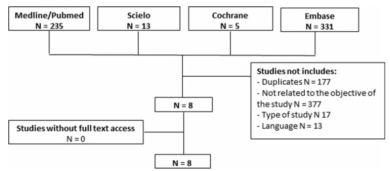
A systematic review was performed following the Preferred Reporting Items for Systematic Reviews and Meta-Analyses (PRISMA) recommendations. The PubMed, SciELO, Cochrane, and Embase databases were consulted, with searches conducted through November 2020, using specific keywords (radial scar OR complex sclerosing lesion, breast cancer, anatomopathological percutaneous biopsy AND/OR surgical biopsy).
Study selection was conducted by two researchers experienced in preparing systematic reviews. The eight selected articles were fully read, and a comparative analysis was performed.
A total of 584 studies was extracted, 8 of which were selected. One of them included women who had undergone a percutaneous biopsy with a histological diagnosis of radial scar/complex sclerosing lesion and subsequently underwent surgical excision; the results were used to assess the underestimation rate of atypical and malignant lesions.
The overall underestimation rate in the 8 studies ranged from 1.3 to 40% and the invasive lesion underestimation rate varied from 0 to 10.5%.
The histopathological diagnosis of a radial scar/complex sclerosing lesion on the breast is not definitive, and it may underestimate atypical andmalignant lesions, which require a different treatment, making surgical excision an important step in diagnostic evaluation.
Summary
Revista Brasileira de Ginecologia e Obstetrícia. 2022;44(1):67-73
02-28-2022
To evaluate the underestimation rate in breast surgical biopsy after the diagnosis of radial scar/complex sclerosing lesion through percutaneous biopsy.
A systematic review was performed following the Preferred Reporting Items for Systematic Reviews and Meta-Analyses (PRISMA) recommendations. The PubMed, SciELO, Cochrane, and Embase databases were consulted, with searches conducted through November 2020, using specific keywords (radial scar OR complex sclerosing lesion, breast cancer, anatomopathological percutaneous biopsy AND/OR surgical biopsy).
Study selection was conducted by two researchers experienced in preparing systematic reviews. The eight selected articles were fully read, and a comparative analysis was performed.
A total of 584 studies was extracted, 8 of which were selected. One of them included women who had undergone a percutaneous biopsy with a histological diagnosis of radial scar/complex sclerosing lesion and subsequently underwent surgical excision; the results were used to assess the underestimation rate of atypical and malignant lesions.
The overall underestimation rate in the 8 studies ranged from 1.3 to 40% and the invasive lesion underestimation rate varied from 0 to 10.5%.
The histopathological diagnosis of a radial scar/complex sclerosing lesion on the breast is not definitive, and it may underestimate atypical andmalignant lesions, which require a different treatment, making surgical excision an important step in diagnostic evaluation.