Summary
Revista Brasileira de Ginecologia e Obstetrícia. 2024;46:e-rbgo30
To evaluate the mode of delivery according to Robson classification (RC) and the perinatal outcomes in fetal growth restriction (FGR) and small for gestational age (SGA) fetuses.
Retrospective cohort study by analyzing medical records of singleton pregnancies from two consecutive years (2018 and 2019). FGR was defined according to Delphi Consensus. The Robson groups were divided into two intervals (1–5.1 and 5.2–10).
Total of 852 cases were included: FGR (n = 85), SGA (n = 20) and control (n=747). FGR showed higher percentages of newborns < 1,500 grams (p<0.001) and higher overall cesarean section (CS) rates (p<0.001). FGR had the highest rates of neonatal resuscitation and neonatal intensive care unit admission (p<0.001). SGA and control presented higher percentage of patients classified in 1 - 5.1 RC groups, while FGR had higher percentage in 5.2 - 10 RC groups (p<0.001). FGR, SGA and control did not differ in the mode of delivery in the 1-5.1 RC groups as all groups showed a higher percentage of vaginal deliveries (p=0.476).
Fetuses with FGR had higher CS rates and worse perinatal outcomes than SGA and control fetuses. Most FGR fetuses were delivered by cesarean section and were allocated in 5.2 to 10 RC groups, while most SGA and control fetuses were allocated in 1 to 5.1 RC groups. Vaginal delivery occurred in nearly 60% of FGR allocated in 1-5.1 RC groups without a significant increase in perinatal morbidity. Therefore, the vaginal route should be considered in FGR fetuses.
Summary
Revista Brasileira de Ginecologia e Obstetrícia. 2016;38(8):373-380
The placenta, translates how the fetus experiences the maternal environment and is a principal influence on birth weight (BW).
To explore the relationship between placental growth measures (PGMs) and BW in a public maternity hospital.
Observational retrospective study of 870 singleton live born infants at Hospital Maternidad Sardá, Universidad de Buenos Aires, Argentina, between January 2011 and August 2012 with complete data of PGMs. Details of history, clinical and obstetrical maternal data, labor and delivery and neonatal outcome data, including placental measures derived from the records, were evaluated. The following manual measurements of the placenta according to standard methods were performed: placental weight (PW, g), larger and smaller diameters (cm), eccentricity, width (cm), shape, area (cm2), BW/PW ratio (BPR) and PW/BW ratio (PBR), and efficiency. Associations between BW and PGMs were examined using multiple linear regression.
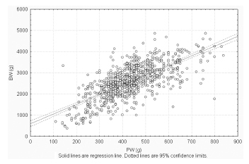
Birth weight was correlated with placental weight (R2 =0.49, p < 0.001), whereas gestational age was moderately correlated with placental weight (R2 =0.64, p < 0.001). By gestational age, there was a positive trend for PW and BPR, but an inverse relationship with PBR (p < 0.001). Placental weight alone accounted for 49% of birth weight variability (p < 0,001), whereas all PGMs accounted for 52% (p < 0,001). Combined, PGMs, maternal characteristics (parity, pre-eclampsia, tobacco use), gestational age and gender explained 77.8% of BW variations (p < 0,001). Among preterm births, 59% of BW variances were accounted for by PGMs, compared with 44% at term. All placental measures except BPR were consistently higher in females than in males, which was also not significant. Indices of placental efficiency showed weakly clinical relevance.
Reliable measures of placental growth estimate 53.6% of BW variances and project this outcome to a greater degree in preterm births than at term. These findings would contribute to the understanding of the maternal-placental programming of chronic diseases.

Summary
Revista Brasileira de Ginecologia e Obstetrícia. 2014;36(12):562-568
DOI 10.1590/SO100-720320140005161
To verify the existence of associations between different maternal ages and the perinatal outcomes of preterm birth and intrauterine growth restriction in the city of São Luís, Maranhão, Northeastern Brazil.
A cross-sectional study using a sample of 5,063 hospital births was conducted in São Luís, from January to December 2010. The participants comprise the birth cohort for the study "Etiological factors of preterm birth and consequences of perinatal factors for infant health: birth cohorts from two Brazilian cities" (BRISA). Frequencies and 95% confidence intervals were used to describe the results. Multiple logistic regression models were applied to assess the adjusted odds ratio (OR) of maternal age associated with the following outcomes: preterm birth and intrauterine growth restriction.
The percentage of early teenage pregnancy (12–15 years old) was 2.2%, and of late (16–19 years old) was 16.4%, while pregnancy at an advanced maternal age (>35 years) was 5.9%. Multivariate analyses showed a statistically significant increase in preterm births among females aged 12–15 years old (OR=1.6; p=0.04) compared with those aged 20–35 years. There was also a higher rate in preterm births among females aged 16–19 years old (OR=1.3; p=0.01). Among those with advanced maternal age (>35 years old), the increase in the prevalence of preterm birth had only borderline statistical significance (OR=1.4; p=0.05). There was no statistically significant association between maternal age and increased prevalence of intrauterine growth restriction.
Summary
Revista Brasileira de Ginecologia e Obstetrícia. 2011;33(3):111-117
DOI 10.1590/S0100-72032011000300002
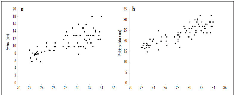
PURPOSE: to assess the distance of the fetal cerebral fissures from the inner edge of the skull by three-dimensional ultrasonography (3DUS). METHODS: this cross-sectional study included 80 women with normal pregnancies between 21st and 34th weeks. The distances between the Sylvian, parieto-occiptal, hippocampus and calcarine fissures and the internal surface of the fetal skull were measured. For the evaluation of the distance of the first three fissures, an axial three-dimensional scan was obtained (at the level of the lateral ventricles). To obtain the calcarine fissure measurement, a coronal scan was used (at the level of the occipital lobes). First degree regressions were performed to assess the correlation between fissure measurements and gestational age, using the determination coefficient (R²) for adjustment. The 5th, 50th and 95th percentiles were calculated for each fissure measurement. Pearson's correlation coefficient (r) was used to assess the correlation between fissure measurements and the biparietal diameter (BPD) and head circumference (HC). RESULTS: all fissure measurements were linearly correlated with gestational age (Sylvian: R²=0.5; parieto-occiptal: R²= 0.7; hippocampus: R²= 0.3 and calcarine: R²= 0.3). Mean fissure measurement ranged from 7.0 to 14.0 mm, 15.9 to 28.7 mm, 15.4 to 25.4 mm and 15.7 to 24.8 mm for the Sylvian, parieto-occiptal, hippocampus and calcarine fissures, respectively. The Sylvian and parieto-occiptal fissure measurements had the highest correlations with the BPD (r=0.8 and 0.7, respectively) and HC (r=0.7 and 0.8, respectively). CONCLUSION: the distance from the fetal cerebral fissures to the inner edge of the skull measured by 3DUS was positively correlated with gestational age.

Summary
Revista Brasileira de Ginecologia e Obstetrícia. 2008;30(12):620-625
DOI 10.1590/S0100-72032008001200006
PURPOSE: to compare delivery and pregnancy follow-up among adolescent and non-adolescent pregnant women whose delivery occurred in a tertiary hospital from Região de Lisboa (Portugal). METHODS: retrospective study with 10,656 deliveries. Pregnancy follow-up, delivery type, need of episiotomy and severe lacerations, Apgar index at the fifth minute and the delivery weight have been evaluated. The pregnant women were divided into two groups, over and under 20 years old. The group with women under 20 was further subdivided in pregnant women under or over 16. The χ2 test has been used for statistical analysis. RESULTS: adolescents presented worse follow-up: first appointment after 12 weeks (46.4 versus 26.3%) and less than four appointments (8.1 versus 3.1%), less dystocia (21.5 versus 35.1%), less caesarian sections (10.6 versus 20.7%), and lower need for inducing labor (16.5 versus 26.5%). There was no significant difference concerning gestational age at delivery and ratio of low weight newborns. Among adolescents, the ones under 16 had more low weight newborns (12 versus 7.4%) and more deliveries between 34 and 37 weeks (10.8 versus 4.2%). CONCLUSIONS: in a hospital attending adolescents with social and psychological support, the fact of them having had a worse follow-up in the pre-natal phase, their performance has not been worse. Nevertheless, special attention might be given to pregnant women under 16.
Summary
Revista Brasileira de Ginecologia e Obstetrícia. 2006;28(3):190-194
DOI 10.1590/S0100-72032006000300009
PURPOSE: to verify the viability of early diagnosis of fetal gender in maternal plasma by the real-time polymerase chain reaction (real-time PCR) starting at the 5th week of pregnancy. METHODS: peripheral blood was collected from pregnant women with single fetus starting at the 5th week of gestation. After centrifugation, 0.4 mL plasma was separated for fetal DNA extraction. The DNA was analyzed in duplicate by real-time PCR for two genomic regions, one of the Y chromosome and the other common to both sexes, through the TaqMan® method, which uses a pair of primers and a fluorescent probe. Patients who aborted were excluded. RESULTS: a total of 79 determinations of fetal DNA in maternal plasma were performed in 52 pregnant women. The results of the determinations were compared to fetal gender after delivery. Accuracy according to gestational age was 92.6% (25 of 27 cases) at 5 weeks with 87% sensitivity, and 95.6% (22 of 23 cases) at 6 weeks with 92% sensitivity. Starting at the 7th week of pregnancy, accuracy was 100% (29 of 29 cases). Specificity was 100% regardless of gestational age. CONCLUSION: real-time PCR for the detection of fetal gender in maternal plasma starting at the 5th week of gestation has good sensitivity and excellent specificity. There was agreement of the results in 100% of the cases in which male gender was diagnosed, regardless of gestational age, and from the 7th week of gestation for female gender diagnosis.
Summary
Revista Brasileira de Ginecologia e Obstetrícia. 2006;28(1):3-9
DOI 10.1590/S0100-72032006000100002
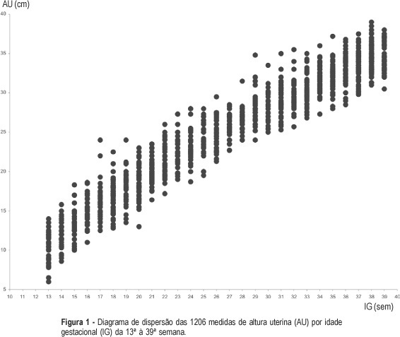
PURPOSE: to build a curve of fundal height according to gestational age among low-risk pregnant women and to compare it with the official standards used in Brazil. METHODS: a prospective observational study was carried out. A sample of 227 low-risk pregnant women with gestational age from 13 to 39 weeks was followed-up in the prenatal care sector of two public health services from João Pessoa, PB. Women with a known gestational age, a single live fetus, without malformation, with no known maternal-fetal pathological condition that could possibly affect fetal growth, with a normal body weight, and non-smokers were included in the study. Their fundal height was measured in a standard way, after a previous ultrasound done to confirm the gestational age. The same investigator performed 1206 measurements and each woman had a mean of 5.3 measurements. Statistical tests were performed with a significance level of 5%. Tables and graphs of fundal height were built according to the gestational age with the 10th, 50th and 90th percentiles. RESULTS: the values of percentiles 10, 50 and 90 of fundal height in each gestational age allowed the construction of a pattern curve of fundal height by gestational age among low-risk pregnant women. A clear visual difference was observed between this new and the official fundal height curve. Statistical analyses showed significant differences between them from the 19th week on. CONCLUSION: the results suggest different normal fundal height and fetal growth patterns among low-risk pregnant women on prenatal assistance compared to the used standard curve, thus with different performances when used for diagnosing fetal growth deviations. Future studies should validate the current fundal height curve by gestational age in order to possibly use it as a reference pattern.

Summary
Revista Brasileira de Ginecologia e Obstetrícia. 2000;22(5):281-286
DOI 10.1590/S0100-72032000000500005
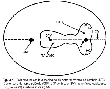
Purpose: to evaluate the effectiveness of the transverse cerebellar diameter (TCD), by ultrasonography, in the evolution of the fetal growth, and to relate it to gestational age, biparietal diameter (BPD), head circumference (HC), abdominal circumference (AC) and femur length (FL). Method: a prospective and longitudinal study was performed on 254 pregnant women considered of low risk, with a gestational age from 20 to 40 weeks. Only 55 pregnant women were included in the study, according to inclusion and exclusion criteria. All the examinations, 217 ultrasonographic evaluations, were done by the author (LN), at least three and at most six examinations for each pregnant woman being accomplished at an interval of one to five weeks. Normality patterns were established between the 10 and 90 percentiles for each gestational age and confirmed postnatally. Results: the transverse cerebellar diameter presented a good correlation with the gestational age either as a dependent variable (R² = 0.90) or as an independent variable (R² = 0.92). A significant relationship was found in the evaluation of the fetal growth between the TCD and the several fetal parameters: BPD and HC (R² = 0.92), FL (R² = 0.90) and AC (R² = 0.89). Conclusions: the transverse cerebellar diameter is a parameter that should be used in the follow-up of development and of fetal growth because of the ascending pattern of its growth curve. Any up- or downward alteration in the growth curve can be useful for the detection of deviations of fetal growth.
