-
Original Article
The Effect of Testosterone Replacement on Intramedullary, Inguinal and Visceral Fat in Ovariectomized Rats
- Lorena Doretto da Silva
 ,
, - Juliana Mora Veridiano ,
- Jussara Celi Conceição Oliveira ,
- Anna Carolina Haddad Sayeg ,
- Ana Maria Amaral Antonio Mader , [ ... ],
- Marcelo Luis Steiner
03-27-2020
Summary
Original ArticleThe Effect of Testosterone Replacement on Intramedullary, Inguinal and Visceral Fat in Ovariectomized Rats
Revista Brasileira de Ginecologia e Obstetrícia. 2020;42(1):43-50
03-27-2020- Lorena Doretto da Silva ,
- Juliana Mora Veridiano ,
- Jussara Celi Conceição Oliveira ,
- Anna Carolina Haddad Sayeg ,
- Ana Maria Amaral Antonio Mader ,
- Giuliana Petri ,
- Bianca Bianco ,
- César Eduardo Fernandes ,
- Olga Maria Szymanski de Toledo ,
- Luciano de Melo Pompei ,
- Marcelo Luis Steiner
Views212See moreAbstract
Objective
The present article aims to evaluate the impact of testosterone treatment on the expansion of visceral, subcutaneous and intramedullary adipose tissue of ovariectomized rats and the visceral and subcutaneous fat expression of peroxisome proliferator-activated receptors (PPARs) gamma.
Methods
In total 48 female Wistar rats were castrated and randomly divided into 6 treatment groups: group E2 was submitted to estradiol 5 μg/day; group T, to testosterone 5 μg/day; group E2+ T, to estradiol 5 μg/day + testosterone 5 μg/day; group TT, to testosterone 30 μg/day; group E2+ TT, to estradiol 5 μg/day+ testosterone 30 μg/day; and placebo was administered to group P. After 5 weeks, the rats were euthanized, the inguinal and visceral adipose tissues were harvested, weighted, and had their PPAR gamma expression evaluated by reverse transcription quantitative polymerase chain reaction (RTqPCR). The right femurs were harvested and histologically prepared to performthe number count of the intramedullary adipocytes.
Results
The expansion of visceral fat tissue was much higher in the TT group when compared with other treated groups (p < 0.001). The TT group also showed a higher expansion of inguinal fat (p < 0.01), and groups E2 +T and E2+ TT presented lower growth compared to the P group (p < 0.01). The number of femur intramedullary adipocytes only showed significant differences between groups TT and E2 + TT (p < 0.05). The expression of PPAR gamma showed no differences among the groups.
Conclusion
The use of testosterone in high doses leads to an important expansion in both visceral and inguinal adipose tissues. Association with estradiol exerts an expansion-repressive effect on the visceral and inguinal adipose tissues.
Views212

This is an Open Access article distributed under the terms of the Creative Commons Attribution License, which permits unrestricted use, distribution, and reproduction in any medium, provided the original work is properly cited. Summary
Original ArticleThe Effect of Testosterone Replacement on Intramedullary, Inguinal and Visceral Fat in Ovariectomized Rats
Revista Brasileira de Ginecologia e Obstetrícia. 2020;42(1):43-50
03-27-2020- Lorena Doretto da Silva ,
- Juliana Mora Veridiano ,
- Jussara Celi Conceição Oliveira ,
- Anna Carolina Haddad Sayeg ,
- Ana Maria Amaral Antonio Mader ,
- Giuliana Petri ,
- Bianca Bianco ,
- César Eduardo Fernandes ,
- Olga Maria Szymanski de Toledo ,
- Luciano de Melo Pompei ,
- Marcelo Luis Steiner
Views212See moreAbstract
Objective
The present article aims to evaluate the impact of testosterone treatment on the expansion of visceral, subcutaneous and intramedullary adipose tissue of ovariectomized rats and the visceral and subcutaneous fat expression of peroxisome proliferator-activated receptors (PPARs) gamma.
Methods
In total 48 female Wistar rats were castrated and randomly divided into 6 treatment groups: group E2 was submitted to estradiol 5 μg/day; group T, to testosterone 5 μg/day; group E2+ T, to estradiol 5 μg/day + testosterone 5 μg/day; group TT, to testosterone 30 μg/day; group E2+ TT, to estradiol 5 μg/day+ testosterone 30 μg/day; and placebo was administered to group P. After 5 weeks, the rats were euthanized, the inguinal and visceral adipose tissues were harvested, weighted, and had their PPAR gamma expression evaluated by reverse transcription quantitative polymerase chain reaction (RTqPCR). The right femurs were harvested and histologically prepared to performthe number count of the intramedullary adipocytes.
Results
The expansion of visceral fat tissue was much higher in the TT group when compared with other treated groups (p < 0.001). The TT group also showed a higher expansion of inguinal fat (p < 0.01), and groups E2 +T and E2+ TT presented lower growth compared to the P group (p < 0.01). The number of femur intramedullary adipocytes only showed significant differences between groups TT and E2 + TT (p < 0.05). The expression of PPAR gamma showed no differences among the groups.
Conclusion
The use of testosterone in high doses leads to an important expansion in both visceral and inguinal adipose tissues. Association with estradiol exerts an expansion-repressive effect on the visceral and inguinal adipose tissues.
This is an Open Access article distributed under the terms of the Creative Commons Attribution License, which permits unrestricted use, distribution, and reproduction in any medium, provided the original work is properly cited. - Lorena Doretto da Silva
-
Original Articles
Estrone and Estradiol Levels in Breast Cancer Patients Using Anastrozole Are Not Related to Body Mass Index
Revista Brasileira de Ginecologia e Obstetrícia. 2017;39(1):14-20
01-01-2017
Summary
Original ArticlesEstrone and Estradiol Levels in Breast Cancer Patients Using Anastrozole Are Not Related to Body Mass Index
Revista Brasileira de Ginecologia e Obstetrícia. 2017;39(1):14-20
01-01-2017Views156See moreABSTRACT
Objective:
Obesity is associated with an increased risk for breast cancer. Recent studies have shown that aromatase inhibitors may be less effective in women with a high body mass index (BMI). The aim of this study was to establish the relationship between the BMI and plasma estrone and estradiol levels in postmenopausal women with hormone receptor-positive breast cancer using anastrozole.
Methods:
In this cohort study, the patients were divided into three groups according to BMI (normal weight, overweight and obese) to compare and correlate plasma hormone levels before starting anastrozole hormone therapy and three months after treatment. Plasma hormone levels were compared for age and use of chemotherapy.
Results:
A statistically significant reduction in estrone and estradiol levels was observed between baseline and three months after starting the anastrozole treatment (p < 0.05). There was no statistically significant difference in plasma estrone and estradiol levels among the BMI groups (p > 0.05), but a significant reduction in plasma estrone levels was observed after three-months' treatment relative to baseline in all groups, as well as a reduction in estradiol in the obese group (p < 0.05). The use of chemotherapy and age > 65 years had no influence on plasma steroid levels.
Conclusion:
Changes in estrone and estradiol levels in the studied groups were not associated with BMI, chemotherapy or age.
Views156This is an Open Access article distributed under the terms of the Creative Commons Attribution License, which permits unrestricted use, distribution, and reproduction in any medium, provided the original work is properly cited. Summary
Original ArticlesEstrone and Estradiol Levels in Breast Cancer Patients Using Anastrozole Are Not Related to Body Mass Index
Revista Brasileira de Ginecologia e Obstetrícia. 2017;39(1):14-20
01-01-2017Views156See moreABSTRACT
Objective:
Obesity is associated with an increased risk for breast cancer. Recent studies have shown that aromatase inhibitors may be less effective in women with a high body mass index (BMI). The aim of this study was to establish the relationship between the BMI and plasma estrone and estradiol levels in postmenopausal women with hormone receptor-positive breast cancer using anastrozole.
Methods:
In this cohort study, the patients were divided into three groups according to BMI (normal weight, overweight and obese) to compare and correlate plasma hormone levels before starting anastrozole hormone therapy and three months after treatment. Plasma hormone levels were compared for age and use of chemotherapy.
Results:
A statistically significant reduction in estrone and estradiol levels was observed between baseline and three months after starting the anastrozole treatment (p < 0.05). There was no statistically significant difference in plasma estrone and estradiol levels among the BMI groups (p > 0.05), but a significant reduction in plasma estrone levels was observed after three-months' treatment relative to baseline in all groups, as well as a reduction in estradiol in the obese group (p < 0.05). The use of chemotherapy and age > 65 years had no influence on plasma steroid levels.
Conclusion:
Changes in estrone and estradiol levels in the studied groups were not associated with BMI, chemotherapy or age.
This is an Open Access article distributed under the terms of the Creative Commons Attribution License, which permits unrestricted use, distribution, and reproduction in any medium, provided the original work is properly cited. 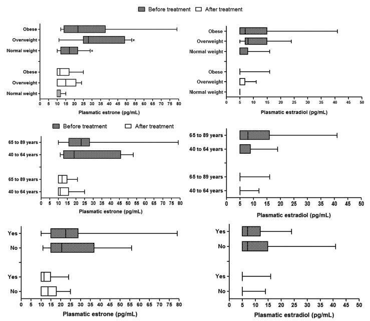
-
Artigos Originais
Cardiomyocytes morphology and collagen quantification in the myocardium of female rats treated with isoflavones or estrogens
Revista Brasileira de Ginecologia e Obstetrícia. 2012;34(10):447-452
12-20-2012
Summary
Artigos OriginaisCardiomyocytes morphology and collagen quantification in the myocardium of female rats treated with isoflavones or estrogens
Revista Brasileira de Ginecologia e Obstetrícia. 2012;34(10):447-452
12-20-2012DOI 10.1590/S0100-72032012001000003
Views99See morePURPOSES: To evaluate the histomorphometry of cardiomyocytes and collagen present in the myocardium of rats treated with a concentrated extract of soy or 17β-estradiol (E2). METHODS: Twenty-eight rats were divided into four groups: GCtrl - estrus phase; GOvx - ovariectomized (Ovx) and receiving vehicle; GIso - Ovx and treated with soy extract (150 mg/kg per day); GE2 - Ovx and treated with E2 (10 µg/kg per day). The drugs and vehicle (0.2 mL propylene glycol) were administered for 30 consecutive days after ovariectomy. On the last day the animals were anesthetized, the hearts removed, submerged in 10% formaldehyde and fragments of the ventricles underwent histological procedures, and the sections were stained with hematoxylin and eosin or picrosirius-red. Histomorphometric analysis (number and volume of nuclei and quantification of collagen) was performed under a light microscope with AxioVision Rel. 4.2 software, and collagen fibers were quantified using IMAGELAB-2000 software. Data were submitted to ANOVA followed by the Tukey test (p<0.05). RESULTS: We observed a higher number of cardiomyocyte nuclei in animals of the Ovx and Iso groups than in GE2 and GCtrl animals (GOvx=121.7±20.2=GIso=92.8±15.4>GE2=70.5±14,8=GCtrl=66.3±9.6; p <0.05), while the nuclear volume was greater in the Ctrl and E2 groups (GE2=35.7±4.8 GCtrl=29.9±3.6=>GIso=26.5±4.5=GOvx=22.4±2.9; p <0.05). Collagen concentration was higher in the Ovx group (GOvx=5.4±0.1>GCtrl=4.0±0.1=GIso=4.4±0.08=GE2=4.3±0.5; p <0.05). CONCLUSIONS: Estrogen may prevent the reduction of the nuclear volume of cardiomyocytes and collagen deposition between heart muscle fibers, while the administration of isoflavones only prevents the deposition of collagen, which can preserve the mechanical properties of cardiac fibers.
Views99This is an Open Access article distributed under the terms of the Creative Commons Attribution License, which permits unrestricted use, distribution, and reproduction in any medium, provided the original work is properly cited. Summary
Artigos OriginaisCardiomyocytes morphology and collagen quantification in the myocardium of female rats treated with isoflavones or estrogens
Revista Brasileira de Ginecologia e Obstetrícia. 2012;34(10):447-452
12-20-2012DOI 10.1590/S0100-72032012001000003
Views99See morePURPOSES: To evaluate the histomorphometry of cardiomyocytes and collagen present in the myocardium of rats treated with a concentrated extract of soy or 17β-estradiol (E2). METHODS: Twenty-eight rats were divided into four groups: GCtrl - estrus phase; GOvx - ovariectomized (Ovx) and receiving vehicle; GIso - Ovx and treated with soy extract (150 mg/kg per day); GE2 - Ovx and treated with E2 (10 µg/kg per day). The drugs and vehicle (0.2 mL propylene glycol) were administered for 30 consecutive days after ovariectomy. On the last day the animals were anesthetized, the hearts removed, submerged in 10% formaldehyde and fragments of the ventricles underwent histological procedures, and the sections were stained with hematoxylin and eosin or picrosirius-red. Histomorphometric analysis (number and volume of nuclei and quantification of collagen) was performed under a light microscope with AxioVision Rel. 4.2 software, and collagen fibers were quantified using IMAGELAB-2000 software. Data were submitted to ANOVA followed by the Tukey test (p<0.05). RESULTS: We observed a higher number of cardiomyocyte nuclei in animals of the Ovx and Iso groups than in GE2 and GCtrl animals (GOvx=121.7±20.2=GIso=92.8±15.4>GE2=70.5±14,8=GCtrl=66.3±9.6; p <0.05), while the nuclear volume was greater in the Ctrl and E2 groups (GE2=35.7±4.8 GCtrl=29.9±3.6=>GIso=26.5±4.5=GOvx=22.4±2.9; p <0.05). Collagen concentration was higher in the Ovx group (GOvx=5.4±0.1>GCtrl=4.0±0.1=GIso=4.4±0.08=GE2=4.3±0.5; p <0.05). CONCLUSIONS: Estrogen may prevent the reduction of the nuclear volume of cardiomyocytes and collagen deposition between heart muscle fibers, while the administration of isoflavones only prevents the deposition of collagen, which can preserve the mechanical properties of cardiac fibers.
This is an Open Access article distributed under the terms of the Creative Commons Attribution License, which permits unrestricted use, distribution, and reproduction in any medium, provided the original work is properly cited. -
Artigos Originais
Modulation by progesterone of pain sensitivity to mechanical and ischemic stimuli in young and healthy women
Revista Brasileira de Ginecologia e Obstetrícia. 2008;30(6):306-311
09-04-2008
Summary
Artigos OriginaisModulation by progesterone of pain sensitivity to mechanical and ischemic stimuli in young and healthy women
Revista Brasileira de Ginecologia e Obstetrícia. 2008;30(6):306-311
09-04-2008DOI 10.1590/S0100-72032008000600007
Views104See morePURPOSE: to investigate the relationship between pain perception (experimental pain threshold and tolerance, in response to ischemia and pressure) in young and healthy young women and female sexual hormone seric levels (estradiol and progesterone). METHODS: 18 volunteers have participated of this study, during three consecutive menstrual cycles. A pressure algometer and a manual dynamometer have been used to measure painful responses to pressure and ischemia algesic stimuli. Blood has been collected for assessment of both hormonal and painful variables, during three menstrual cycles, whose characterization was based on daily oral temperature record, a diary of the menstrual cycles with the onset and end of each cycle, and on estradiol and progesterone plasmatic levels. The average for the algesic variables measured has been compared by analysis of variance (ANOVA) and the Tukey-Kramer's post-test, among the menstrual cycle phases (follicular, periovulatory, early luteal, late luteal and menstrual). The Pearson's test has been used for correlation analysis between algesic and hormonal variables. Statistical significance has been defined as p<0.05. RESULTS: no significant change in pain parameters among the menstrual cycle phases has been observed. Nevertheless, there have been significant negative correlations between progesterone and ischemic threshold (r=-0.23; p<0.01), and pressure tolerance (r=-0.23; p<0.01) at the early luteal phase. CONCLUSIONS: these results indicate that the increase in progesterone levels correlates with a decrease of ischemic threshold and pressure tolerance, suggesting that progesterone plays a role in the pain modulation during the early luteal phase.
Views104This is an Open Access article distributed under the terms of the Creative Commons Attribution License, which permits unrestricted use, distribution, and reproduction in any medium, provided the original work is properly cited. Summary
Artigos OriginaisModulation by progesterone of pain sensitivity to mechanical and ischemic stimuli in young and healthy women
Revista Brasileira de Ginecologia e Obstetrícia. 2008;30(6):306-311
09-04-2008DOI 10.1590/S0100-72032008000600007
Views104See morePURPOSE: to investigate the relationship between pain perception (experimental pain threshold and tolerance, in response to ischemia and pressure) in young and healthy young women and female sexual hormone seric levels (estradiol and progesterone). METHODS: 18 volunteers have participated of this study, during three consecutive menstrual cycles. A pressure algometer and a manual dynamometer have been used to measure painful responses to pressure and ischemia algesic stimuli. Blood has been collected for assessment of both hormonal and painful variables, during three menstrual cycles, whose characterization was based on daily oral temperature record, a diary of the menstrual cycles with the onset and end of each cycle, and on estradiol and progesterone plasmatic levels. The average for the algesic variables measured has been compared by analysis of variance (ANOVA) and the Tukey-Kramer's post-test, among the menstrual cycle phases (follicular, periovulatory, early luteal, late luteal and menstrual). The Pearson's test has been used for correlation analysis between algesic and hormonal variables. Statistical significance has been defined as p<0.05. RESULTS: no significant change in pain parameters among the menstrual cycle phases has been observed. Nevertheless, there have been significant negative correlations between progesterone and ischemic threshold (r=-0.23; p<0.01), and pressure tolerance (r=-0.23; p<0.01) at the early luteal phase. CONCLUSIONS: these results indicate that the increase in progesterone levels correlates with a decrease of ischemic threshold and pressure tolerance, suggesting that progesterone plays a role in the pain modulation during the early luteal phase.
This is an Open Access article distributed under the terms of the Creative Commons Attribution License, which permits unrestricted use, distribution, and reproduction in any medium, provided the original work is properly cited. -
Artigos Originais
Relationship of serum anti-Müllerian hormone, inhibin B, estradiol and FSH on day 3 with ovarian follicular status
Revista Brasileira de Ginecologia e Obstetrícia. 2007;29(4):186-191
07-30-2007
Summary
Artigos OriginaisRelationship of serum anti-Müllerian hormone, inhibin B, estradiol and FSH on day 3 with ovarian follicular status
Revista Brasileira de Ginecologia e Obstetrícia. 2007;29(4):186-191
07-30-2007DOI 10.1590/S0100-72032007000400004
Views61See morePURPOSE: to examine the hypothesis that serum anti-Müllerian hormone (AMH) levels reflect the ovarian follicular status. METHODS: Design: prospective study. Patients: we studied 101 IVF-ET candidates undergoing controlled ovarian hyperstimulation with GnRH agonist and FSH. After the achievement of pituitary suppression and before FSH administration (baseline), serum AMH, inhibin B, and FSH levels were measured. The number of antral follicles was determined by ultrasound at baseline (early antral follicles; 3-10 mm). RESULTS: at baseline, median serum levels of AMH, inhibin B, E2, P4 and FSH were 3.42±0.14 ng/mL, 89±4.8 pg/mL, 34±2.7 pg/mL, 0.22±0.23 ng/mL and 6.6±0.1 mIU/mL, respectively, and the mean number of early antral follicles was 17±0.39. Serum levels of AMH were negatively correlated with age (r=-0.19, p<0.04), and positively correlated with number of antral follicles (r=0.65, p<0.0001), but this did not apply to serum levels of either inhibin B, E2 or FSH. CONCLUSION: the data demonstrate an association between AMH and antral follicular counts. Therefore, AMH is probable a biomarker of ovarian follicular status.
Views61This is an Open Access article distributed under the terms of the Creative Commons Attribution License, which permits unrestricted use, distribution, and reproduction in any medium, provided the original work is properly cited. Summary
Artigos OriginaisRelationship of serum anti-Müllerian hormone, inhibin B, estradiol and FSH on day 3 with ovarian follicular status
Revista Brasileira de Ginecologia e Obstetrícia. 2007;29(4):186-191
07-30-2007DOI 10.1590/S0100-72032007000400004
Views61See morePURPOSE: to examine the hypothesis that serum anti-Müllerian hormone (AMH) levels reflect the ovarian follicular status. METHODS: Design: prospective study. Patients: we studied 101 IVF-ET candidates undergoing controlled ovarian hyperstimulation with GnRH agonist and FSH. After the achievement of pituitary suppression and before FSH administration (baseline), serum AMH, inhibin B, and FSH levels were measured. The number of antral follicles was determined by ultrasound at baseline (early antral follicles; 3-10 mm). RESULTS: at baseline, median serum levels of AMH, inhibin B, E2, P4 and FSH were 3.42±0.14 ng/mL, 89±4.8 pg/mL, 34±2.7 pg/mL, 0.22±0.23 ng/mL and 6.6±0.1 mIU/mL, respectively, and the mean number of early antral follicles was 17±0.39. Serum levels of AMH were negatively correlated with age (r=-0.19, p<0.04), and positively correlated with number of antral follicles (r=0.65, p<0.0001), but this did not apply to serum levels of either inhibin B, E2 or FSH. CONCLUSION: the data demonstrate an association between AMH and antral follicular counts. Therefore, AMH is probable a biomarker of ovarian follicular status.
This is an Open Access article distributed under the terms of the Creative Commons Attribution License, which permits unrestricted use, distribution, and reproduction in any medium, provided the original work is properly cited. -
Trabalhos Originais
Hormonal and serum marker evaluation in patients with abortion
Revista Brasileira de Ginecologia e Obstetrícia. 1998;20(2):91-95
04-16-1998
Summary
Trabalhos OriginaisHormonal and serum marker evaluation in patients with abortion
Revista Brasileira de Ginecologia e Obstetrícia. 1998;20(2):91-95
04-16-1998DOI 10.1590/S0100-72031998000200006
Views77See morePredicting pregnancy outcome from one or more maternal serum factors has been the subject of numerous investigations with controversial results. The aim of this study was to evaluate the serum levels of CA-125, CA-19.9, CA-15.3, beta-hCG, estradiol, progesterone, alpha-fetoprotein and CEA in women with abortion (n=18) and with pregnancy complicated by bleeding (n=6), in comparison to the serum levels of the control group (n=7). The results showed that the serum levels of CA-125 were significantly increased in the abortion group (153.9 ± 43.3 IU/ml), but no difference was detected in pregnancy complicated by bleeding (17.4 ± 2.6 IU/ml), as compared to control (24.7 ± 13.4 IU/ml). However, high serum levels of CA-19.9 were found in the group with pregnancy complicated by bleeding in comparison with the abortion group (20.2 ± 11.4 IU/ml versus 6.6 ± 1.4 IU/ml, respectively). In relation to hormone serum levels, both, the abortion (17.38 ± 9.4 ng/ml) and bleeding (18.3 ± 8.9 ng/ml) groups showed lower serum levels of progesterone, as compared to control (60.4 ± 26.8 ng/ml). Besides, women with abortion had additional low estradiol serum levels, when compared to controls (1,327 ± 1,015 ng/ml versus 10,774 ± 9,244 ng/ml). It was concluded that the serum levels of progesterone, CA-19.9 and beta-hCG seem to add valuable information to the evaluation of a pregnancy complicated by bleeding.
Views77This is an Open Access article distributed under the terms of the Creative Commons Attribution License, which permits unrestricted use, distribution, and reproduction in any medium, provided the original work is properly cited. Summary
Trabalhos OriginaisHormonal and serum marker evaluation in patients with abortion
Revista Brasileira de Ginecologia e Obstetrícia. 1998;20(2):91-95
04-16-1998DOI 10.1590/S0100-72031998000200006
Views77See morePredicting pregnancy outcome from one or more maternal serum factors has been the subject of numerous investigations with controversial results. The aim of this study was to evaluate the serum levels of CA-125, CA-19.9, CA-15.3, beta-hCG, estradiol, progesterone, alpha-fetoprotein and CEA in women with abortion (n=18) and with pregnancy complicated by bleeding (n=6), in comparison to the serum levels of the control group (n=7). The results showed that the serum levels of CA-125 were significantly increased in the abortion group (153.9 ± 43.3 IU/ml), but no difference was detected in pregnancy complicated by bleeding (17.4 ± 2.6 IU/ml), as compared to control (24.7 ± 13.4 IU/ml). However, high serum levels of CA-19.9 were found in the group with pregnancy complicated by bleeding in comparison with the abortion group (20.2 ± 11.4 IU/ml versus 6.6 ± 1.4 IU/ml, respectively). In relation to hormone serum levels, both, the abortion (17.38 ± 9.4 ng/ml) and bleeding (18.3 ± 8.9 ng/ml) groups showed lower serum levels of progesterone, as compared to control (60.4 ± 26.8 ng/ml). Besides, women with abortion had additional low estradiol serum levels, when compared to controls (1,327 ± 1,015 ng/ml versus 10,774 ± 9,244 ng/ml). It was concluded that the serum levels of progesterone, CA-19.9 and beta-hCG seem to add valuable information to the evaluation of a pregnancy complicated by bleeding.
This is an Open Access article distributed under the terms of the Creative Commons Attribution License, which permits unrestricted use, distribution, and reproduction in any medium, provided the original work is properly cited. -
Trabalhos Originais
A study of hormone activity in premenopausal tamoxifen-treated women
Revista Brasileira de Ginecologia e Obstetrícia. 1998;20(9):533-536
04-12-1998
Summary
Trabalhos OriginaisA study of hormone activity in premenopausal tamoxifen-treated women
Revista Brasileira de Ginecologia e Obstetrícia. 1998;20(9):533-536
04-12-1998DOI 10.1590/S0100-72031998000900007
Views126See morePurpose: to evaluate the effects of tamoxifen (TAM) on plasma levels of estradiol, progesterone, prolactin, luteinizing hormone (LH), follicle-stimulating hormone (FSH) and steroid hormone-binding globulin (SHBG) when given to premenopausal women in the doses of 10 and 20 mg/day for 22 days. Patients and Methods: a randomized double-blind study was performed with 43 premenopausal eumenorrheic women. The patients were divided into three groups: A (N = 15, placebo); B (N = 15, TAM 10 mg/day) and C (N = 13, 20 mg/day). They started taking an oral dose of TAM or placebo on the very first day of the menstrual cycle. Two hormone determinations were performed, both on the 22nd day of the menstrual cycle: the first in the cycle that preceded the use of the drug and the second, in the following cycle, after 22 days of using the medication. We used the Levine and Student tests in order to evaluate the homogeneity of the sample and the variation of the hormone determinations respectively. Results:serum levels of estradiol, progesterone and SHBG increased significantly in groups B and C. In group C, we also observed increase in serum level of FSH (p < 0.0045) and a fall in prolactin level (p < 0.0055). Conclusions: TAM promoted a significant increase in serum concentrations of estradiol, progesterone and SHBG either in the doses of 10 or 20 mg/day. However, significant increase in FSH and decrease in prolactin were obtained only with the dose of 20 mg/day.
Views126This is an Open Access article distributed under the terms of the Creative Commons Attribution License, which permits unrestricted use, distribution, and reproduction in any medium, provided the original work is properly cited. Summary
Trabalhos OriginaisA study of hormone activity in premenopausal tamoxifen-treated women
Revista Brasileira de Ginecologia e Obstetrícia. 1998;20(9):533-536
04-12-1998DOI 10.1590/S0100-72031998000900007
Views126See morePurpose: to evaluate the effects of tamoxifen (TAM) on plasma levels of estradiol, progesterone, prolactin, luteinizing hormone (LH), follicle-stimulating hormone (FSH) and steroid hormone-binding globulin (SHBG) when given to premenopausal women in the doses of 10 and 20 mg/day for 22 days. Patients and Methods: a randomized double-blind study was performed with 43 premenopausal eumenorrheic women. The patients were divided into three groups: A (N = 15, placebo); B (N = 15, TAM 10 mg/day) and C (N = 13, 20 mg/day). They started taking an oral dose of TAM or placebo on the very first day of the menstrual cycle. Two hormone determinations were performed, both on the 22nd day of the menstrual cycle: the first in the cycle that preceded the use of the drug and the second, in the following cycle, after 22 days of using the medication. We used the Levine and Student tests in order to evaluate the homogeneity of the sample and the variation of the hormone determinations respectively. Results:serum levels of estradiol, progesterone and SHBG increased significantly in groups B and C. In group C, we also observed increase in serum level of FSH (p < 0.0045) and a fall in prolactin level (p < 0.0055). Conclusions: TAM promoted a significant increase in serum concentrations of estradiol, progesterone and SHBG either in the doses of 10 or 20 mg/day. However, significant increase in FSH and decrease in prolactin were obtained only with the dose of 20 mg/day.
This is an Open Access article distributed under the terms of the Creative Commons Attribution License, which permits unrestricted use, distribution, and reproduction in any medium, provided the original work is properly cited. -
Artigos Originais
FSH, LH, estradiol, progesterone, and histamine concentrations in serum, peritoneal fluid and follicular fluid of women with and without endometriosis
Revista Brasileira de Ginecologia e Obstetrícia. 2006;28(11):643-651
02-12-2006
Summary
Artigos OriginaisFSH, LH, estradiol, progesterone, and histamine concentrations in serum, peritoneal fluid and follicular fluid of women with and without endometriosis
Revista Brasileira de Ginecologia e Obstetrícia. 2006;28(11):643-651
02-12-2006DOI 10.1590/S0100-72032006001100003
Views117PURPOSE: literature reports show that there are no conclusive data about the association between endometriosis and the concentrations of hormones involved in the control of reproduction. Thus, the present study was undertaken to determine FSH, LH, estradiol (E), progesterone (P), and histamine (Hi) concentrations in serum, peritoneal fluid and follicular fluid of women with and without endometriosis. METHODS: the extent of the disease was staged according to the revised American Fertility Society classification (1997). For the collection of serum and peritoneal fluid, 28 women with endometriosis undergoing diagnostic laparoscopy were selected (18 infertile women with endometriosis I-II and ten infertile women with endometriosis III-IV). For the control group, 21 fertile women undergoing laparoscopy for tubal sterilization were selected. Follicular fluid was obtained from 39 infertile women undergoing in vitro fertilization (21 women with endometriosis and 18 women without endometriosis). RESULTS: FSH and LH levels in serum, peritoneal fluid and follicular fluid did not differ significantly between groups. On the other hand, E and P concentrations in the peritoneal fluid were significantly lower in infertile women with endometriosis (E: 154.2±15.3 for stages I-II and 89.3 ng/mL±9.8 ng/mL for stages III-IV; P: 11.2±1.5 for stages I-II and 7.6 ng/mL±0.8 for stages III-IV) in comparison with control women (E: 289.1 ng/mL±30.1; P: 32.8±4.1 ng/mL) (Kruskal-Wallis/Dunn tests; p<0.05). In serum, estradiol and progesterone concentrations followed the same pattern. In the follicular fluid, E and Hi concentrations were significantly lower in women with endometriosis (E: 97.4±11.1 pg/mL; Hi: 6.6±0.9 ng/mL) in comparison to women without endometriosis (E: 237.5±28.5 pg/mL; Hi: 13.8±1.3 ng/mL) (Student t-test; p<0.05), while progesterone levels revealed no significant difference between groups. CONCLUSIONS: our results indicate ovary dysfunction in women with endometriosis, with reduction on E, P and Hi concentrations, which may contribute to the subfertility often associated with the disease.
Key-words EndometriosisEstradiolFertilization in vitroFollicle stimulating hormoneHistamineInfertility, femaleLuteinizing hormoneProgesteroneSee moreViews117This is an Open Access article distributed under the terms of the Creative Commons Attribution License, which permits unrestricted use, distribution, and reproduction in any medium, provided the original work is properly cited. Summary
Artigos OriginaisFSH, LH, estradiol, progesterone, and histamine concentrations in serum, peritoneal fluid and follicular fluid of women with and without endometriosis
Revista Brasileira de Ginecologia e Obstetrícia. 2006;28(11):643-651
02-12-2006DOI 10.1590/S0100-72032006001100003
Views117PURPOSE: literature reports show that there are no conclusive data about the association between endometriosis and the concentrations of hormones involved in the control of reproduction. Thus, the present study was undertaken to determine FSH, LH, estradiol (E), progesterone (P), and histamine (Hi) concentrations in serum, peritoneal fluid and follicular fluid of women with and without endometriosis. METHODS: the extent of the disease was staged according to the revised American Fertility Society classification (1997). For the collection of serum and peritoneal fluid, 28 women with endometriosis undergoing diagnostic laparoscopy were selected (18 infertile women with endometriosis I-II and ten infertile women with endometriosis III-IV). For the control group, 21 fertile women undergoing laparoscopy for tubal sterilization were selected. Follicular fluid was obtained from 39 infertile women undergoing in vitro fertilization (21 women with endometriosis and 18 women without endometriosis). RESULTS: FSH and LH levels in serum, peritoneal fluid and follicular fluid did not differ significantly between groups. On the other hand, E and P concentrations in the peritoneal fluid were significantly lower in infertile women with endometriosis (E: 154.2±15.3 for stages I-II and 89.3 ng/mL±9.8 ng/mL for stages III-IV; P: 11.2±1.5 for stages I-II and 7.6 ng/mL±0.8 for stages III-IV) in comparison with control women (E: 289.1 ng/mL±30.1; P: 32.8±4.1 ng/mL) (Kruskal-Wallis/Dunn tests; p<0.05). In serum, estradiol and progesterone concentrations followed the same pattern. In the follicular fluid, E and Hi concentrations were significantly lower in women with endometriosis (E: 97.4±11.1 pg/mL; Hi: 6.6±0.9 ng/mL) in comparison to women without endometriosis (E: 237.5±28.5 pg/mL; Hi: 13.8±1.3 ng/mL) (Student t-test; p<0.05), while progesterone levels revealed no significant difference between groups. CONCLUSIONS: our results indicate ovary dysfunction in women with endometriosis, with reduction on E, P and Hi concentrations, which may contribute to the subfertility often associated with the disease.
Key-words EndometriosisEstradiolFertilization in vitroFollicle stimulating hormoneHistamineInfertility, femaleLuteinizing hormoneProgesteroneSee moreThis is an Open Access article distributed under the terms of the Creative Commons Attribution License, which permits unrestricted use, distribution, and reproduction in any medium, provided the original work is properly cited.