Summary
Revista Brasileira de Ginecologia e Obstetrícia. 2021;43(12):894-903
01-24-2021
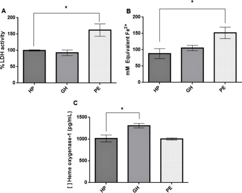
Gestational hypertension (GH) is characterized by increased blood pressure after the 20th gestational week; the presence of proteinuria and/or signs of end-organ damage indicate preeclampsia (PE). Heme oxygenase-1 (HO-1) is an antioxidant enzyme with an important role in maintaining endothelial function, and induction of HO-1 by certain molecules shows potential in attenuating the condition’s effects over endothelial tissue. HO-1 production can also be stimulated by potassium iodide (KI). Therefore, we evaluated the effects of KI over HO-1 expression in human umbilical vein endothelial cells (HUVECs) incubated with plasma from women diagnosed with GH or PE.
Human umbilical vein endothelial cells were incubated with a pool of plasma of healthy pregnant women (n = 12), pregnant women diagnosed with GH (n = 10) or preeclamptic women (n = 11)with or without the addition of KI for 24 hours to evaluate its effect on this enzyme expression. Analysis of variance was performed followed by Dunnet’s test for multiple comparisons between groups only or between groups with addition of KI (p ≤ 0.05).
KI solution (1,000 µM) reduced HO-1 in the gestational hypertension group (p = 0.0018) and cytotoxicity in the preeclamptic group (p = 0.0143); treatment with KI reduced plasma cytotoxicity but did not affect the preeclamptic group’s HO-1 expression.
Our findings suggest that KI alleviates oxidative stress leading to decreased HO-1 expression; plasma from preeclamptic women did not induce the enzyme’s expression in HUVECs, and we hypothesize that this is possibly due to inhibitory post-transcriptional mechanisms in response to overexpression of this enzyme during early pregnancy.

Summary
Revista Brasileira de Ginecologia e Obstetrícia. 2009;31(7):342-348
10-09-2009
DOI 10.1590/S0100-72032009000700004
PURPOSE: to compare echographical cardiovascular risk factors between obese and non-obese patients with micropolycystic ovarian syndrome (MPOS). METHODS: in this transversal study, 30 obese (Body Mass Index, BMI>30 kg/m²) and 60 non-obese (BMI<30 kg/m²) MPOS patients, aging between 18 and 35 years old, were included. The following variables were measured: flow-mediated dilatation (FMD) of the brachial artery, thickness of the intima-media of the carotid artery (IMT), anthropometric data, systolic arterial pressure (SAP) and diastolic arterial pressure (DAP). The women had no previous medical treatment and no comorbidity besides MPOS and obesity. For statistical analysis, the non-paired tand Mann-Whitney's tests were used. RESULTS: obese weighted more than non-obese patients (92.1±11.7 kg versus 61.4±10.7 kg, p<0.0001) and had a larger waist circumference (105.0±10.4 cm versus 78.5±9.8 cm, p<0.0001). The SBP of obese patients was higher than that of the non-obese ones (126.1±10.9 mmHg versus 115.8±9.0 mmHg, p<0.0001) and the IMT was also bigger (0.51±0.07 mm versus 0.44±0.09 mm, p<0.0001). There was no significant difference between the groups as to FMD and carotid rigidity index (β). CONCLUSIONS: obesity in young women with MPOS is associated with higher blood pressure and alteration of arterial structure, represented by a thicker intima-media of the carotid artery.
Summary
Revista Brasileira de Ginecologia e Obstetrícia. 2009;31(3):111-116
06-08-2009
DOI 10.1590/S0100-72032009000300002
PURPOSE: to evaluate whether the presence of insulin resistance (IR) alters cardiovascular risk factors in women with polycystic ovary syndrome (POS). METHODS: transversal study where 60 POS women with ages from 18 to 35 years old, with no hormone intake, were evaluated. IR was assessed through the quantitative insulin sensitivity check index (QUICKI) and defined as QUICKI <0.33. The following variables have been compared between the groups with or without IR: anthropometric (weight, height, waist circumference, arterial blood pressure, cardiac frequency), laboratorial (homocysteine, interleucines-6, factor of tumoral-α necrosis, testosterone, fraction of free androgen, total cholesterol and fractions, triglycerides, C reactive protein, insulin, glucose), and ultrasonographical (distensibility and carotid intima-media thickness, dilation mediated by the brachial artery flux). RESULTS: Eighteen women (30%) presented IR and showed significant differences in the following anthropometric markers, as compared to the women without IR (POS with and without IR respectively): body mass index (35.56±5.69 kg/m² versus 23.90±4.88 kg/m², p<0.01), waist (108.17±11.53 versus 79.54±11.12 cm, p<0.01), systolic blood pressure (128.00±10.80 mmHg versus 114.07±8.97 mmHg, p<0.01), diastolic blood pressure (83.67±9.63 mmHg versus 77.07±7.59 mmHg, p=0.01). It has also been observed significant differences in the following laboratorial markers: triglycerides (120.00±56.53 mg/dL versus 77.79±53.46 mg/dL, p=0.01), HDL (43.06±6.30 mg/dL versus 40.45±10.82 mg/dL, p=0.01), reactive C protein (7.98±10.54 mg/L versus 2.61±3.21 mg/L, p<0.01), insulin (28.01±18.18 µU/mL versus 5.38±2.48 µU/mL, p<0.01), glucose (93.56±10.00 mg/dL versus 87.52±8.75 mg/dL, p=0.02). Additionally, two out of the three ultrasonographical markers of cardiovascular risk were also different between the groups: carotid distensibility (0.24±0.05 mmHg-1 versus 0.30±0.08 mmHg-1, p<0.01) and carotid intima-media thickness (0.52±0.08 mm versus 0.43±0.09, p<0.01). Besides, the metabolic syndrome ratio was higher in women with IR (nine cases=50% versus three cases=7.1%, p<0.01). CONCLUSIONS: POS and IR women present significant differences in several ultrasonographical, seric and anthropometric markers, which point out to higher cardiovascular risk, as compared to women without POS and IR. In face of that, the systematic IR evaluation in POS women may help to identify patients with cardiovascular risk.