Summary
Revista Brasileira de Ginecologia e Obstetrícia. 2022;44(3):287-294
To evaluate the association between polycystic ovary syndrome (PCOS) and metabolic syndrome (MetS), adding liver assessment through elastography and ultrasound, for correlation with non-alcoholic fatty liver disease (NAFLD). Metabolic syndrome occurs in~43% of women with PCOS, and NAFLD is the hepatic expression of MetS.
One hundred women, 50 with PCOS and 50 controls, matched by age (18- 35 years) and body mass index (BMI) were included, restricted to patients with overweight and obesity grade 1, at the Assis Chateaubrian Maternity School, Universidade Federal do Ceará, Brazil. For the diagnosis of PCOS, we adopted the Rotterdam criteria, and for the diagnosis of MetS, the criteria of the National Cholesterol Education Program (NCEP/ATP III). Hepatic elastography and ultrasound were performed to assess liver stiffness and echotexture, respectively.
The average ages were 29.1 (±5.3) and 30.54 (±4.39) years, for the PCOS and the control group, respectively. Patients with PCOS had a risk 4 times higher of having MetS, odds ratio (95% confidence interval)=4.14, than those in the control group. Women with PCOS had higher average of abdominal circumference (100.9±9.08 cm vs 94.96±6.99 cm) and triglycerides (162±54.63 mg/dL vs 137.54±36.91mg/dL) and lower average of HDL cholesterol (45.66±6.88 mg/dL vs 49.78±7.05 mg/dL), with statistically significant difference. Hepatic steatosis was observed on ultrasound in women with PCOS; however, with no statistically significant difference. There was no change to NAFLD at elastography in any group.
Women with PCOS had 4-fold higher frequency of MetS andmore hepatic steatosis, with no statistically significant difference. There was no change in liver stiffness between the groups at elastography. The results can be extended only to populations of overweight and obesity grade 1, with PCOS or not. They cannot be generalized to other untested groups.
Summary
Revista Brasileira de Ginecologia e Obstetrícia. 2017;39(2):72-79
To evaluate the diagnostic accuracy of elastography for breast cancer identification in patients with indeterminate lesions on ultrasound.
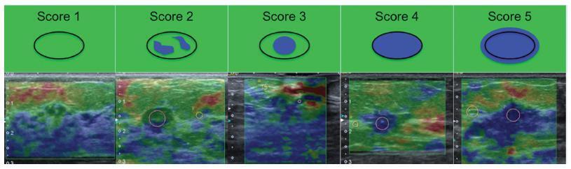
This prospective, descriptive study included patients with indeterminate breast lesions in the ultrasound and with indication for percutaneous or surgical biopsy. The elastography was evaluated by qualitative analysis and by two methods for the semi quantitative analysis.
We evaluated 125 female patients with 159 lesions, with a mean age of 47 years, and a range of 20-85 years. Ultrasound has shown to be a method with good sensitivity (98.1%), but with a lower specificity (40.6%). On the elastography qualitative analysis, the specificity and accuracy were of 80.2% and 81.8% respectively. The mean size of the lesions showed no difference in classification by elastography. For the semiquantitative elastography, the mean values of the malignant lesions were statistically higher when compared with the subcutaneous tissue or the adjacent fibroglandular tissue. The analysis of the receiver operating characteristic (ROC) curves for these two semiquantitativemethods showed that both are considered satisfactory, with an area under the curve above 0.75 and statistical significance (p < 0.0001). The best results were obtained when using the findings of combined conventional ultrasound and qualitative elastography, with 100% sensitivity and 63.2% specificity.
Elastography can be a useful complementary method, increasing the specificity and diagnostic accuracy of conventional ultrasound for the diagnosis of breast cancer in patients with indeterminate breast lesions.
Summary
Revista Brasileira de Ginecologia e Obstetrícia. 2017;39(2):72-79
To evaluate the diagnostic accuracy of elastography for breast cancer identification in patients with indeterminate lesions on ultrasound.
This prospective, descriptive study included patients with indeterminate breast lesions in the ultrasound and with indication for percutaneous or surgical biopsy. The elastography was evaluated by qualitative analysis and by two methods for the semi quantitative analysis.
We evaluated 125 female patients with 159 lesions, with a mean age of 47 years, and a range of 20-85 years. Ultrasound has shown to be a method with good sensitivity (98.1%), but with a lower specificity (40.6%). On the elastography qualitative analysis, the specificity and accuracy were of 80.2% and 81.8% respectively. The mean size of the lesions showed no difference in classification by elastography. For the semiquantitative elastography, the mean values of the malignant lesions were statistically higher when compared with the subcutaneous tissue or the adjacent fibroglandular tissue. The analysis of the receiver operating characteristic (ROC) curves for these two semiquantitativemethods showed that both are considered satisfactory, with an area under the curve above 0.75 and statistical significance (p < 0.0001). The best results were obtained when using the findings of combined conventional ultrasound and qualitative elastography, with 100% sensitivity and 63.2% specificity.
Elastography can be a useful complementary method, increasing the specificity and diagnostic accuracy of conventional ultrasound for the diagnosis of breast cancer in patients with indeterminate breast lesions.