Summary
Revista Brasileira de Ginecologia e Obstetrícia. 09-08-2023;45(7):393-400
Endometriosis causes a decrease in oocyte quality. However, this mechanism is not fully understood. The present study aimed to analyze the effect of endometriosis on cumulus cell adenosine triphosphate ATP level, the number of mitochondria, and the oocyte maturity level.
A true experimental study with a post-test only control group design on experimental animals. Thirty-two mice were divided into control and endometriosis groups. Cumulus oocyte complex (COC) was obtained from all groups. Adenosine triphosphate level on cumulus cells was examined using the Elisa technique, the number of mitochondria was evaluated with a confocal laser scanning microscope and the oocyte maturity level was evaluated with an inverted microscope.
The ATP level of cumulus cells and the number of mitochondria in the endometriosis group increased significantly (p < 0.05; p < 0.05) while the oocyte maturity level was significantly lower (p < 0.05). There was a significant relationship between ATP level of cumulus cells and the number of mitochondrial oocyte (p < 0.01). There was no significant relationship between cumulus cell ATP level and the number of mitochondrial oocytes with oocyte maturity level (p > 0.01; p > 0.01). The ROC curve showed that the number of mitochondrial oocytes (AUC = 0.672) tended to be more accurate than cumulus cell ATP level (AUC = 0.656) in determining the oocyte maturity level.
In endometriosis model mice, the ATP level of cumulus cells and the number of mitochondrial oocytes increased while the oocyte maturity level decreased. There was a correlation between the increase in ATP level of cumulus cells and an increase in the number of mitochondrial oocytes.
Summary
Revista Brasileira de Ginecologia e Obstetrícia. 12-01-2018;40(12):763-770
The aim of the present study was to provide a better understanding of the specific action of two follicle-stimulating hormone (FSH) isoforms (β-follitropin and sheep FSH) on the membrane potential of human cumulus cells.
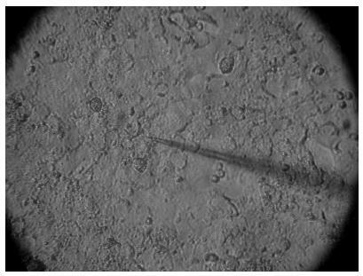
Electrophysiological data were associated with the characteristics of the patient, such as age and cause of infertility. The membrane potential of cumulus cells was recorded with borosilicate microelectrodes filled with KCl (3 M) with tip resistance of 15 to 25 MΩ. Sheep FSH and β-follitropin were topically administered onto the cells after stabilization of the resting potential for at least 5 minutes.
In cumulus cells, the mean resting membrane potential was - 34.02 ± 2.04 mV (n = 14). The mean membrane resistance was 16.5 ± 1.8 MΩ (n = 14). Sheep FSH (4 mUI/mL) and β-follitropin (4 mUI/mL) produced depolarization in the membrane potential 180 and 120 seconds after the administration of the hormone, respectively.
Both FSH isoforms induced similar depolarization patterns, but β-follitropin presented a faster response. A better understanding of the differences of the effects of FSH isoforms on cell membrane potential shall contribute to improve the use of gonadotrophins in fertility treatments.
