-
Original Articles
Gene expression profile of ABC transporters and cytotoxic effect of ibuprofen and acetaminophen in an epithelial ovarian cancer cell line in vitro
Revista Brasileira de Ginecologia e Obstetrícia. 2015;37(6):283-290
06-01-2015
Summary
Original ArticlesGene expression profile of ABC transporters and cytotoxic effect of ibuprofen and acetaminophen in an epithelial ovarian cancer cell line in vitro
Revista Brasileira de Ginecologia e Obstetrícia. 2015;37(6):283-290
06-01-2015DOI 10.1590/SO100-720320150005292
Views136PURPOSES:
To determine the basic expression of ABC transporters in an epithelial ovarian cancer cell line, and to investigate whether low concentrations of acetaminophen and ibuprofen inhibited the growth of this cell line in vitro.
METHODS:
TOV-21 G cells were exposed to different concentrations of acetaminophen (1.5 to 15 μg/mL) and ibuprofen (2.0 to 20 μg/mL) for 24 to 48 hours. The cellular growth was assessed using a cell viability assay. Cellular morphology was determined by fluorescence microscopy. The gene expression profile of ABC transporters was determined by assessing a panel including 42 genes of the ABC transporter superfamily.
RESULTS:
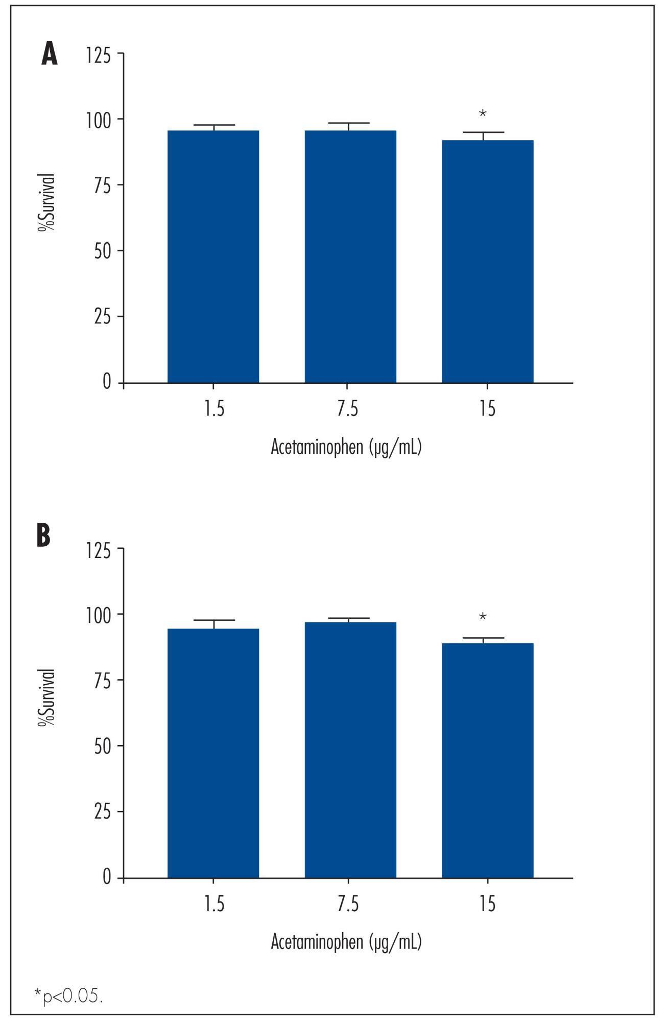
We observed a significant decrease in TOV-21 G cell growth after exposure to 15 μg/mL of acetaminophen for 24 (p=0.02) and 48 hours (p=0.01), or to 20 μg/mL of ibuprofen for 48 hours (p=0.04). Assessing the morphology of TOV-21 G cells did not reveal evidence of extensive apoptosis. TOV-21 G cells had a reduced expression of the genes ABCA1, ABCC3, ABCC4, ABCD3, ABCD4 and ABCE1 within the ABC transporter superfamily.
CONCLUSIONS:
This study provides in vitro evidence of inhibitory effects of growth in therapeutic concentrations of acetaminophen and ibuprofen on TOV-21 G cells. Additionally, TOV-21 G cells presented a reduced expression of the ABCA1, ABCC3, ABCC4, ABCD3, ABCD4 and ABCE1 transporters.
Key-words AcetaminophenApoptosisATP-binding cassette transportersCell proliferationChemopreventionDrug therapyIbuprofenOvarian neoplasmsSee more
-
Artigos Originais
Expression of Bax protein in normal tissue of premenopausal women treated with raloxifene
Revista Brasileira de Ginecologia e Obstetrícia. 2008;30(4):177-181
07-02-2008
Summary
Artigos OriginaisExpression of Bax protein in normal tissue of premenopausal women treated with raloxifene
Revista Brasileira de Ginecologia e Obstetrícia. 2008;30(4):177-181
07-02-2008DOI 10.1590/S0100-72032008000400004
Views96See morePURPOSE: to evaluate the expression of Bax antigen in the normal mammary epithelium of premenopausal women treated with raloxifene. METHODS: a randomized double-blind study was conducted in 33 ovulatory premenopausal women with fibroadenoma. Patients were divided into two groups: Placebo, (n=18) and Raloxifene 60 mg, (n=15). The medication was used for 22 days, beginning on the first day of the menstrual cycle. An excisional biopsy was carried out on the 23rd day of the menstrual cycle and a sample of normal breast tissue adjacent to the fibroadenoma was collected and submitted to immunohistochemical study using anti-Bax polyclonal antibody to evaluate the expression of Bax protein. Immunoreaction for Bax was evaluated taking into consideration intensity and fraction of stained cells, whose combination resulted in a final score ranging from 0 to 6. Cases with a final score >3 were classified as positive for Bax. The c2 test was used for statistical analysis (p<0.05). RESULTS: the percentage of positivity of Bax protein expression was 66.7 and 73.3% in Groups A and B, respectively. There was no significant difference in Bax expression between the two groups (p=0.678). CONCLUSIONS: raloxifene, administered for 22 days in the dose of 60 mg/day, did not alter the expression of Bax protein in the breast normal tissue of premenopausal women.


