Summary
Revista Brasileira de Ginecologia e Obstetrícia. 2017;39(9):443-452
09-01-2017
To define transvaginal ultrasound reference ranges for uterine cervix measurements according to gestational age (GA) in low-risk pregnancies.
Cohort of low-risk pregnantwomen undergoing transvaginal ultrasound exams every 4 weeks, comprisingmeasurements of the cervical length and volume, the transverse and anteroposterior diameters of the cervix, and distance fromthe entrance of the uterine artery into the cervix until the internal os. The inter- and intraobserver variabilities were assessed with the linear correlation coefficient and the Student t-test. Within each period of GA, 2.5, 10, 50, 90 and 97.5 percentiles were estimated, and the variation by GA was assessed with analysis of variance for dependent samples. Mean values and Student t-test were used to compare the values stratified by control variables.
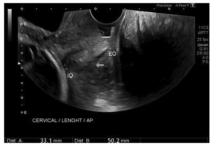
After confirming the high reproducibility of the method, 172 women followed in this cohort presented a reduction in cervical length, with an increase in volume and in the anteroposterior and transverse diameters during pregnancy. Smaller cervical lengths were associated with younger age, lower parity, and absence of previous cesarean section (C-section).
In the studied population, we observed cervical length shortening throughout pregnancy, suggesting a physiological reduction mainly in the vaginal portion of the cervix. In order to better predict pretermbirth, cervical insufficiency and premature rupture of membranes, reference curves and specific cut-off values need to be validated.
Summary
Revista Brasileira de Ginecologia e Obstetrícia. 2017;39(9):443-452
09-01-2017
To define transvaginal ultrasound reference ranges for uterine cervix measurements according to gestational age (GA) in low-risk pregnancies.
Cohort of low-risk pregnantwomen undergoing transvaginal ultrasound exams every 4 weeks, comprisingmeasurements of the cervical length and volume, the transverse and anteroposterior diameters of the cervix, and distance fromthe entrance of the uterine artery into the cervix until the internal os. The inter- and intraobserver variabilities were assessed with the linear correlation coefficient and the Student t-test. Within each period of GA, 2.5, 10, 50, 90 and 97.5 percentiles were estimated, and the variation by GA was assessed with analysis of variance for dependent samples. Mean values and Student t-test were used to compare the values stratified by control variables.
After confirming the high reproducibility of the method, 172 women followed in this cohort presented a reduction in cervical length, with an increase in volume and in the anteroposterior and transverse diameters during pregnancy. Smaller cervical lengths were associated with younger age, lower parity, and absence of previous cesarean section (C-section).
In the studied population, we observed cervical length shortening throughout pregnancy, suggesting a physiological reduction mainly in the vaginal portion of the cervix. In order to better predict pretermbirth, cervical insufficiency and premature rupture of membranes, reference curves and specific cut-off values need to be validated.
Summary
Revista Brasileira de Ginecologia e Obstetrícia. 2003;25(2):115-121
07-04-2003
DOI 10.1590/S0100-72032003000200007
PURPOSE: to establish a normality curve of cervical length during pregnancy measured by transvaginal ultrasonography. METHODS: we conducted a prospective, longitudinal study on 82 healthy pregnant women who were followed up from the beginning of pregnancy to delivery at four-week intervals, of whom 49 concluded the study. Patients were divided according to parity into nulliparous women and women with one or more previous deliveries. Cervical length was measured in a sagittal view by transvaginal ultrasonography, as the linear distance between internal and external cervical os. RESULTS: no significant difference was observed in mean cervical length or the 5th, 25, 50th, 75th, or 95th percentile according to gestational age between groups (p>0.05). Between the 20thand 24th gestacional week, the 5th, 50th and 95th percentiles of cervical length were 28, 35 and 47.2 mm, respectively. Cervical length decreased progressively during normal pregnancy, with a significant shortening observed after 20 weeks of gestation and being more marked after 28 weeks (p<0.05). CONCLUSION: the pattern of cervical length behavior does not seem to differ between nulliparous women and women with one or more previous deliveries. The numerical values of the normality curve of cervical length according to gestational age reflect the variability in the peculiar characteristics of the studied sample, thus emphasizing the value of the parameters established for different populations.