-
Original Article
Thalidomide Reduces Cell Proliferation in Endometriosis Experimentally Induced in Rats
Revista Brasileira de Ginecologia e Obstetrícia. 2019;41(11):668-672
12-20-2019
Summary
Original ArticleThalidomide Reduces Cell Proliferation in Endometriosis Experimentally Induced in Rats
Revista Brasileira de Ginecologia e Obstetrícia. 2019;41(11):668-672
12-20-2019Views145See moreAbstract
Objective
To analyze the effect of thalidomide on the progression of endometriotic lesions experimentally induced in rats and to characterize the pattern of cell proliferation by immunohistochemical Proliferating Cell Nuclear Antigen (PCNA) labeling of eutopic and ectopic endometrium.
Methods
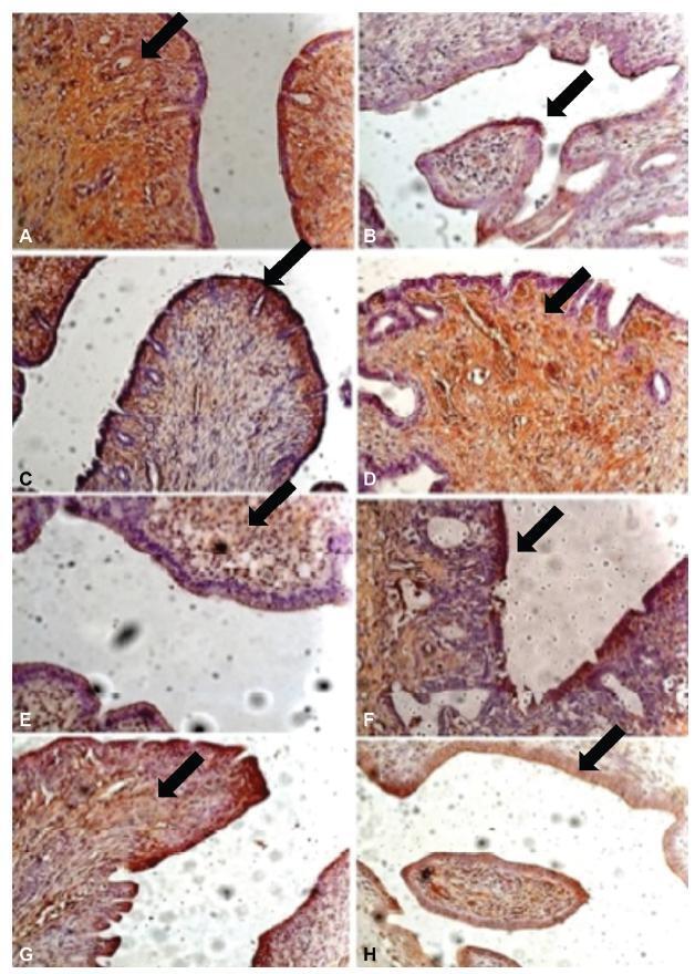
Fifteen female Wistar rats underwent laparotomy for endometriosis induction by resection of one uterine horn, isolation of the endometrium and fixation of a tissue segment to the pelvic peritoneum. Four weeks after, the animals were divided into 3 groups: control (I), 10mg/kg/day (II) and 1mg/kg/day (III) intraperitoneal thalidomide for 10 days. The lesion was excised together with the opposite uterine horn for endometrial gland and stroma analysis. Eutopic and ectopic endometrial tissue was submitted to immunohistochemistry for analysis of cell proliferation by PCNA labeling and the cell proliferation index (CPI) was calculated as the number of labeled cells per 1,000 cells.
Results
Group I showed a mean CPI of 0.248 ± 0.0513 in the gland and of 0.178 ± 0.046 in the stroma. In contrast, Groups II and III showed a significantly lower CPI, that is, 0.088 ± 0.009 and 0.080 ± 0.021 for the gland (p < 0.001) and 0.0945 ± 0.0066 and 0.075 ± 0.018 for the stroma (p < 0.001), respectively. Also, the mean lesion area of Group I was 69.2mm2, a significantly higher value compared with Group II (49.4mm2, p = 0.023) and Group III (48.6mm2, p = 0.006). No significant difference was observed between Groups II and III.
Conclusion
Thalidomide proved to be effective in reducing the lesion area and CPI of the experimental endometriosis implants both at the dose of 1mg/kg/day and at the dose of 10 mg/kg/day.
-
Original Articles
Increased Expression Levels of Metalloprotease, Tissue Inhibitor of Metalloprotease, Metallothionein, and p63 in Ectopic Endometrium: An Animal Experimental Study
Revista Brasileira de Ginecologia e Obstetrícia. 2018;40(11):705-712
11-01-2018
Summary
Original ArticlesIncreased Expression Levels of Metalloprotease, Tissue Inhibitor of Metalloprotease, Metallothionein, and p63 in Ectopic Endometrium: An Animal Experimental Study
Revista Brasileira de Ginecologia e Obstetrícia. 2018;40(11):705-712
11-01-2018Views137See moreAbstract
Objective
To characterize the patterns of cell differentiation, proliferation, and tissue invasion in eutopic and ectopic endometrium of rabbits with induced endometriotic lesions via a well- known experimental model, 4 and 8 weeks after the endometrial implantation procedure.
Methods
Twenty-nine female New Zealand rabbits underwent laparotomy for endometriosis induction through the resection of one uterine horn, isolation of the endometrium, and fixation of tissue segment to the pelvic peritoneum. Two groups of animals (one with 14 animals, and the other with15) were sacrificed 4 and 8 weeks after endometriosis induction. The lesion was excised along with the opposite uterine horn for endometrial gland and stroma determination. Immunohistochemical reactions were performed in eutopic and ectopic endometrial tissues for analysis of the following markers: metalloprotease (MMP-9) and tissue inhibitor of metalloprotease (TIMP-2), which are involved in the invasive capacity of the endometrial tissue; and metallothionein (MT) and p63, which are involved in cell differentiation and proliferation.
Results
The intensity of the immunostaining for MMP9, TIMP-2, MT, and p63 was higher in ectopic endometria than in eutopic endometria. However, when the ectopic lesions were compared at 4 and 8 weeks, no significant difference was observed, with the exception of the marker p63, which was more evident after 8 weeks of evolution of the ectopic endometrial tissue.
Conclusion
Ectopic endometrial lesions seem to express greater power for cell differentiation and tissue invasion, compared with eutopic endometria, demonstrating a potentially invasive, progressive, and heterogeneous presentation of endometriosis.

-
Original Articles
Gene expression profile of ABC transporters and cytotoxic effect of ibuprofen and acetaminophen in an epithelial ovarian cancer cell line in vitro
Revista Brasileira de Ginecologia e Obstetrícia. 2015;37(6):283-290
06-01-2015
Summary
Original ArticlesGene expression profile of ABC transporters and cytotoxic effect of ibuprofen and acetaminophen in an epithelial ovarian cancer cell line in vitro
Revista Brasileira de Ginecologia e Obstetrícia. 2015;37(6):283-290
06-01-2015DOI 10.1590/SO100-720320150005292
Views124PURPOSES:
To determine the basic expression of ABC transporters in an epithelial ovarian cancer cell line, and to investigate whether low concentrations of acetaminophen and ibuprofen inhibited the growth of this cell line in vitro.
METHODS:
TOV-21 G cells were exposed to different concentrations of acetaminophen (1.5 to 15 μg/mL) and ibuprofen (2.0 to 20 μg/mL) for 24 to 48 hours. The cellular growth was assessed using a cell viability assay. Cellular morphology was determined by fluorescence microscopy. The gene expression profile of ABC transporters was determined by assessing a panel including 42 genes of the ABC transporter superfamily.
RESULTS:
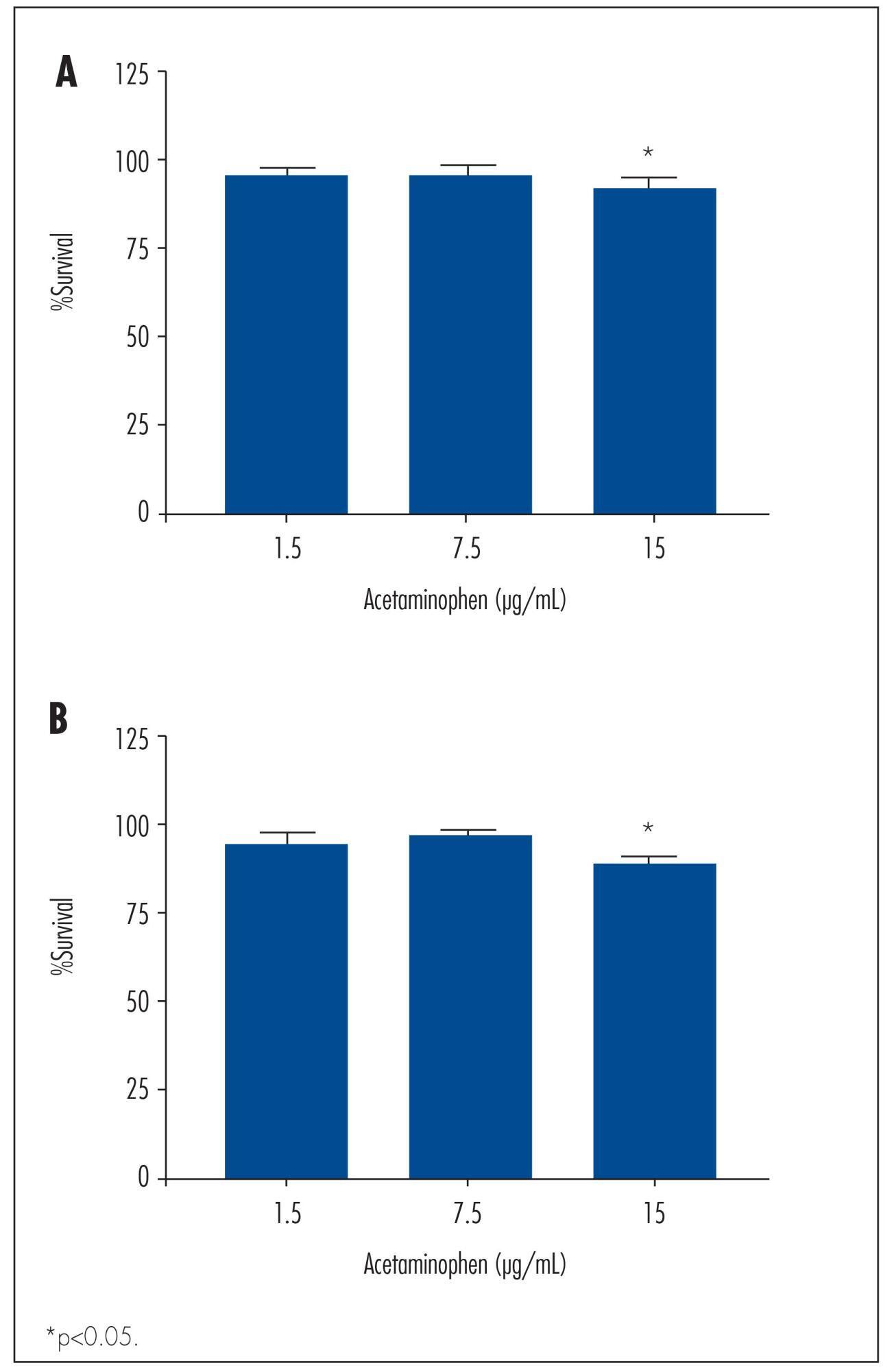
We observed a significant decrease in TOV-21 G cell growth after exposure to 15 μg/mL of acetaminophen for 24 (p=0.02) and 48 hours (p=0.01), or to 20 μg/mL of ibuprofen for 48 hours (p=0.04). Assessing the morphology of TOV-21 G cells did not reveal evidence of extensive apoptosis. TOV-21 G cells had a reduced expression of the genes ABCA1, ABCC3, ABCC4, ABCD3, ABCD4 and ABCE1 within the ABC transporter superfamily.
CONCLUSIONS:
This study provides in vitro evidence of inhibitory effects of growth in therapeutic concentrations of acetaminophen and ibuprofen on TOV-21 G cells. Additionally, TOV-21 G cells presented a reduced expression of the ABCA1, ABCC3, ABCC4, ABCD3, ABCD4 and ABCE1 transporters.
Key-words AcetaminophenApoptosisATP-binding cassette transportersCell proliferationChemopreventionDrug therapyIbuprofenOvarian neoplasmsSee more
-
Artigos Originais
Effects of tibolone on the breast parenchyma: experimental study
Revista Brasileira de Ginecologia e Obstetrícia. 2015;37(5):233-240
05-01-2015
Summary
Artigos OriginaisEffects of tibolone on the breast parenchyma: experimental study
Revista Brasileira de Ginecologia e Obstetrícia. 2015;37(5):233-240
05-01-2015DOI 10.1590/SO100-720320150005333
Views84OBJECTIVE:
To assess the effect of tibolone on mammary tissue of castrated rats over 3
different periods of time.METHODS:
Sixty virgin female Wistar rats were submitted to oophorectomy. Twenty-one days
after surgery, with hypoestrogenism confirmed, the experimental rats were randomly
assigned to six groups: Tibolone 1 (n=10) received tibolone 1 mg/day for 23 days,
tibolone 2 (n=10) for 59 days and tibolone 3 (n=10) for 118 days. The groups
control 1 (n=8), control 2 (n=7) and control 2 (n=10) received distilled water for
23, 59 and 118 days, respectively. After treatment, all six pairs of mammary
glands were removed and stained with hematoxylin and eosin (HE) for histological
analysis after euthanasia. The histological parameters evaluated were: epithelial
cell proliferation and secretory activity. The variables were analyzed
statistically, with the level of significance set at 0.05.RESULTS:
Histological changes were observed in 20/55 rats, mild epithelial hyperplasia in
7/55, moderate epithelial hyperplasia in 5/55, alveolar-nodular hyperplasia in
7/55, atypia without epithelial proliferation in 1/55, and no cases of severe
epithelial hyperplasia were found. Secretory activity was observed in 31/55 rats.
The secretory activity was significantly higher in the tibolone groups compared to
control at all the time points assessed (p=0,001). The histological changes were
did not show significance when the control and tibolone groups were compared. The
time of exposure to tibolone did not show significance when the three different
periods of evaluation were compared.CONCLUSION:
No relation between histological modification and tibolone treatment was verified
after short-, medium- and long-term treatment.Key-words Breast neoplasmsCell proliferationEstrogens/therapeutic useHormone replacement therapyHyperplasiaRatsTiboloneSee more


