-
Original Article
Bacteriological characteristics of primary breast abscesses in patients from the community in the era of microbial resistance
- Vicente Sperb Antonello
 ,
, - Jessica Dallé ,
- Mirela Foresti Jimenez ,
- Patrícia Tramontini ,
- Andrei Gustavo Reginatto
04-09-2024
Summary
Original ArticleBacteriological characteristics of primary breast abscesses in patients from the community in the era of microbial resistance
Revista Brasileira de Ginecologia e Obstetrícia. 2024;46:e-rbgo34
04-09-2024- Vicente Sperb Antonello ,
- Jessica Dallé ,
- Mirela Foresti Jimenez ,
- Patrícia Tramontini ,
- Andrei Gustavo Reginatto
Views330See moreAbstract
Objective:
The aim of this study is to evaluate the etiological profile and antimicrobial resistance in breast abscess cultures from patients from the community, treated at a public hospital located in Porto Alegre, Brazil.
Methods:
This is an retrospective cross-sectional study that evaluated the medical records of patients with bacterial isolates in breast abscess secretion cultures and their antibiograms, from January 2010 to August 2022.
Results:
Based on 129 positive cultures from women from the community diagnosed with breast abscesses and treated at Fêmina Hospital, 99 (76.7%) of the patients had positive cultures for Staphylococcus sp, 91 (92%) of which were cases of Staphylococcus aureus. Regarding the resistance profile of S. aureus, 32% of the strains were resistant to clindamycin, 26% to oxacillin and 5% to trimethoprim-sulfamethoxazole. The antimicrobials vancomycin, linezolid and tigecycline did not show resistance for S. aureus.
Conclusion:
Staphylococcus aureus was the most common pathogen found in the breast abscess isolates during the study period. Oxacillin remains a good option for hospitalized patients. The use of sulfamethoxazole plus trimethoprim should be considered as a good option for use at home, due to its low bacterial resistance, effectiveness and low cost.
Views330

This is an Open Access article distributed under the terms of the Creative Commons Attribution License, which permits unrestricted use, distribution, and reproduction in any medium, provided the original work is properly cited. Summary
Original ArticleBacteriological characteristics of primary breast abscesses in patients from the community in the era of microbial resistance
Revista Brasileira de Ginecologia e Obstetrícia. 2024;46:e-rbgo34
04-09-2024- Vicente Sperb Antonello ,
- Jessica Dallé ,
- Mirela Foresti Jimenez ,
- Patrícia Tramontini ,
- Andrei Gustavo Reginatto
Views330See moreAbstract
Objective:
The aim of this study is to evaluate the etiological profile and antimicrobial resistance in breast abscess cultures from patients from the community, treated at a public hospital located in Porto Alegre, Brazil.
Methods:
This is an retrospective cross-sectional study that evaluated the medical records of patients with bacterial isolates in breast abscess secretion cultures and their antibiograms, from January 2010 to August 2022.
Results:
Based on 129 positive cultures from women from the community diagnosed with breast abscesses and treated at Fêmina Hospital, 99 (76.7%) of the patients had positive cultures for Staphylococcus sp, 91 (92%) of which were cases of Staphylococcus aureus. Regarding the resistance profile of S. aureus, 32% of the strains were resistant to clindamycin, 26% to oxacillin and 5% to trimethoprim-sulfamethoxazole. The antimicrobials vancomycin, linezolid and tigecycline did not show resistance for S. aureus.
Conclusion:
Staphylococcus aureus was the most common pathogen found in the breast abscess isolates during the study period. Oxacillin remains a good option for hospitalized patients. The use of sulfamethoxazole plus trimethoprim should be considered as a good option for use at home, due to its low bacterial resistance, effectiveness and low cost.
This is an Open Access article distributed under the terms of the Creative Commons Attribution License, which permits unrestricted use, distribution, and reproduction in any medium, provided the original work is properly cited. - Vicente Sperb Antonello
-
Review Article
Underestimation Rate in the Percutaneous Diagnosis of Radial Scar/Complex Sclerosing Lesion of the Breast: Systematic Review
- Ana Beatrice Bonganha Zanon ,
- Jonathan Yugo Maesaka ,
- Bruna Bello Chequin ,
- Ana Gabriela de Siqueira Santos ,
- Edmund Chada Baracat , [ ... ],
- José Roberto Filassi
02-28-2022
Summary
Review ArticleUnderestimation Rate in the Percutaneous Diagnosis of Radial Scar/Complex Sclerosing Lesion of the Breast: Systematic Review
Revista Brasileira de Ginecologia e Obstetrícia. 2022;44(1):67-73
02-28-2022- Ana Beatrice Bonganha Zanon ,
- Jonathan Yugo Maesaka ,
- Bruna Bello Chequin ,
- Ana Gabriela de Siqueira Santos ,
- Edmund Chada Baracat ,
- José Roberto Filassi
Views113See moreAbstract
Objective
To evaluate the underestimation rate in breast surgical biopsy after the diagnosis of radial scar/complex sclerosing lesion through percutaneous biopsy.
Data Sources
A systematic review was performed following the Preferred Reporting Items for Systematic Reviews and Meta-Analyses (PRISMA) recommendations. The PubMed, SciELO, Cochrane, and Embase databases were consulted, with searches conducted through November 2020, using specific keywords (radial scar OR complex sclerosing lesion, breast cancer, anatomopathological percutaneous biopsy AND/OR surgical biopsy).
Data collection
Study selection was conducted by two researchers experienced in preparing systematic reviews. The eight selected articles were fully read, and a comparative analysis was performed.
Study selection
A total of 584 studies was extracted, 8 of which were selected. One of them included women who had undergone a percutaneous biopsy with a histological diagnosis of radial scar/complex sclerosing lesion and subsequently underwent surgical excision; the results were used to assess the underestimation rate of atypical and malignant lesions.
Data synthesis
The overall underestimation rate in the 8 studies ranged from 1.3 to 40% and the invasive lesion underestimation rate varied from 0 to 10.5%.
Conclusion
The histopathological diagnosis of a radial scar/complex sclerosing lesion on the breast is not definitive, and it may underestimate atypical andmalignant lesions, which require a different treatment, making surgical excision an important step in diagnostic evaluation.
Views113This is an Open Access article distributed under the terms of the Creative Commons Attribution License, which permits unrestricted use, distribution, and reproduction in any medium, provided the original work is properly cited. Summary
Review ArticleUnderestimation Rate in the Percutaneous Diagnosis of Radial Scar/Complex Sclerosing Lesion of the Breast: Systematic Review
Revista Brasileira de Ginecologia e Obstetrícia. 2022;44(1):67-73
02-28-2022- Ana Beatrice Bonganha Zanon ,
- Jonathan Yugo Maesaka ,
- Bruna Bello Chequin ,
- Ana Gabriela de Siqueira Santos ,
- Edmund Chada Baracat ,
- José Roberto Filassi
Views113See moreAbstract
Objective
To evaluate the underestimation rate in breast surgical biopsy after the diagnosis of radial scar/complex sclerosing lesion through percutaneous biopsy.
Data Sources
A systematic review was performed following the Preferred Reporting Items for Systematic Reviews and Meta-Analyses (PRISMA) recommendations. The PubMed, SciELO, Cochrane, and Embase databases were consulted, with searches conducted through November 2020, using specific keywords (radial scar OR complex sclerosing lesion, breast cancer, anatomopathological percutaneous biopsy AND/OR surgical biopsy).
Data collection
Study selection was conducted by two researchers experienced in preparing systematic reviews. The eight selected articles were fully read, and a comparative analysis was performed.
Study selection
A total of 584 studies was extracted, 8 of which were selected. One of them included women who had undergone a percutaneous biopsy with a histological diagnosis of radial scar/complex sclerosing lesion and subsequently underwent surgical excision; the results were used to assess the underestimation rate of atypical and malignant lesions.
Data synthesis
The overall underestimation rate in the 8 studies ranged from 1.3 to 40% and the invasive lesion underestimation rate varied from 0 to 10.5%.
Conclusion
The histopathological diagnosis of a radial scar/complex sclerosing lesion on the breast is not definitive, and it may underestimate atypical andmalignant lesions, which require a different treatment, making surgical excision an important step in diagnostic evaluation.
This is an Open Access article distributed under the terms of the Creative Commons Attribution License, which permits unrestricted use, distribution, and reproduction in any medium, provided the original work is properly cited. 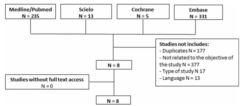
- Ana Beatrice Bonganha Zanon
-
Artigos Originais
Association between breast arterial calcifications and cardiovascular risk factors in menopausal women
Revista Brasileira de Ginecologia e Obstetrícia. 2014;36(7):315-319
07-29-2014
Summary
Artigos OriginaisAssociation between breast arterial calcifications and cardiovascular risk factors in menopausal women
Revista Brasileira de Ginecologia e Obstetrícia. 2014;36(7):315-319
07-29-2014DOI 10.159/S0100-720320140004977
Views132PURPOSE:
To analyze associations between mammographic arterial mammary calcifications in menopausal women and risk factors for cardiovascular disease.
METHODS:
This was a cross-sectional retrospective study, in which we analyzed the mammograms and medical records of 197 patients treated between 2004 and 2005. Study variables were: breast arterial calcifications, stroke, acute coronary syndrome, age, obesity, diabetes mellitus, smoking, and hypertension. For statistical analysis, we used the Mann-Whitney, χ2 and Cochran-Armitage tests, and also evaluated the prevalence ratios between these variables and mammary artery calcifications. Data were analyzed with the SAS version 9.1 software.
RESULTS:
In the group of 197 women, there was a prevalence of 36.6% of arterial calcifications on mammograms. Among the risk factors analyzed, the most frequent were hypertension (56.4%), obesity (31.9%), smoking (15.2%), and diabetes (14.7%). Acute coronary syndrome and stroke presented 5.6 and 2.0% of prevalence, respectively. Among the mammograms of women with diabetes, the odds ratio of mammary artery calcifications was 2.1 (95%CI 1.0-4.1), with p-value of 0.02. On the other hand, the mammograms of smokers showed the low occurrence of breast arterial calcification, with an odds ratio of 0.3 (95%CI 0.1-0.8). Hypertension, obesity, diabetes mellitus, stroke and acute coronary syndrome were not significantly associated with breast arterial calcification.
CONCLUSION:
The occurrence of breast arterial calcification was associated with diabetes mellitus and was negatively associated with smoking. The presence of calcification was independent of the other risk factors for cardiovascular disease analyzed.
Key-words Breast diseasesCalcinosis/pathologyCardiovascular diseasesMammographyMenopauseRisk factorsSee moreViews132This is an Open Access article distributed under the terms of the Creative Commons Attribution License, which permits unrestricted use, distribution, and reproduction in any medium, provided the original work is properly cited. Summary
Artigos OriginaisAssociation between breast arterial calcifications and cardiovascular risk factors in menopausal women
Revista Brasileira de Ginecologia e Obstetrícia. 2014;36(7):315-319
07-29-2014DOI 10.159/S0100-720320140004977
Views132PURPOSE:
To analyze associations between mammographic arterial mammary calcifications in menopausal women and risk factors for cardiovascular disease.
METHODS:
This was a cross-sectional retrospective study, in which we analyzed the mammograms and medical records of 197 patients treated between 2004 and 2005. Study variables were: breast arterial calcifications, stroke, acute coronary syndrome, age, obesity, diabetes mellitus, smoking, and hypertension. For statistical analysis, we used the Mann-Whitney, χ2 and Cochran-Armitage tests, and also evaluated the prevalence ratios between these variables and mammary artery calcifications. Data were analyzed with the SAS version 9.1 software.
RESULTS:
In the group of 197 women, there was a prevalence of 36.6% of arterial calcifications on mammograms. Among the risk factors analyzed, the most frequent were hypertension (56.4%), obesity (31.9%), smoking (15.2%), and diabetes (14.7%). Acute coronary syndrome and stroke presented 5.6 and 2.0% of prevalence, respectively. Among the mammograms of women with diabetes, the odds ratio of mammary artery calcifications was 2.1 (95%CI 1.0-4.1), with p-value of 0.02. On the other hand, the mammograms of smokers showed the low occurrence of breast arterial calcification, with an odds ratio of 0.3 (95%CI 0.1-0.8). Hypertension, obesity, diabetes mellitus, stroke and acute coronary syndrome were not significantly associated with breast arterial calcification.
CONCLUSION:
The occurrence of breast arterial calcification was associated with diabetes mellitus and was negatively associated with smoking. The presence of calcification was independent of the other risk factors for cardiovascular disease analyzed.
Key-words Breast diseasesCalcinosis/pathologyCardiovascular diseasesMammographyMenopauseRisk factorsSee moreThis is an Open Access article distributed under the terms of the Creative Commons Attribution License, which permits unrestricted use, distribution, and reproduction in any medium, provided the original work is properly cited. -
Artigos Originais
Training and standardized criteria improve the diagnosis of premalignant breast lesions
Revista Brasileira de Ginecologia e Obstetrícia. 2008;30(11):550-555
01-12-2008
Summary
Artigos OriginaisTraining and standardized criteria improve the diagnosis of premalignant breast lesions
Revista Brasileira de Ginecologia e Obstetrícia. 2008;30(11):550-555
01-12-2008DOI 10.1590/S0100-72032008001100004
Views113PURPOSE: to analyze interobserver variability in the histopathological diagnosis of premalignant breast lesions before and after training with diagnostic standardized criteria. METHODS: Slides containing histological sections representative of three kinds of breast lesions (atypical ductal hyperplasia, ductal carcinoma in situ and ductal carcinoma in situ with microinvasion), revised by an international specialist in breast pathology whose diagnoses were considered as golden standard, have been used. The same slides have been evaluated at two different times by five pathologists from the community according to a specific protocol for classifying the lesions. In the first evaluation, the cases were analyzed and classified according to the specific criteria adopted in each service. At the second time, the pathologists were given a tutorial containing diagnostic criteria and representative images, and the lesions were classified again, employing the standardized criteria. Interobserver analysis using percent agreement and weighted Kappa index has been performed. RESULTS: There has been a large diagnostic variation among the pathologists in the initial analysis without the use of standardized diagnostic criteria concerning the diagnostic, nuclear grade and histological grade (weighted Kappa indexes related to diagnosis varied from 0.15 to 0.40). In the second evaluation using standardized criteria, there has been a significant improvement in the diagnostic concordance among the five pathologists concerning the diagnosis, nuclear grade and histological grade (weighted Kappa indexes related to diagnosis have varied from 0.42 to 0.80). CONCLUSIONS: interobserver concordance related to diagnosis and classification of breast premalignant lesions may be improved with specific training and the use of standardized histopathological criteria.
Key-words BreastBreast diseasesCarcinomaductalHyperplasiaintraductalNeoplasm invasivenessnoninfiltratingSee moreViews113This is an Open Access article distributed under the terms of the Creative Commons Attribution License, which permits unrestricted use, distribution, and reproduction in any medium, provided the original work is properly cited. Summary
Artigos OriginaisTraining and standardized criteria improve the diagnosis of premalignant breast lesions
Revista Brasileira de Ginecologia e Obstetrícia. 2008;30(11):550-555
01-12-2008DOI 10.1590/S0100-72032008001100004
Views113PURPOSE: to analyze interobserver variability in the histopathological diagnosis of premalignant breast lesions before and after training with diagnostic standardized criteria. METHODS: Slides containing histological sections representative of three kinds of breast lesions (atypical ductal hyperplasia, ductal carcinoma in situ and ductal carcinoma in situ with microinvasion), revised by an international specialist in breast pathology whose diagnoses were considered as golden standard, have been used. The same slides have been evaluated at two different times by five pathologists from the community according to a specific protocol for classifying the lesions. In the first evaluation, the cases were analyzed and classified according to the specific criteria adopted in each service. At the second time, the pathologists were given a tutorial containing diagnostic criteria and representative images, and the lesions were classified again, employing the standardized criteria. Interobserver analysis using percent agreement and weighted Kappa index has been performed. RESULTS: There has been a large diagnostic variation among the pathologists in the initial analysis without the use of standardized diagnostic criteria concerning the diagnostic, nuclear grade and histological grade (weighted Kappa indexes related to diagnosis varied from 0.15 to 0.40). In the second evaluation using standardized criteria, there has been a significant improvement in the diagnostic concordance among the five pathologists concerning the diagnosis, nuclear grade and histological grade (weighted Kappa indexes related to diagnosis have varied from 0.42 to 0.80). CONCLUSIONS: interobserver concordance related to diagnosis and classification of breast premalignant lesions may be improved with specific training and the use of standardized histopathological criteria.
Key-words BreastBreast diseasesCarcinomaductalHyperplasiaintraductalNeoplasm invasivenessnoninfiltratingSee moreThis is an Open Access article distributed under the terms of the Creative Commons Attribution License, which permits unrestricted use, distribution, and reproduction in any medium, provided the original work is properly cited. -
Artigos Originais
Evaluation of breast microcalcifications according to Breast Imaging Reporting and Data System (BI-RADS TM) and Le Gal’s classifications
Revista Brasileira de Ginecologia e Obstetrícia. 2008;30(2):75-79
06-03-2008
Summary
Artigos OriginaisEvaluation of breast microcalcifications according to Breast Imaging Reporting and Data System (BI-RADS TM) and Le Gal’s classifications
Revista Brasileira de Ginecologia e Obstetrícia. 2008;30(2):75-79
06-03-2008DOI 10.1590/S0100-72032008000200005
Views55See morePURPOSE: the aim of this study is to evaluate the accuracy of mammography in the diagnosis of suspicious breast microcalcifications, using BI-RADS TM and Le Gal's classifications. METHODS: one hundred and thirty cases were selected with mammograms contain only microcalcifications of file and initially classified as suspicious (categories 4 and 5) without lesions clinical detectable and reclassified by two examiners, getting a consensus diagnosis. The biopsies were reviewed by two pathologists getting also a consensus diagnosis. Both, mammogram and histopathologic analysis were double blinded reviewed. Qui-square test, Fleiss-square statistic and EPI-INFO 6.0 were used in this study. RESULTS: the correlation between histopathological and mammographic analysis using BI-RADS TM and Le Gal classification showed the same sensitivity of 96.4%, specificity of 55.9 and 30.3%, positive predictive value (PPV) of 37.5 and 27.5%, and accuracy of 64.6 and 44.6% respectively. The PPV by BI-RADS TM categories was: category 2, 0%; category 3, 1.8%; category 4, 30.8%; and category 5, 60%. The PPV by Le Gal classification was: category 2, 3.1%; category 3, 18.1%; category 4, 26.4%;category 5, 66.7%, and non classified 5.2%. CONCLUSIONS: the results were better for the classification of BI-RADS™, but it did not get to reduce the ambiguity in assessment of breast microcalcifications.
Views55This is an Open Access article distributed under the terms of the Creative Commons Attribution License, which permits unrestricted use, distribution, and reproduction in any medium, provided the original work is properly cited. Summary
Artigos OriginaisEvaluation of breast microcalcifications according to Breast Imaging Reporting and Data System (BI-RADS TM) and Le Gal’s classifications
Revista Brasileira de Ginecologia e Obstetrícia. 2008;30(2):75-79
06-03-2008DOI 10.1590/S0100-72032008000200005
Views55See morePURPOSE: the aim of this study is to evaluate the accuracy of mammography in the diagnosis of suspicious breast microcalcifications, using BI-RADS TM and Le Gal's classifications. METHODS: one hundred and thirty cases were selected with mammograms contain only microcalcifications of file and initially classified as suspicious (categories 4 and 5) without lesions clinical detectable and reclassified by two examiners, getting a consensus diagnosis. The biopsies were reviewed by two pathologists getting also a consensus diagnosis. Both, mammogram and histopathologic analysis were double blinded reviewed. Qui-square test, Fleiss-square statistic and EPI-INFO 6.0 were used in this study. RESULTS: the correlation between histopathological and mammographic analysis using BI-RADS TM and Le Gal classification showed the same sensitivity of 96.4%, specificity of 55.9 and 30.3%, positive predictive value (PPV) of 37.5 and 27.5%, and accuracy of 64.6 and 44.6% respectively. The PPV by BI-RADS TM categories was: category 2, 0%; category 3, 1.8%; category 4, 30.8%; and category 5, 60%. The PPV by Le Gal classification was: category 2, 3.1%; category 3, 18.1%; category 4, 26.4%;category 5, 66.7%, and non classified 5.2%. CONCLUSIONS: the results were better for the classification of BI-RADS™, but it did not get to reduce the ambiguity in assessment of breast microcalcifications.
This is an Open Access article distributed under the terms of the Creative Commons Attribution License, which permits unrestricted use, distribution, and reproduction in any medium, provided the original work is properly cited. -
Artigos Originais
What characteristics proposed by BIRADS ultrasound better distinguish between benign and malignant nodes?
Revista Brasileira de Ginecologia e Obstetrícia. 2007;29(12):625-632
03-11-2007
Summary
Artigos OriginaisWhat characteristics proposed by BIRADS ultrasound better distinguish between benign and malignant nodes?
Revista Brasileira de Ginecologia e Obstetrícia. 2007;29(12):625-632
03-11-2007DOI 10.1590/S0100-72032007001200005
Views74See morePURPOSE: to analyze which characteristics proposed by the BIRADS lexicon for ultrasound have the greatest impact on distinguishing between benign and malignant lesions. METHODS: ultrasonography features from the third edition of the BIRADS were studied in 384 nodes submitted to percutaneous biopsy from February 2003 to December 2006, at the Medical School of Botucatu. For the ultrasonography, the equipment Logic 5 with a 7.5-12 MHz multifrequential linear transducer was used. The ultrasonography analysis of the node considered the features proposed by the BIRADS lexicon for ultrasound. The data were submitted to statistical analysis by the logistic regression model. RESULTS: the benign lesions represented 42.4% and the malignant, 57.6%. The logistic regression analysis found an odds ratio (OR) for cancer of 7.69 times when the surrounding tissue was altered, of 6.25 times when there were microcalcifications in the lesions interior, of 1.95 when the acoustic effect is shadowing, of 25.0 times when there was the echogenic halo, and of 7.14 times when the orientation was non-parallel. CONCLUSIONS: among the features studied, the lesion limit, represented by the presence or not of the halogenic halo, is the most important differentiator of the benign from the malignant masses.
Views74This is an Open Access article distributed under the terms of the Creative Commons Attribution License, which permits unrestricted use, distribution, and reproduction in any medium, provided the original work is properly cited. Summary
Artigos OriginaisWhat characteristics proposed by BIRADS ultrasound better distinguish between benign and malignant nodes?
Revista Brasileira de Ginecologia e Obstetrícia. 2007;29(12):625-632
03-11-2007DOI 10.1590/S0100-72032007001200005
Views74See morePURPOSE: to analyze which characteristics proposed by the BIRADS lexicon for ultrasound have the greatest impact on distinguishing between benign and malignant lesions. METHODS: ultrasonography features from the third edition of the BIRADS were studied in 384 nodes submitted to percutaneous biopsy from February 2003 to December 2006, at the Medical School of Botucatu. For the ultrasonography, the equipment Logic 5 with a 7.5-12 MHz multifrequential linear transducer was used. The ultrasonography analysis of the node considered the features proposed by the BIRADS lexicon for ultrasound. The data were submitted to statistical analysis by the logistic regression model. RESULTS: the benign lesions represented 42.4% and the malignant, 57.6%. The logistic regression analysis found an odds ratio (OR) for cancer of 7.69 times when the surrounding tissue was altered, of 6.25 times when there were microcalcifications in the lesions interior, of 1.95 when the acoustic effect is shadowing, of 25.0 times when there was the echogenic halo, and of 7.14 times when the orientation was non-parallel. CONCLUSIONS: among the features studied, the lesion limit, represented by the presence or not of the halogenic halo, is the most important differentiator of the benign from the malignant masses.
This is an Open Access article distributed under the terms of the Creative Commons Attribution License, which permits unrestricted use, distribution, and reproduction in any medium, provided the original work is properly cited. -
Artigo de Revisão
Breast-conserving surgery for breast cancer
Revista Brasileira de Ginecologia e Obstetrícia. 2007;29(8):428-434
11-01-2007
Summary
Artigo de RevisãoBreast-conserving surgery for breast cancer
Revista Brasileira de Ginecologia e Obstetrícia. 2007;29(8):428-434
11-01-2007DOI 10.1590/S0100-72032007000800008
Views44See moreThe surgical strategy for breast cancer treatment has changed considerably over the last decade. The breast conserving surgery (BCS) is the standard treatment for early stage breast cancer nowadays. With the current population breast cancer screening programs and the emerging use of systemic neoadjuvant therapy, an increasing number of patients have been eligible to BCS. However, several specific factors must be considered for the therapeutic planning for these patients. This review provides a surgical methodology overview for the BCS in breast carcinoma.
Views44This is an Open Access article distributed under the terms of the Creative Commons Attribution License, which permits unrestricted use, distribution, and reproduction in any medium, provided the original work is properly cited. Summary
Artigo de RevisãoBreast-conserving surgery for breast cancer
Revista Brasileira de Ginecologia e Obstetrícia. 2007;29(8):428-434
11-01-2007DOI 10.1590/S0100-72032007000800008
Views44See moreThe surgical strategy for breast cancer treatment has changed considerably over the last decade. The breast conserving surgery (BCS) is the standard treatment for early stage breast cancer nowadays. With the current population breast cancer screening programs and the emerging use of systemic neoadjuvant therapy, an increasing number of patients have been eligible to BCS. However, several specific factors must be considered for the therapeutic planning for these patients. This review provides a surgical methodology overview for the BCS in breast carcinoma.
This is an Open Access article distributed under the terms of the Creative Commons Attribution License, which permits unrestricted use, distribution, and reproduction in any medium, provided the original work is properly cited. -
Artigos Originais
Multiple bilateral fibroadenomas after kidney transplantation and immunossuppression with cyclosporine A
Revista Brasileira de Ginecologia e Obstetrícia. 2007;29(7):366-369
10-29-2007
Summary
Artigos OriginaisMultiple bilateral fibroadenomas after kidney transplantation and immunossuppression with cyclosporine A
Revista Brasileira de Ginecologia e Obstetrícia. 2007;29(7):366-369
10-29-2007DOI 10.1590/S0100-72032007000700007
Views74Fibroadenoma is the most frequent benign neoplasia in the female breast and it is considered a mixed tumor, constituted by variable amounts of connective and epithelial tissue. Cyclosporine A seems to be related with the development of mamary fibroadenomas in patients who underwent kidney transplantation in reproductive age. We reported the case in which the patient, in therapeutic use of cyclosporine A, after kidney transplantation, presented several bilateral lumps. The imaging and palpable findings suggested fibroadenoma, confirmed after biopsy.
Key-words Breast diseasesBreast neoplasmsCyclosporineDiagnostic imagingFibroadenomaImmune toleranceImmunosuppressive agentsKidney transplantationSee moreViews74This is an Open Access article distributed under the terms of the Creative Commons Attribution License, which permits unrestricted use, distribution, and reproduction in any medium, provided the original work is properly cited. Summary
Artigos OriginaisMultiple bilateral fibroadenomas after kidney transplantation and immunossuppression with cyclosporine A
Revista Brasileira de Ginecologia e Obstetrícia. 2007;29(7):366-369
10-29-2007DOI 10.1590/S0100-72032007000700007
Views74Fibroadenoma is the most frequent benign neoplasia in the female breast and it is considered a mixed tumor, constituted by variable amounts of connective and epithelial tissue. Cyclosporine A seems to be related with the development of mamary fibroadenomas in patients who underwent kidney transplantation in reproductive age. We reported the case in which the patient, in therapeutic use of cyclosporine A, after kidney transplantation, presented several bilateral lumps. The imaging and palpable findings suggested fibroadenoma, confirmed after biopsy.
Key-words Breast diseasesBreast neoplasmsCyclosporineDiagnostic imagingFibroadenomaImmune toleranceImmunosuppressive agentsKidney transplantationSee moreThis is an Open Access article distributed under the terms of the Creative Commons Attribution License, which permits unrestricted use, distribution, and reproduction in any medium, provided the original work is properly cited.