-
Review Article
Sentinel Lymph Node Biopsy in Endometrial Cancer – A Systematic Review and Quality Assessment of Meta-Analyses
Revista Brasileira de Ginecologia e Obstetrícia. 2022;44(8):785-789
06-20-2022
Summary
Review ArticleSentinel Lymph Node Biopsy in Endometrial Cancer – A Systematic Review and Quality Assessment of Meta-Analyses
Revista Brasileira de Ginecologia e Obstetrícia. 2022;44(8):785-789
06-20-2022Views103See moreAbstract
Objective
To assess the quality of recent meta-analyses reviewing the diagnostic utility of sentinel node biopsy in endometrial cancer.
Methods
With the MeSH terms endometrial neoplasms and sentinel lymph node biopsy, PubMed and Embase databases were searched on October 21, 2020, and again on November 10, 2021, with meta-analysis and publication date filters set to since 2015. The articles included were classified with the A Measurement Tool to Assess Systematic Reviews (AMSTAR 2) assessment tool.
Results
The database searches found 17, 7 of which, after the screening, were selected for full review by the author, finally extracting six meta-analyzes for quality analysis. The rating with the AMSTAR 2 assessment tool found that overall confidence in their results was critically low.
Conclusion
This study found that the quality of recent meta-analyses on the utility of the staging of endometrial cancer with sentinel node biopsy, evaluated by the AMSTAR 2 assessment tool, is classified as critically low, and, therefore, these meta-analyses are not reliable in the summary of their studies.
-
Case Report
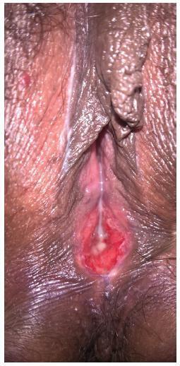
Vulvar Hemangioma: Case Report
Revista Brasileira de Ginecologia e Obstetrícia. 2018;40(6):369-371
06-01-2018
Summary
Case ReportVulvar Hemangioma: Case Report
Revista Brasileira de Ginecologia e Obstetrícia. 2018;40(6):369-371
06-01-2018Views137See moreAbstract
Hemangioma is a benign neoplasm that may affect the vulva, and it can cause functional or emotional disability. This article reports the case of a 52-year-old female patient with a history of a genital ulcer for the past 3 years and who had undergone various treatments with creams and ointments. The patient was biopsied and diagnosed with vulvar hemangioma and was subsequently submitted to surgical excision of the lesion. We emphasize the importance of following the steps of the differential diagnosis and proceeding with a surgical approach only if necessary.

-
Original Article
Underdiagnosis of cervical intraepithelial neoplasia (CIN) 2 orWorse Lesion inWomenwith a Previous Colposcopy-Guided Biopsy Showing CIN 1
Revista Brasileira de Ginecologia e Obstetrícia. 2017;39(3):123-127
03-01-2017
Summary
Original ArticleUnderdiagnosis of cervical intraepithelial neoplasia (CIN) 2 orWorse Lesion inWomenwith a Previous Colposcopy-Guided Biopsy Showing CIN 1
Revista Brasileira de Ginecologia e Obstetrícia. 2017;39(3):123-127
03-01-2017Views135Abstract
Objective
Expectant follow-up for biopsy-proven cervical intraepithelial neoplasia (CIN) 1 is the current recommendation for the management of this lesion. Nevertheless, the performance of the biopsy guided by colposcopy might not be optimal. Therefore, this study aimed to calculate the rate of underdiagnoses of more severe lesions in women with CIN 1 diagnosis and to evaluate whether age, lesion extent and biopsy site are factors associated with diagnostic failure.
Methods
Eighty women with a diagnosis of CIN 1 obtained by colposcopy-guided biopsy were selected for this study. These women were herein submitted to large loop excision of the transformation zone (LLETZ). The prevalence of lesions more severe than CIN 1 was calculated, and the histological diagnoses of the LLETZ specimens were grouped into two categories: "CIN 1 or less" and "CIN 2 or worse."
Results
The prevalence of lesions diagnosed as CIN 2 or worse in the LLETZ specimens was of 19% (15/80). Three women revealed CIN 3, and 1 woman revealed a sclerosing adenocarcinoma stage I-a, a rare type of malignant neoplasia of low proliferation, which was not detected by either colposcopy or previous biopsy. The underdiagnosis of CIN 2 was not associated with the women's age, lesion extension and biopsy site.
Conclusions
The standard methods used for the diagnosis of CIN 1 may underestimate the severity of the true lesion and, therefore, women undergoing expectant management must have an adequate follow-up.
Key-words BiopsyCervical intraepithelial neoplasiacolposcopic surgical proceduresColposcopyNeoplasmsuterine cervicalSee more -
Artigos Originais
Axillary lymph node aspiration guided by ultrasound is effective as a method of predicting lymph node involvement in patients with breast cancer?
Revista Brasileira de Ginecologia e Obstetrícia. 2014;36(3):118-123
03-01-2014
Summary
Artigos OriginaisAxillary lymph node aspiration guided by ultrasound is effective as a method of predicting lymph node involvement in patients with breast cancer?
Revista Brasileira de Ginecologia e Obstetrícia. 2014;36(3):118-123
03-01-2014DOI 10.1590/S0100-72032014000300005
Views97See morePURPOSE:
To assess the feasibility and diagnostic accuracy of preoperative ultrasound combined with ultrasound-guided fine-needle aspiration (US-FNA) cytology and clinical examination of axillary lymph node in patients with breast cancer.
METHODS:
In this prospective study, 171 axillae of patients with breast cancer were evaluated by clinical examination and ultrasonography (US) with and without fine needle aspiration (FNA). Lymph nodes with maximum ultrasonographic cortical thickness > 2.3 mm were considered suspicious and submitted to US-FNA.
RESULTS:
Logistic regression analysis showed no statistically significant correlation between clinical examination and pathologically positive axillae. However, in axillae considered suspicious by ultrasonography, the risk of positive anatomopathological findings increased 12.6-fold. Cohen's Kappa value was 0.12 for clinical examination, 0.48 for US, and 0.80 for US-FNA. Accuracy was 61.4% for clinical examination, 73.1% for US and 90.1% for US-FA. Receiver Operating Characteristics (ROC) analysis demonstrated that a cortical thickness of 2.75 mm corresponded to the highest sensitivity and specificity in predicting axillary metastasis (82.7 and 82.2%, respectively).
CONCLUSIONS:
Ultrasonography combined with fine-needle aspiration is more accurate than clinical examination in assessing preoperative axillary status in women with breast cancer. Those who are US-FNA positive can be directed towards axillary lymph node dissection straight away, and only those who are US-FNA negative should be considered for sentinel lymph node biopsy.
-
Original Articles
Accuracy of sonography and hysteroscopy in the diagnosis of premalignant and malignant polyps in postmenopausal women
Revista Brasileira de Ginecologia e Obstetrícia. 2013;35(6):243-248
08-02-2013
Summary
Original ArticlesAccuracy of sonography and hysteroscopy in the diagnosis of premalignant and malignant polyps in postmenopausal women
Revista Brasileira de Ginecologia e Obstetrícia. 2013;35(6):243-248
08-02-2013DOI 10.1590/S0100-72032013000600002
Views104See morePURPOSE: To evaluate the accuracy of sonographic endometrial thickness and hysteroscopic characteristics in predicting malignancy in postmenopausal women undergoing surgical resection of endometrial polyps. METHODS: Five hundred twenty-one (521) postmenopausal women undergoing hysteroscopic resection of endometrial polyps between January 1998 and December 2008 were studied. For each value of sonographic endometrial thickness and polyp size on hysteroscopy, the sensitivity, specificity, positive predictive value (PPV) and negative predictive value (NPV) were calculated in relation to the histologic diagnosis of malignancy. The best values of sensitivity and specificity for the diagnosis of malignancy were determined by the Receiver Operating Characteristic (ROC) curve. RESULTS: Histologic diagnosis identified the presence of premalignancy or malignancy in 4.1% of cases. Sonographic measurement revealed a greater endometrial thickness in cases of malignant polyps when compared to benign and premalignant polyps. On surgical hysteroscopy, malignant endometrial polyps were also larger. An endometrial thickness of 13 mm showed a sensitivity of 69.6%, specificity of 68.5%, PPV of 9.3%, and NPV of 98% in predicting malignancy in endometrial polyps. Polyp measurement by hysteroscopy showed that for polyps 30 mm in size, the sensitivity was 47.8%, specificity was 66.1%, PPV was 6.1%, and NPV was 96.5% for predicting cancer. CONCLUSIONS: Sonographic endometrial thickness showed a higher level of accuracy than hysteroscopic measurement in predicting malignancy in endometrial polyps. Despite this, both techniques showed low accuracy for predicting malignancy in endometrial polyps in postmenopausal women. In suspected cases, histologic evaluation is necessary to exclude malignancy.
-
Artigos Originais
Underestimation of malignancy of core needle biopsy for nonpalpable breast lesions
Revista Brasileira de Ginecologia e Obstetrícia. 2011;33(7):123-131
10-11-2011
Summary
Artigos OriginaisUnderestimation of malignancy of core needle biopsy for nonpalpable breast lesions
Revista Brasileira de Ginecologia e Obstetrícia. 2011;33(7):123-131
10-11-2011DOI 10.1590/S0100-72032011000700002
Views80PURPOSE: To determine the rate of underestimation of an image-guided core biopsy of nonpalpable breast lesions, with validation by histologic examination after surgical excision. METHODS: We retrospectively reviewed 352 biopsies from patients who were submitted to surgery from February 2000 to December 2005, and whose histopathologic findings were recorded in the database system. Results were compared to surgical findings and underestimation rate was determined by dividing the number of lesions that proved to be carcinomas at surgical excision by the total number of lesions evaluated with excisional biopsy. Clinical, imaging, core biopsy and pathologic features were analyzed to identify factors that affect the rate of underestimation. The degree of agreement between the results was obtained by the percentage of agreement and Cohen's kappa coefficient. The association of variables with the underestimation of the diagnosis was determined by the chi-square, Fisher exact, ANOVA and Mann-Whitney U tests. The risk of underestimation was measured by the relative risk (RR) together with the respective 95% confidence intervals (95%CI). RESULTS: Inconclusive core biopsy findings occurred in 15.6% of cases. The histopathological result was benign in 26.4%, a high-risk lesion in 12.8% and malignant in 45.2%. There was agreement between core biopsy and surgery in 82.1% of cases (kappa=0.75). The false-negative rate was 5.4% and the lesion was completely removed in 3.4% of cases. The underestimation rate was 9.1% and was associated with BI-RADS® category 5 (p=0,01), microcalcifications (p CONCLUSIONS: The core breast biopsy under image guidance is a reliable procedure but the recommendation of surgical excision of high-risk lesions detected in the core biopsy remains since it was not possible to assess clinical, imaging, core biopsy and pathologic features that could predict underestimation and avoid excision. Representative samples are much more important than number of fragments.
Key-words BiopsyBreast neoplasmsSee more -
Relato de Caso
Lymphoma of the uterine cervix: report of two cases and review of the literature
Revista Brasileira de Ginecologia e Obstetrícia. 2008;30(12):626-630
02-04-2008
Summary
Relato de CasoLymphoma of the uterine cervix: report of two cases and review of the literature
Revista Brasileira de Ginecologia e Obstetrícia. 2008;30(12):626-630
02-04-2008DOI 10.1590/S0100-72032008001200007
Views52See moreThe occurrence of primary lymphomas of the female genital tract is rare. The diagnosis is usually not possible by the cytological examination; a tissue biopsy is necessary. The present paper reports two patients referred to our service with a diagnosis of cervical lymphoma. In one of them, the diagnostic difficulties are demonstrated. Both patients were submitted to chemotherapy with satisfactory post-operatory period. There is no standard treatment for uterine lymphomas. Exclusive local treatment is supported by some reports in the literature in clinical stage IE, while others prefer systemic treatment irrespective of clinical stage.
-
Artigo de Revisão
Histeroscopy in menopause: analysis of the techniques and accuracy of the method
Revista Brasileira de Ginecologia e Obstetrícia. 2008;30(10):524-530
11-27-2008
Summary
Artigo de RevisãoHisteroscopy in menopause: analysis of the techniques and accuracy of the method
Revista Brasileira de Ginecologia e Obstetrícia. 2008;30(10):524-530
11-27-2008DOI 10.1590/S0100-72032008001000008
Views58See moreDetection of endometrial cancer in asymptomatic women has not proved to be a cost-effective procedure. Studies on this matter have shown that ultrasonography as a detecting method presents a high ratio of false-positive results and a negligible effect on the mortality rate. This way, the assistance strategy should be based on earlier diagnosis and appropriate treatment in women who present postmenopause bleeding. Being a non-invasive method, largely available and with high sensitivity, the transvaginal ultrasonography should be the initial investigative method. Though there is no consensus about the echographical endometrial thickness, above which the investigation is to proceed, diagnostic hysteroscopy should be the next step. The risk of neoplasia in endometriums with thickness under or equal to 3 mm is low enough to limit hysteroscopy to exceptional cases. Biopsy must be a necessary part of the hysteroscopy, because the diagnosis, made on visual basis, alone may lead to false results. Outpatient hysteroscopy can be done in more than 95% of the cases, even in menopausal women, rarely with severe complications. The adoption of "non-contact" examination techniques and the progressive reduction of the hysteroscope diameter have decreased the discomfort associated to small outpatient procedures.


