Summary
Revista Brasileira de Ginecologia e Obstetrícia. 2018;40(3):147-155
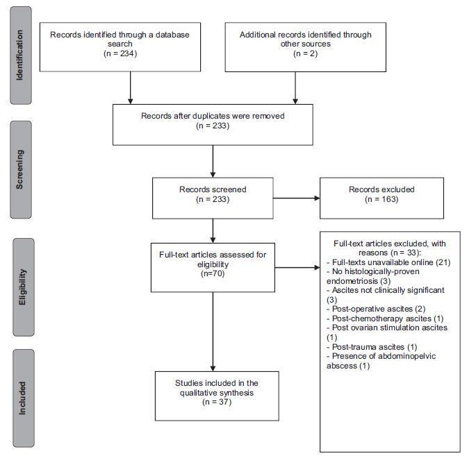
Endometriosis can have several different presentations, including overt ascites and peritonitis; increased awareness can improve diagnostic accuracy and patient outcomes. We aimto provide a systematic review and report a case of endometriosis with this unusual clinical presentation. The PubMed/MEDLINE database was systematically reviewed until October 2016. Women with histologically-proven endometriosis presenting with clinically significant ascites and/or frozen abdomen and/or encapsulating peritonitis were included; thosewith potentially confounding conditionswere excluded.Our search yielded 37 articles describing 42 women, all of reproductive age. Ascites was mostly hemorrhagic, recurrent and not predicted by cancer antigen 125 (CA-125) levels. In turn, dysmenorrhea, dyspareunia and infertility were not consistently reported. The treatment choices and outcomes were different across the studies, and are described in detail. Endometriosis should be a differential diagnosis of massive hemorrhagic ascites in women of reproductive age.

Summary
Revista Brasileira de Ginecologia e Obstetrícia. 2013;35(7):331-335
DOI 10.1590/S0100-72032013000700008
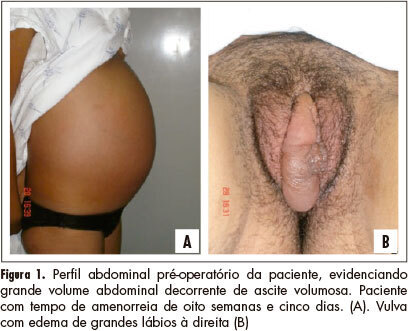
The sclerosing stromal tumor of the ovary is an extremely rare benign tumor more common in young women and without specific symptoms in most cases. Less than 150 cases have been described, of which 8 were diagnosed during pregnancy. In this report, we describe the association between sclerosing stromal tumor of the ovary, Meigs' syndrome and elevated levels of CA-125 in term pregnancy.

Summary
Revista Brasileira de Ginecologia e Obstetrícia. 1999;21(6):353-357
DOI 10.1590/S0100-72031999000600009
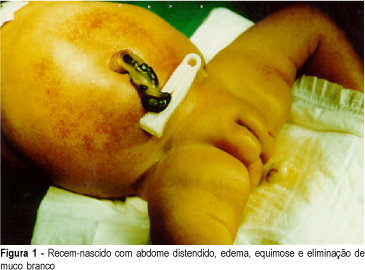
Introduction: meconium peritonitis as result of fetal intestinal perforation has a low incidence (1:30,000 deliveries) and high mortality (50% or more). Prenatal ultrasound findings include fetal ascites and intra-abdominal calcifications. Evidence suggests that prenatal diagnosis can improve postnatal prognosis. Case Report: R.C.M.S., 22 years, II pregnancy O para, presented ultrasound (12/02/98) with diagnosis of fetal ascites. Investigation for hydrops fetalis was performed and immune and nonimmune causes were excluded. Severe fetal ascites persisted on subsequent ultrasound examinations, without calcifications. Vaginal delivery occurred at 36 weeks (01/02/99), with polyhydramnios. Female neonate weighing 2,670 g, with signs of respiratory distress, abdominal distension and petechiae. Abdominal distension worsened progressively, with palpation of a petrous tumor in the right upper quadrant and elimination of white mucus at rectal examination. Radiological findings (01/04/99) were disseminated abdominal calcifications, intestinal dilatation and absence of gas at rectal ampulla. Exploratory laparotomy was indicated with diagnosis of meconium peritonitis. A giant meconium cyst and ileal atresia were observed and lysis of adhesions and ileostomy were performed. Initial postoperative evolution was satisfactory but was subsequently complicated by sepsis and neonatal death occurred (01/09/99). Conclusion: meconium peritonitis should be remembered at differential diagnosis of fetal ascites. In the present case, surgical indication could be anticipated if prenatal diagnosis were established, with improvement of neonatal evolution.
