You searched for:"Edmund Chada Baracat"
We found (71) results for your search.Summary
Rev Bras Ginecol Obstet. 2009;31(8):385-390
DOI 10.1590/S0100-72032009000800003
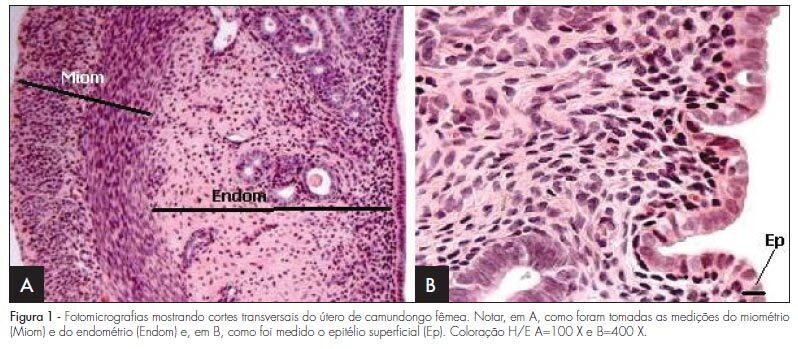
PURPOSE: to evaluate the effect of hyperprolactinemia induced by metoclopramide on the endometrium and myometrium of female mice in the proestrus phase. METHODS: 24 female mice were randomly divided in two groups: CtrG/control and ExpG/treated with metoclopramide (6.7 mg/g daily). After 50 days, the animals were sacrificed in the proestrus phase, and the blood was collected to determine the levels of estradiol, progesterone and prolactin. The uterine horns were removed, fixed in 10% formaldehyde and processed before being included in paraffin. Slices of 4 µm were stained by hematoxylin and eosin (H/E). In the morphological analysis, a Carl Zeiss light microscope, with objectives varying from 4 to 400 X was used for each histological slice characterization. In the morphometrical analysis, the superficial epithelium, the lamina propria and the myometrium thickness were evaluated, with the help of an image analyzer (AxionVision - Carl Zeiss) attached to the light microscope (Carl Zeiss). The statistical analysis was done by ANOVA, followed by the Wilcoxon test. P-value was considered as significant, when <0.05. RESULTS: our findings have shown an increase in the seric levels of prolactin (295.6±38.0 ng/mL) and significant decrease in the progesterone levels (11.3±0.9 ng/mL) in the ExpG, as compared to the CtrG (45.5±5.2 ng/mL and 18.2±1.6 ng/mL, respectively; p<0.001). Concerning the seric level of estradiol, significant differences between the groups were not obtained (ExpG=119.1±12.3 pg/mL and CtrG=122.7±8.4 pg/mL; p=0.418). The morphological study has shown that the uterus from the ExpG presented the endometrium with more developed superficial epithelium and lamina propria, as compared to the CtrG, the same happening with the myometrium. The thickness morphometrical values of the luminal epithelium (8.0±1.1 µm) and endometrium (116.2±21.1x10² µm) from the CtrG were lower than the ones from the ExpG (10.2±0.8 µm and 163.2±23.3x10² µm, respectively) with p<0.05. Nevertheless, data obtained in the myometrium have not shown significant differences between the groups (CtrG=152.2±25.2x10² µm and ExpG=140.8±18.0x10² µm). CONCLUSIONS: data have shown that hyperprolactinemia induced by metoclopramide determines endometrial proliferation and interferes with the ovarian function, mainly in the progesterone production.
Summary
Rev Bras Ginecol Obstet. 2009;31(8):385-390
DOI 10.1590/S0100-72032009000800003
PURPOSE: to evaluate the effect of hyperprolactinemia induced by metoclopramide on the endometrium and myometrium of female mice in the proestrus phase. METHODS: 24 female mice were randomly divided in two groups: CtrG/control and ExpG/treated with metoclopramide (6.7 mg/g daily). After 50 days, the animals were sacrificed in the proestrus phase, and the blood was collected to determine the levels of estradiol, progesterone and prolactin. The uterine horns were removed, fixed in 10% formaldehyde and processed before being included in paraffin. Slices of 4 µm were stained by hematoxylin and eosin (H/E). In the morphological analysis, a Carl Zeiss light microscope, with objectives varying from 4 to 400 X was used for each histological slice characterization. In the morphometrical analysis, the superficial epithelium, the lamina propria and the myometrium thickness were evaluated, with the help of an image analyzer (AxionVision - Carl Zeiss) attached to the light microscope (Carl Zeiss). The statistical analysis was done by ANOVA, followed by the Wilcoxon test. P-value was considered as significant, when <0.05. RESULTS: our findings have shown an increase in the seric levels of prolactin (295.6±38.0 ng/mL) and significant decrease in the progesterone levels (11.3±0.9 ng/mL) in the ExpG, as compared to the CtrG (45.5±5.2 ng/mL and 18.2±1.6 ng/mL, respectively; p<0.001). Concerning the seric level of estradiol, significant differences between the groups were not obtained (ExpG=119.1±12.3 pg/mL and CtrG=122.7±8.4 pg/mL; p=0.418). The morphological study has shown that the uterus from the ExpG presented the endometrium with more developed superficial epithelium and lamina propria, as compared to the CtrG, the same happening with the myometrium. The thickness morphometrical values of the luminal epithelium (8.0±1.1 µm) and endometrium (116.2±21.1x10² µm) from the CtrG were lower than the ones from the ExpG (10.2±0.8 µm and 163.2±23.3x10² µm, respectively) with p<0.05. Nevertheless, data obtained in the myometrium have not shown significant differences between the groups (CtrG=152.2±25.2x10² µm and ExpG=140.8±18.0x10² µm). CONCLUSIONS: data have shown that hyperprolactinemia induced by metoclopramide determines endometrial proliferation and interferes with the ovarian function, mainly in the progesterone production.
Summary
Rev Bras Ginecol Obstet. 2003;25(6):396-401
DOI 10.1590/S0100-72032003000600003
PURPOSE: to evaluate the significance of neoangiogenesis for the prognosis of endometrial carcinoma, by quantifying and comparing the vessels with the grade of histologic differentiation and tumor staging. METHODS: the 56 studied cases consisted of 11 atrophic endometria, 10 proliferative endometria, 10 GI, 13 GII and 12 GIII adenocarcinomas. Two histologic sections were obtained for each case: one was stained with hematoxylin-eosin and the other was sent for a immunohistochemical study with anti-CD34. The utilized histometric method was vessel counting at the tumoral growth interface with the adjacent stroma, and in the control group, at the endometrial gland interface with the stroma. Couting was done by a KS300, evaluating 10 fields at 100X magnification. RESULTS: the counted vessel means were 11.6 for atrophic endometria, 13.2 for proliferative endometria, 15.3 for GI adenocarcinoma, 19 for GII adenocarcinoma, and 22.7 for GIII adenocarcinoma. In the group of stage I patients, it was observed that the mean number of vessels (18.6) was similar to that observed in stages II, III and IV (20.9) computed together. CONCLUSION: less differentiated adenocarcinomas were more angiogenic than well-differentiated carcinomas and normal endometrium. Vessel counting was not influenced by the disease stage as an isolated factor.
Summary
Rev Bras Ginecol Obstet. 2001;23(1):41-45
DOI 10.1590/S0100-72032001000100006
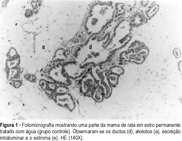
Purpose: the morphologic and morphometric aspects of the breasts of rats in permanent estrus submitted to danazol were studied. Methods: the animals were divided into three groups: group A (n = 12) received water and was used as control, group B (n = 13) was exposed to 20 mg danazol kg-1 day-1, and group C (n = 10) was exposed to 80 mg danazole kg-1 day-1 for 35 consecutive days. The microscopic study evaluated the ductal and acinar distribution. Histometry of the relationship duct/stroma was based on the principles of stereology with a Zeiss k-10X ocular, with Integrationsplatte I reticulum of Weibel of 25 hits, with 100X magnification. For each studied section, 10 aleatoric fields were counted, with a total of 250 points. The variance analysis test (Kruskal-Wallis) was applied to compare the three groups in relation to the mean number of alveoli and ducts (alpha = 0.05). Results: when submitted to morphological study, all groups presented lobules with alveoli lined with cubic cells with nuclei in their central or basal portion. Small amounts of eosinophilic material were observed in some cases in the lumen, with no differences between the groups. At morphometry, with a magnification of 100X, a mean number of 28.6 ducts/10 fields was found in group A, 28.4 in group B and 29.2 in group C (Kruskal-Wallis test: Hcrit = 0.1). The mean number of alveoli in 10 fields was 5.9, 9.3 and 6.5 in groups A, B and C, respectively (Kruskal-Wallis test: Hcrit = 2.9), with no significant differences between the groups. Conclusion: danazol did not cause any changes in the morphology and morphometry of the permanent estrus mammary epithelium.
Summary
Rev Bras Ginecol Obstet. 2001;23(1):41-45
DOI 10.1590/S0100-72032001000100006
Purpose: the morphologic and morphometric aspects of the breasts of rats in permanent estrus submitted to danazol were studied. Methods: the animals were divided into three groups: group A (n = 12) received water and was used as control, group B (n = 13) was exposed to 20 mg danazol kg-1 day-1, and group C (n = 10) was exposed to 80 mg danazole kg-1 day-1 for 35 consecutive days. The microscopic study evaluated the ductal and acinar distribution. Histometry of the relationship duct/stroma was based on the principles of stereology with a Zeiss k-10X ocular, with Integrationsplatte I reticulum of Weibel of 25 hits, with 100X magnification. For each studied section, 10 aleatoric fields were counted, with a total of 250 points. The variance analysis test (Kruskal-Wallis) was applied to compare the three groups in relation to the mean number of alveoli and ducts (alpha = 0.05). Results: when submitted to morphological study, all groups presented lobules with alveoli lined with cubic cells with nuclei in their central or basal portion. Small amounts of eosinophilic material were observed in some cases in the lumen, with no differences between the groups. At morphometry, with a magnification of 100X, a mean number of 28.6 ducts/10 fields was found in group A, 28.4 in group B and 29.2 in group C (Kruskal-Wallis test: Hcrit = 0.1). The mean number of alveoli in 10 fields was 5.9, 9.3 and 6.5 in groups A, B and C, respectively (Kruskal-Wallis test: Hcrit = 2.9), with no significant differences between the groups. Conclusion: danazol did not cause any changes in the morphology and morphometry of the permanent estrus mammary epithelium.
Summary
Rev Bras Ginecol Obstet. 2005;27(7):415-420
DOI 10.1590/S0100-72032005000700008
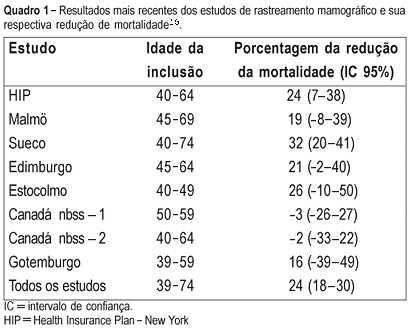
PURPOSE: to evaluate the cost of preventive mammographic screening in climacteric women, as compared to the cost of breast cancer treatment in more advanced stages. METHODS: one thousand and fourteen patients attended at the Climacteric outpatient service of the Gynecology Department, Federal University of São Paulo Paulista School of Medicine, were included in the study and submitted to mammographic test. All mammographic test's were analyzed by the same two physicians and classified according to the BI-RADS (Breast Imaging Reporting and Data System American College of Radiology) categories. The detected lesions were submitted to cytological and histological examination. RESULTS: the final diagnostic impression of the 1014 examinations, according to the classification of BI-RADS categories was: 1=261, 2=671, 3=59, 4=22 and 5=1. The invasive procedures were performed through a needle guided by ultrasound or stereotactic examinations: 33 fine-needle aspiration biopsies, 6 core biopsies guided by ultrasound and 20 core biopsies guided by stereotactic examination. Five cancer diagnoses were established. The total cost of this screening based on Brazilian procedure values was R$ 76,593.79 (25,534 dollars). Therefore, the cost of the diagnosis of the five cases of cancer in this screening was R$ 15,318.75 (5,106 dollars) each. However, the average cost per patient screened was R$ 75.53 (25 dollars). CONCLUSIONS: considering that the total treatment cost of only one case of breast cancer in advanced stage including hospital costs, surgery, chemotherapy, radiotherapy and hormonal treatment is similar to the cost of 1,000 mammographic screenings in climacteric women, it may be concluded that the cost of the early cancer diagnosis program is worth it and should be included in the public health program, as a way of lowering the public health expense.
Summary
Rev Bras Ginecol Obstet. 2005;27(7):415-420
DOI 10.1590/S0100-72032005000700008
PURPOSE: to evaluate the cost of preventive mammographic screening in climacteric women, as compared to the cost of breast cancer treatment in more advanced stages. METHODS: one thousand and fourteen patients attended at the Climacteric outpatient service of the Gynecology Department, Federal University of São Paulo Paulista School of Medicine, were included in the study and submitted to mammographic test. All mammographic test's were analyzed by the same two physicians and classified according to the BI-RADS (Breast Imaging Reporting and Data System American College of Radiology) categories. The detected lesions were submitted to cytological and histological examination. RESULTS: the final diagnostic impression of the 1014 examinations, according to the classification of BI-RADS categories was: 1=261, 2=671, 3=59, 4=22 and 5=1. The invasive procedures were performed through a needle guided by ultrasound or stereotactic examinations: 33 fine-needle aspiration biopsies, 6 core biopsies guided by ultrasound and 20 core biopsies guided by stereotactic examination. Five cancer diagnoses were established. The total cost of this screening based on Brazilian procedure values was R$ 76,593.79 (25,534 dollars). Therefore, the cost of the diagnosis of the five cases of cancer in this screening was R$ 15,318.75 (5,106 dollars) each. However, the average cost per patient screened was R$ 75.53 (25 dollars). CONCLUSIONS: considering that the total treatment cost of only one case of breast cancer in advanced stage including hospital costs, surgery, chemotherapy, radiotherapy and hormonal treatment is similar to the cost of 1,000 mammographic screenings in climacteric women, it may be concluded that the cost of the early cancer diagnosis program is worth it and should be included in the public health program, as a way of lowering the public health expense.
Summary
Rev Bras Ginecol Obstet. 2000;22(7):429-433
DOI 10.1590/S0100-72032000000700005
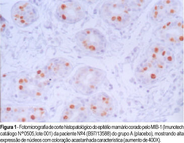
Purpose: to study the monoclonal antibody MIB-1 in the normal breast epithelium adjacent to a fibroadenoma in women in the luteal phase of the menstrual cycle treated with tamoxifen. Patients and methods: the proliferative activity of the mammary epithelium adjacent to the fibroadenoma was studied by immunohistochemistry based on immunoexpression of the monoclonal antibody MIB-1. The study was randomized and double blind and was conducted on 44 women with fibroadenomas, divided into 3 groups: A (n = 16; placebo), B (n = 15; tamoxifen, 10 mg), and C (n = 13; tamoxifen, 20 mg). Tamoxifen was administered for 22 days starting on the 2nd day of the menstrual cycle and a biopsy was taken on the 23rd day. Results: the mean percentage of stained nuclei per 1000 cells was 9.2 in group A, 4.5 in group B, and 3.2 in group C. Fisher's test revealed that tamoxifen significantly reduced the immunoexpression of MIB-1 at the doses of 10 and 20 mg compared to the placebo group (p<0.0001), with no significant differences between doses in terms of proliferative activity (p = 0.21). Conclusion: we conclude that tamoxifen significantly reduced the proliferative activity of the mammary epithelium at the doses of 10 and 20 mg/day.
Summary
Rev Bras Ginecol Obstet. 2000;22(7):429-433
DOI 10.1590/S0100-72032000000700005
Purpose: to study the monoclonal antibody MIB-1 in the normal breast epithelium adjacent to a fibroadenoma in women in the luteal phase of the menstrual cycle treated with tamoxifen. Patients and methods: the proliferative activity of the mammary epithelium adjacent to the fibroadenoma was studied by immunohistochemistry based on immunoexpression of the monoclonal antibody MIB-1. The study was randomized and double blind and was conducted on 44 women with fibroadenomas, divided into 3 groups: A (n = 16; placebo), B (n = 15; tamoxifen, 10 mg), and C (n = 13; tamoxifen, 20 mg). Tamoxifen was administered for 22 days starting on the 2nd day of the menstrual cycle and a biopsy was taken on the 23rd day. Results: the mean percentage of stained nuclei per 1000 cells was 9.2 in group A, 4.5 in group B, and 3.2 in group C. Fisher's test revealed that tamoxifen significantly reduced the immunoexpression of MIB-1 at the doses of 10 and 20 mg compared to the placebo group (p<0.0001), with no significant differences between doses in terms of proliferative activity (p = 0.21). Conclusion: we conclude that tamoxifen significantly reduced the proliferative activity of the mammary epithelium at the doses of 10 and 20 mg/day.
Summary
Rev Bras Ginecol Obstet. 2008;30(9):432-436
DOI 10.1590/S0100-72032008000900002
PURPOSE: sentinel lymph node biopsy in early-stage breast cancer patients has been substituting the total axillary lymph node is presented dissection. The technique of processing the sentinel lymph node and the aim of this study was to investigate the efficacy of occult metastasis identification based on the standard histological and immunohistochemical examination. METHODS: between 2002 and 2005, 266 sentinel lymph nodes were harvested from axillary biopsy of 170 patients with early stage breast cancer. All lymph nodes were considered to be negative according to standard intra-operative cytological assessment. Lymph nodes were transversally sectioned in four or five slices and embedded in paraffin blocks. Two paraffin-embedded tissue sections with 4 µm in thickness were mounted on glass slides and stained with hematoxylin-eosin and immunoperoxidase (cytokeratin AE1/AE3) techniques. RESULTS: standard histological examination identified metastasis in 22 patients (12.9%) and micrometastatic disease was observed in six of these patients (3.5%). The immunohistochemical examination identified metastatic disease in 16 patients (9.4%). Among them, isolated tumor cells were observed in 11 (6.5%) and micrometastases were identified in five (2.9%). CONCLUSIONS: the association of the standard histological examination and immunohistochemical technique increases the chances of sentinel lymph node metastasis identification.
Summary
Rev Bras Ginecol Obstet. 2002;24(7):433-438
DOI 10.1590/S0100-72032002000700002
Purpose: to analyze the correlation between Valsalva leak point pressure and maximum urethral closure pressure and clinical symptoms in women with stress urinary incontinence. Methods: we analyzed retrospectively 164 patients with urodynamic diagnosis of stress and mixed urinary incontinence established by the Urogynecology and Vaginal Surgery Sector of UNIFESP/EPM. All patients were submmited to medical interview, physical examination and urodynamic study. Patients were divided into groups according to the subjective degree of stress urinary incontinence. Valsalva leak point pressure (VLPP) was measured with a vesical volume of 200 mL. Urethral profile was determined using a flow catheter number 8 with measurement of maximum urethral closure pressure (MUCP). Data were compared by chi², ANOVA and Tukey tests. Results: mean age was 51.2 years (19-82), 76 women (47.2%) were in menacme and 85 (52.8%) in postmenopausal status. Mean parity was 3.9 (0-18). The exact test for trend demonstrated a statistically significant (p<0.0001) correlation between the number of patients with VLPP of 60 cmH2O or less and clinical complaints. The group with severe leakage had mean VLPP of 69.1 cmH2O. The group with moderated leakage had mean VLPP of 84.6 cmH2O and the group with mild leakage had mean VLPP of 90.6 cmH2O. Conclusions: VLPP correlated with the subjective degree of stress urinary incontinence. Higher grades of stress urinary incontinence had a higher likelyhood of a low VLPP. MUCP did not correlate with clinical complaints.
Summary
Rev Bras Ginecol Obstet. 2004;26(6):441-447
DOI 10.1590/S0100-72032004000600004
OBJECTIVE: to compare Baden and Walker's (BW) classification system to the International Continence Society (ICS) standardization of terminology of female pelvic organ prolapse. METHODS: information about urogynecological investigation on 101 women, performed by the Urogynecology and Vaginal Surgery Sector of UNIFESP/EPM, was retrospectively analyzed. Only patients who had undergone the standard ICS exam which quantifies the pelvic prolapse were selected. According to ICS, the prolapse is analyzed through a standard reference system relating the hymen to the anatomic position of six vaginal points: two in the anterior vaginal wall, two in the vaginal apex and other two in the posterior vaginal wall. The maximum amount of pelvic organ prolapse was viewed and recorded during a Valsalva's maneuver. The measurement of the most distal point of the prolapse was performed and it was compared to the BW classification system. The data were analyzed by kappa statistics, to assess the concordance between the two terminologies. RESULTS: There was total correspondence only for the posterior vaginal prolapse stage IV (one patient) and for the uterus prolapse stage 0 (29 patients) with severe rectocele and absence of prolapse, respectively, according to BW. In the three types of prolapses evaluated, the values of kappa statistics were below 0.4, indicating a weak concordance between the two terminologies. There is an extensive variation in the measurement of the most distal point of prolapse when the BW classification is perfomed. CONCLUSIONS: there is a weak concordance between the BW classification system and the ICS standardization of terminology of female pelvic organ prolapse.