Summary
Revista Brasileira de Ginecologia e Obstetrícia. 2019;41(3):137-141
Summary
Revista Brasileira de Ginecologia e Obstetrícia. 2019;41(3):137-141
Summary
Revista Brasileira de Ginecologia e Obstetrícia. 2021;43(2):137-144
The present study aims to evaluate the profile of endometrial carcinomas and uterine sarcomas attended in a Brazilian cancer center in the period from 2001 to 2016 and to analyze the impact of time elapsed fromsymptoms to diagnoses or treatment in cancer stage and survival.
This observational study with 1,190 cases evaluated the year of diagnosis, age-group, cancer stage and histological type. A subgroup of 185 women with endometrioid histology attended in the period from 2012 to 2017 was selected to assess information about initial symptoms, diagnosticmethods, overall survival, and to evaluate the influence of the time elapsed from symptoms to diagnosis and treatment on staging and survival. The statistics used were descriptive, trend test, and the Kaplan- Meier method, with p-values < 0.05 for significance.
A total of 1,068 (89.7%) carcinomas (77.2% endometrioid and 22.8% nonendometrioid) and 122 (10.3%) sarcomas were analyzed, with an increasing trend in the period (p < 0.05). Histologies of non-endometrioid carcinomas, G3 endometrioid, and carcinosarcomas constituted 30% of the cases. Non-endometrioid carcinomas and sarcomas weremore frequently diagnosed in patients over 70 years of age and those on stage IV (p < 0.05). The endometrioid subgroup with 185 women reported 92% of abnormal uterine bleeding and 43% diagnosis after curettage. The average time elapsed between symptoms to diagnosis was 244 days, and between symptoms to treatment was 376 days, all without association with staging (p = 0.976) and survival (p = 0.160). Only 12% of the patients started treatment up to 60 days after diagnosis.
The number of uterine carcinoma and sarcoma cases increased over the period of 2001 to 2016. Aggressive histology comprised 30% of the patients and, for endometrioid carcinomas, the time elapsed between symptoms and diagnosis or treatment was long, although without association with staging or survival.
Summary
Revista Brasileira de Ginecologia e Obstetrícia. 2002;24(2):137-137
Summary
Revista Brasileira de Ginecologia e Obstetrícia. 2002;24(2):137-137
Summary
Revista Brasileira de Ginecologia e Obstetrícia. 2002;24(2):137-137
DOI 10.1590/S0100-72032002000200013
Summary
Revista Brasileira de Ginecologia e Obstetrícia. 2002;24(2):137-137
DOI 10.1590/S0100-72032002000200013
Summary
Revista Brasileira de Ginecologia e Obstetrícia. 2002;24(2):137-138
DOI 10.1590/S0100-72032002000200015
Summary
Revista Brasileira de Ginecologia e Obstetrícia. 2002;24(2):137-138
DOI 10.1590/S0100-72032002000200015
Summary
Revista Brasileira de Ginecologia e Obstetrícia. 2005;27(3):137-142
DOI 10.1590/S0100-72032005000300007
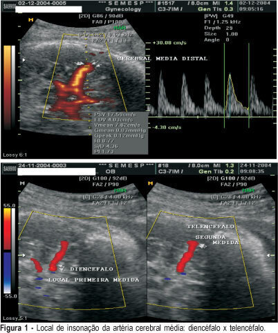
PURPOSE: to evaluate if there is any difference between Doppler indexes in the middle cerebral artery in two different sites of insonation in healthy patients and in patients with diseases. METHODS: a random prospective survey, in the period from June 2003 to March 2004 that analyzed the Doppler indexes of 100 patients: patient group (n = 50) included patients admitted to Clemente Farias University Hospital, which is part of UNIMONTES-MG, havinfg as inclusion criteria: to be in the 28th to 34th gestational week, diagnosis of chronic arterial hypertension, pre-eclampsia, intrauterine growth restriction. As control group, 50 healthy pregnant patients between the 28th and the 34th week, originary from SEMESP's clinic. The Doppler variables were the resistance index (RI), the pulsatility index (PI) and the relation systole/diastole (SD). All three Doppler indexes were assessed at two different sites of the cerebral artery: the first measurement in the diencephalons region, soon after the beginning of the middle cerebral artery and the second on a distal location in the telencephalon. The median Doppler indexes in the patient group in the first and second measurements were 1.55 and 1.69 for the PI, 0.77 and 0.79 for RI and 4.29 and 4.86 for SD, respectively. In the control group, the values were 1.73 and 1.86 for the PI; 0.83 and 0.79 for RI and 5.83 and 5.46 for SD. There were no differences between sites with a p value of 0.38, 0.29 and 0.39 for PI, RI and SD, respectively. In 15t fetuses with centralization (brain sparing effects), in the diencephalon the median indexes were 1.02 for PI, 0.63 for RI and 2.68 for SD. In the epencephalon the median indexes were 0.95 for IP, 0.62 for RI and 2.44 for SD. There were no differences between sites, with a p value of 0.53 for PI; 0.56 for IR and 0.31 for SD. The Doppler index site of assessment in the middle cerebral arteries does not interfere with the results.
Summary
Revista Brasileira de Ginecologia e Obstetrícia. 2005;27(3):137-142
DOI 10.1590/S0100-72032005000300007
PURPOSE: to evaluate if there is any difference between Doppler indexes in the middle cerebral artery in two different sites of insonation in healthy patients and in patients with diseases. METHODS: a random prospective survey, in the period from June 2003 to March 2004 that analyzed the Doppler indexes of 100 patients: patient group (n = 50) included patients admitted to Clemente Farias University Hospital, which is part of UNIMONTES-MG, havinfg as inclusion criteria: to be in the 28th to 34th gestational week, diagnosis of chronic arterial hypertension, pre-eclampsia, intrauterine growth restriction. As control group, 50 healthy pregnant patients between the 28th and the 34th week, originary from SEMESP's clinic. The Doppler variables were the resistance index (RI), the pulsatility index (PI) and the relation systole/diastole (SD). All three Doppler indexes were assessed at two different sites of the cerebral artery: the first measurement in the diencephalons region, soon after the beginning of the middle cerebral artery and the second on a distal location in the telencephalon. The median Doppler indexes in the patient group in the first and second measurements were 1.55 and 1.69 for the PI, 0.77 and 0.79 for RI and 4.29 and 4.86 for SD, respectively. In the control group, the values were 1.73 and 1.86 for the PI; 0.83 and 0.79 for RI and 5.83 and 5.46 for SD. There were no differences between sites with a p value of 0.38, 0.29 and 0.39 for PI, RI and SD, respectively. In 15t fetuses with centralization (brain sparing effects), in the diencephalon the median indexes were 1.02 for PI, 0.63 for RI and 2.68 for SD. In the epencephalon the median indexes were 0.95 for IP, 0.62 for RI and 2.44 for SD. There were no differences between sites, with a p value of 0.53 for PI; 0.56 for IR and 0.31 for SD. The Doppler index site of assessment in the middle cerebral arteries does not interfere with the results.
Summary
Revista Brasileira de Ginecologia e Obstetrícia. 2001;23(3):137-143
DOI 10.1590/S0100-72032001000300002
Purpose: to study, in high risk pregnancies with cerebral redistribution of blood flow, the fetal surveillance and perinatal outcome, according to umbilical artery dopplervelocimetry. Methods: a total of 717 high-risk pregnancies attended at the Fetal Surveillance Unit were included. The last examination performed until 72 h prior to delivery was taken into account. Multiple gestations and fetal anomalies were excluded. The redistribution of blood flow was diagnosed if the pulsatility index of middle cerebral artery was below the 5th percentile for gestational age. The umbilical artery dopplervelocimetry was abnormal when A/B ratio was more than the 95th p. Results: in the group with normal umbilical artery dopplervelocimetry (560 cases -- 78.1%), significant correlation was found only between redistribution of blood flow and suspected or abnormal cardiotocography (17.1%). In the group with abnormal umbilical artery dopplervelocimetry (157 cases -- 21.9%) we found significant correlation between redistribution of blood flow (105 cases -- 66.9%) and cardiotocography abnormalities (57.2%), abnormal 1st(43.8%) and 5th (12.4%) minute Apgar scores. In these cases, the mean values of gestational age at delivery (34.4 ± 3.6 weeks), birth weight (1,810.5 ± 769.3 g), and pH at birth (7.20 ± 0.1) were significantly lower. Conclusion: The redistribution of fetal blood flow characterized by means of middle cerebral artery dopplervelocimetry is related to perinatal results when some level of placental insufficiency occurs, and does not present association to perinatal outcome when pregnancy shows normal fetal-placental blood flow.
Summary
Revista Brasileira de Ginecologia e Obstetrícia. 1998;20(3):137-144
DOI 10.1590/S0100-72031998000300003
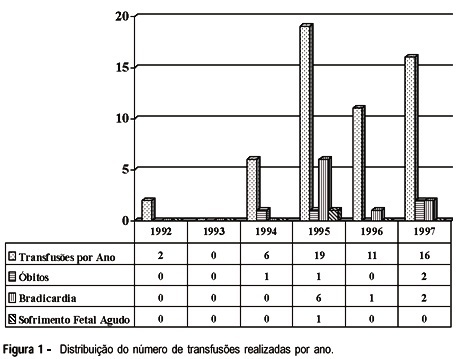
Objective: to report 54 intrauterine intravascular transfusions (IITs), describing procedure related complications and associated perinatal morbidity and mortality. Methods: fetuses undergoing IITs at Clínica Materno-Fetal and Maternidade Carmela Dutra, Florianópolis, SC, between January 1992 and August 1997 were included in the study. Patients demographics, procedure and newborn related data were tabulated for analysis and presented in descriptive form, using percentage, mean, standard deviation, median, range and relative risk (RR) with 95% confidence interval as appropriate. Results: fifty IITs and four exchange transfusions were performed in twenty-one fetuses. There were four deaths (20%), three of which occurred (75%) in hydropic fetuses. Mean gestational age at the time of the first IIT was 29.1 weeks, the mean hemoglobin concentration was 7.1 mg/dl and the mean rise in hemoglobin level per procedure was 5.69 mg/dl. Procedure related mortality rate was 7.4%. Mean gestational age at birth was 33.9 weeks and mean birth weight was 2,437 grams. Sixty-five percent of the newborns received complementary exchange transfusions. Conclusion: the procedure related mortality rate was 7.4%, similar to the mortality rate reported in the world literature. The perinatal mortality rate (20%) was higher than that reported in other countries but lower than the perinatal mortality rate reported in a study conducted in Brazil, with a similar prevalence of hydropic fetuses.
Summary
Revista Brasileira de Ginecologia e Obstetrícia. 1998;20(3):137-144
DOI 10.1590/S0100-72031998000300003
Objective: to report 54 intrauterine intravascular transfusions (IITs), describing procedure related complications and associated perinatal morbidity and mortality. Methods: fetuses undergoing IITs at Clínica Materno-Fetal and Maternidade Carmela Dutra, Florianópolis, SC, between January 1992 and August 1997 were included in the study. Patients demographics, procedure and newborn related data were tabulated for analysis and presented in descriptive form, using percentage, mean, standard deviation, median, range and relative risk (RR) with 95% confidence interval as appropriate. Results: fifty IITs and four exchange transfusions were performed in twenty-one fetuses. There were four deaths (20%), three of which occurred (75%) in hydropic fetuses. Mean gestational age at the time of the first IIT was 29.1 weeks, the mean hemoglobin concentration was 7.1 mg/dl and the mean rise in hemoglobin level per procedure was 5.69 mg/dl. Procedure related mortality rate was 7.4%. Mean gestational age at birth was 33.9 weeks and mean birth weight was 2,437 grams. Sixty-five percent of the newborns received complementary exchange transfusions. Conclusion: the procedure related mortality rate was 7.4%, similar to the mortality rate reported in the world literature. The perinatal mortality rate (20%) was higher than that reported in other countries but lower than the perinatal mortality rate reported in a study conducted in Brazil, with a similar prevalence of hydropic fetuses.