You searched for:"Geraldo Rodrigues de Lima"
We found (29) results for your search.Summary
Rev Bras Ginecol Obstet. 2004;26(8):613-617
DOI 10.1590/S0100-72032004000800004
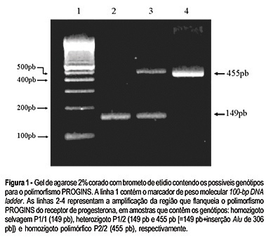
OBJECTIVE: the aim of the present study was to verify whether there is a correlation between the prevalence of the polymorphism in the progesterone receptor gene named PROGINS and pelvic endometriosis at different stages. METHODS: a case-control study carried out from November 2003 to May 2004. The genotypes of 104 women were analyzed 66 women had had surgically confirmed endometriosis (26 women at stages I-II and 40 at stages III-IV), and 38 were healthy women. The 306-base pair Alu insertion polymorphism in the intron G of the progesterone receptor gene was detected by polymerase chain reaction and analyzed on 2% agarose gel stained with ethidium bromide. ANOVA analysis was performed in order to make comparisons between among the studied groups. RESULTS: the groups of women with endometriosis stages I-II (EndoI group) and stages III-IV (EndoII group) showed statistically significant increased incidence of PROGINS polymorphic allele as compared with the control group: 27% in the EndoI group, 38% in EndoII and 18% in the control group (p < 0.001. In the analyses, a high frequency of the PROGINS insertion was observed in women with endometriosis as compared to healthy women, disregarding the clinical stage of the disease (p = 0.0385). CONCLUSION: there is a significant statistical association between the PROGINS polymorphism and pelvic endometriosis.
Summary
Rev Bras Ginecol Obstet. 2004;26(8):613-617
DOI 10.1590/S0100-72032004000800004
OBJECTIVE: the aim of the present study was to verify whether there is a correlation between the prevalence of the polymorphism in the progesterone receptor gene named PROGINS and pelvic endometriosis at different stages. METHODS: a case-control study carried out from November 2003 to May 2004. The genotypes of 104 women were analyzed 66 women had had surgically confirmed endometriosis (26 women at stages I-II and 40 at stages III-IV), and 38 were healthy women. The 306-base pair Alu insertion polymorphism in the intron G of the progesterone receptor gene was detected by polymerase chain reaction and analyzed on 2% agarose gel stained with ethidium bromide. ANOVA analysis was performed in order to make comparisons between among the studied groups. RESULTS: the groups of women with endometriosis stages I-II (EndoI group) and stages III-IV (EndoII group) showed statistically significant increased incidence of PROGINS polymorphic allele as compared with the control group: 27% in the EndoI group, 38% in EndoII and 18% in the control group (p < 0.001. In the analyses, a high frequency of the PROGINS insertion was observed in women with endometriosis as compared to healthy women, disregarding the clinical stage of the disease (p = 0.0385). CONCLUSION: there is a significant statistical association between the PROGINS polymorphism and pelvic endometriosis.
Summary
Rev Bras Ginecol Obstet. 2006;28(11):658-663
DOI 10.1590/S0100-72032006001100005
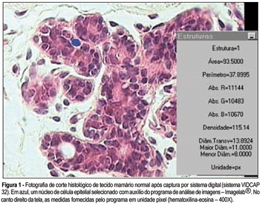
PURPOSE: to analyze breast tissue of postmenopausal women before and after six months of continuous combined estrogen-progestin replacement therapy (0.625 mg conjugated equine estrogens associated with 2.5 mg medroxyprogesterone acetate). METHODS: all patients were evaluated before treatment and considered eligible to receive the drug. The material was obtained from the upper outer left quadrant, through a percutaneous large-core breast biopsy. Epithelial density and nuclear volume on hematoxylin-eosin-stained plates were evaluated for the morphological study. Morphometry was graphically analyzed by optical microscopy (400X) after acquisition of image by a digital image-capturing system (Vidcap 32) and image analysis system (Imagelab 2000 Software®). RESULTS: after six months of estrogen-progestin replacement therapy, there was a significant increase in nuclear volume in late postmenopausal women (103.6 to 138.1 µm³). There was no difference in epithelial density with the treatment (before 0.08 and later 0.10). CONCLUSIONS: estrogen-progestin combined replacement therapy for six months induced an enhacement in nuclear volume of breast epithelial cells, suggesting an increase in their metabolic activity. However, it is important to emphasize that this finding was observed only in late postmenopausal women. The increased nuclear volume could precede other events that confirm the stimulation of cellular proliferation by these hormones.
Summary
Rev Bras Ginecol Obstet. 2006;28(11):658-663
DOI 10.1590/S0100-72032006001100005
PURPOSE: to analyze breast tissue of postmenopausal women before and after six months of continuous combined estrogen-progestin replacement therapy (0.625 mg conjugated equine estrogens associated with 2.5 mg medroxyprogesterone acetate). METHODS: all patients were evaluated before treatment and considered eligible to receive the drug. The material was obtained from the upper outer left quadrant, through a percutaneous large-core breast biopsy. Epithelial density and nuclear volume on hematoxylin-eosin-stained plates were evaluated for the morphological study. Morphometry was graphically analyzed by optical microscopy (400X) after acquisition of image by a digital image-capturing system (Vidcap 32) and image analysis system (Imagelab 2000 Software®). RESULTS: after six months of estrogen-progestin replacement therapy, there was a significant increase in nuclear volume in late postmenopausal women (103.6 to 138.1 µm³). There was no difference in epithelial density with the treatment (before 0.08 and later 0.10). CONCLUSIONS: estrogen-progestin combined replacement therapy for six months induced an enhacement in nuclear volume of breast epithelial cells, suggesting an increase in their metabolic activity. However, it is important to emphasize that this finding was observed only in late postmenopausal women. The increased nuclear volume could precede other events that confirm the stimulation of cellular proliferation by these hormones.
Summary
Rev Bras Ginecol Obstet. 2003;25(9):667-672
DOI 10.1590/S0100-72032003000900008
PURPOSE: to compare the results of hysterosonography with those of hysteroscopy and the histopathologic study in postmenopausal women. METHODS: hysterosonography, hysteroscopy and endometrial biopsy were performed in 59 women who had an endometrial echo over 4 mm, age above 40 years and amenorrhea over one year, and whose follicle-stimulating hormone levels were over 35 mIU/mL. Patients using hormones were excluded, as well those in whom it was impossible to perform histerosonography, histeroscopy or endometrial biopsy. The statistical analysis was performed using the nonparametric "G"-Cochran and McNemar tests. In addition, sensitivity and specificity, as well as positive and negative predictive values were determined. The value of 0.05 or 5% for rejection level of the null hypothesis was applied. RESULTS: the agreement rates of hysterosonographic results compared to hysteroscopy and histopatology were 94.8 ande 77.6%, respectively. Sensitivity and specificity of hysterosonographic evaluation of the abnormal endometrial cavity were 98 and 75%, respectively, when compared to hysteroscopy. In addittion, positive and negative predictive values of hysterosonography were 96 and 86%, respectively. When the histopathologic study was used as the gold standard, sensitivity and specificity were 98 and 33%, with positive predictive value of 76% and negative predictive value of 86%, for the detection of the endometrial cavitary changes. One great concern were the histopathologic results of two patients with uterine synechia who showed endometrial hyperplasia. Also, one patient was diagnosed as normal using histerosonography and the histopatological result showed simple hyperplasia. CONCLUSIONS: our data suggest that hysterosonography presented good sensitivity as compared with hysteroscopy. However, uterine synechia is the great limitation of this method as compared with histopathology.
Summary
Rev Bras Ginecol Obstet. 2003;25(10):705-709
DOI 10.1590/S0100-72032003001000002
PURPOSE: to evaluate patients who presented post-hysterectomy vaginal vault prolapse and were treated surgically by abdominal sacropexy (ASP) during the period of 1995-2000 at the São Paulo Hospital (EPM-UNIFESP). METHODS: we studied retrospectively 21 patients with post-hysterectomy vaginal vault prolapse with previous correction of cystocele and rectocele. An analysis was made taking into account the average age of the patients, number of parturitions, weight, body mass index (BMI), time between the appearance of the prolapse and the hysterectomy, duration of surgery, blood loss and recurrences. The patients underwent surgery using the abdominal sacropexy technique with or without the interposition of a synthetic prosthesis between the vaginal wall and the sacrum. RESULTS: of the patients attended in our service, 15 used the ASP technique and in one case, due to intra-operational difficulties, the Te Linde correction was used. The average age of the patients was 63.7 (47-95 years), parity of 4.6 and BMI of 26.9. ASP was performed on average 18 years after total abdominal hysterectomy and 3 years after vaginal hysterectomy. The average surgical time was 2 h and 15 min, without the need of a blood transfusion. There were no recurrences of the prolapse or preoperative symptoms (follow-up of 1-5 years). CONCLUSIONS: surgical treatment of the vaginal vault prolapse can be done by vaginal access (colpocleisis or the fixation to the sacrospinal ligament) or abdominal approach (sacropexy). The latter has the advantage of restoring the vaginal axis, preserving its depth, which apart from improving the prolapse, allows the restoration of sexual, intestinal and urinary functions (especially when associated with colpofixation - Burch). When diagnosis and treatment are adequate and the surgical team has complete knowledge of the pelvic anatomy, we can affirm that ASP reaches its objective in the treatment of vaginal vault prolapse with excellent correction and minimum morbidity.
Summary
Rev Bras Ginecol Obstet. 2002;24(2):87-91
DOI 10.1590/S0100-72032002000200003
Purpose: to analyze the prevalence of urogynecological symptoms and their relationship with final urodynamic diagnosis, and to compare the clinical sign of stress urinary incontinence with urodynamic diagnosis. Methods: a total of 114 patients were included in a retrospective study from June 2000 to January 2001. All patients were evaluated through medical interview, physical examination and urodynamic study. They were classified according to clinical symptom, presence of clinical sign of urine loss and urodynamic study. The data analysis was performed using a test to determine sensitivity, specificity, and positive and negative predictive values. Results: the mean age was 51 years (19-80), 61 patients (53.5%) were in menacme and 53 (46.5%) in postmenopausal stage. Ten (18.8%) were using hormone replacement therapy and 25 (21.9%) had been submitted to surgery for incontinence. The isolated clinical symptom of urine loss was reported in 41 (36.0%) patients, the isolated urgency/urgency-incontinence in 13 (11.4%) and mixed symptoms in 60 (52.6%). In the urodynamic study, of all patients with symptom of isolated urine loss, 34 (83%) had stress urinary incontinence (SUI), no patient had detrusor instability (DI), 2 (4.9%) had mixed incontinence (MI) and 5 (12.1%) had a normal result. Of all patients with isolated urgency/urgency-incontinence, in the urodynamic study, none had SUI, 5 (38.5%) had ID, 1 (7.7%) had MI and 7 (53.8%) had a normal result. Of the patients with mixed symptoms, we identified, on the urodynamic evaluation, 25 (41.6%) who had SUI, 10 (16.7%) ID, 10 (16.7%) MI and 15 (25.0%) a normal result. The clinical sign of urine loss was identified in 50 (43.9%) patients. A total of 35 (70%) had SUI on urodynamic study, 6 (12%) had SUI and another diagnosis and 9 (18%) did not have SUI. Urine loss was absent in 64 (56.1%) women. Of those 23 (35.9%) had SUI on urodynamic study, 7 (11%) had SUI and another diagnosis and 34 (53.1%) did not have SUI. Conclusions: clinical history and physical examination are important in the management of urinary incontinence, although they should not be used as the only diagnostic method. Objective tests are available and should be used together with clinical data.