Summary
Revista Brasileira de Ginecologia e Obstetrícia. 2021;43(1):72-73
Summary
Revista Brasileira de Ginecologia e Obstetrícia. 2021;43(1):72-73
Summary
Revista Brasileira de Ginecologia e Obstetrícia. 2018;40(2):72-78
To analyze the reaction of women after reading the Informed Consent Form (ICF) before undergoing elective gynecological/urogynecological surgeries.
A qualitative study with 53 women was conducted between September 2014 and May 2015. The analysis of the content was conducted after a scripted interview was made in a reserved room and transcribed verbatim.We read the ICF once more in front of the patient, and then she was interviewed according to a script of questions about emotions and reactions that occurred about the procedure and her expectations about the intra- and postoperative period.
The women had a mean age of 52 years, they were multiparous, and most had only a few years of schooling (54.7%). The majority (60.4%) of them had undergone urogynecological surgeries. Hysterectomy and colpoperineoplasty were themost frequent procedures. Ten women had not undergone any previous abdominal surgery. Fear (34.6%) was the feeling that emerged most frequently from the interviews after reading the ICF, followed by indifference (30.8%) and resignation (13.5%). Nine women considered their reaction unexpected after reading the ICF. Three patients did not consider the information contained in the ICF to be sufficient, and 3 had questions about the surgery after reading the document.
Reading the ICF generates fear in most women; however, they believe this feeling did not interfere in their decision-making process.
Summary
Revista Brasileira de Ginecologia e Obstetrícia. 2017;39(2):72-79
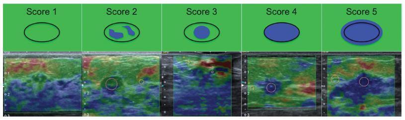
To evaluate the diagnostic accuracy of elastography for breast cancer identification in patients with indeterminate lesions on ultrasound.
This prospective, descriptive study included patients with indeterminate breast lesions in the ultrasound and with indication for percutaneous or surgical biopsy. The elastography was evaluated by qualitative analysis and by two methods for the semi quantitative analysis.
We evaluated 125 female patients with 159 lesions, with a mean age of 47 years, and a range of 20-85 years. Ultrasound has shown to be a method with good sensitivity (98.1%), but with a lower specificity (40.6%). On the elastography qualitative analysis, the specificity and accuracy were of 80.2% and 81.8% respectively. The mean size of the lesions showed no difference in classification by elastography. For the semiquantitative elastography, the mean values of the malignant lesions were statistically higher when compared with the subcutaneous tissue or the adjacent fibroglandular tissue. The analysis of the receiver operating characteristic (ROC) curves for these two semiquantitativemethods showed that both are considered satisfactory, with an area under the curve above 0.75 and statistical significance (p < 0.0001). The best results were obtained when using the findings of combined conventional ultrasound and qualitative elastography, with 100% sensitivity and 63.2% specificity.
Elastography can be a useful complementary method, increasing the specificity and diagnostic accuracy of conventional ultrasound for the diagnosis of breast cancer in patients with indeterminate breast lesions.
Summary
Revista Brasileira de Ginecologia e Obstetrícia. 2017;39(2):72-79
To evaluate the diagnostic accuracy of elastography for breast cancer identification in patients with indeterminate lesions on ultrasound.
This prospective, descriptive study included patients with indeterminate breast lesions in the ultrasound and with indication for percutaneous or surgical biopsy. The elastography was evaluated by qualitative analysis and by two methods for the semi quantitative analysis.
We evaluated 125 female patients with 159 lesions, with a mean age of 47 years, and a range of 20-85 years. Ultrasound has shown to be a method with good sensitivity (98.1%), but with a lower specificity (40.6%). On the elastography qualitative analysis, the specificity and accuracy were of 80.2% and 81.8% respectively. The mean size of the lesions showed no difference in classification by elastography. For the semiquantitative elastography, the mean values of the malignant lesions were statistically higher when compared with the subcutaneous tissue or the adjacent fibroglandular tissue. The analysis of the receiver operating characteristic (ROC) curves for these two semiquantitativemethods showed that both are considered satisfactory, with an area under the curve above 0.75 and statistical significance (p < 0.0001). The best results were obtained when using the findings of combined conventional ultrasound and qualitative elastography, with 100% sensitivity and 63.2% specificity.
Elastography can be a useful complementary method, increasing the specificity and diagnostic accuracy of conventional ultrasound for the diagnosis of breast cancer in patients with indeterminate breast lesions.
Summary
Revista Brasileira de Ginecologia e Obstetrícia. 2022;44(7):721-722
Summary
Revista Brasileira de Ginecologia e Obstetrícia. 2022;44(7):721-722
Summary
Revista Brasileira de Ginecologia e Obstetrícia. 2004;26(9):721-725
DOI 10.1590/S0100-72032004000900008
PURPOSE: to analyze the association between bacterial vaginosis (BV), high-risk HPV DNA, and Pap smear abnormalities in women submitted to diathermic conization for the treatment of high-grade cervical intraepithelial neoplasia (CIN 2 or 3). METHODS: a descriptive clinical study with 81 women submitted to diathermic conization for the treatment of CIN 2 or 3. Initial Pap smear was performed by the time of the biopsy and was also used to verify the presence of BV. Prior to conization, samples for the detection of high-risk HPV DNA through hybrid capture II (HC II) were collected. A control visit was scheduled for four months after the conization to repeat these tests. Twenty-seven women were found to have BV and 54 were not. Statistical analysis comprised odds ratios (OR) to assess the correlations between BV and HPV detection before and after diathermic conization and cytological abnormalities. All analyses were performed with a 95% confidence interval (95% CI). RESULTS: high-risk HPV DNA detection before conization was identical in both groups (89%). After conization, HPV DNA detection decreased to 26 and 18% in the groups with and without BV, respectively (OR=1.5; 95% CI 0.5 to 4.6). In addition, 41% of the women with BV and 20% without BV showed Pap smear abnormalities (OR=2.7; 95% CI 1.0 to 7.4). Regarding these 22 women with Pap smear abnormalities approximately four months after the diathermic conization, 83% of the BV group tested positive for HPV DNA compared with 50% in the group without BV (OR=5.0; IC 95% 0.5 a 52.9). CONCLUSION: women with BV presented more Pap smear abnormalities after conization when compared to the women without BV, although this was not statistically significant. This association was not related to high-risk HPV DNA.
Summary
Revista Brasileira de Ginecologia e Obstetrícia. 2006;28(12):721-727
DOI 10.1590/S0100-72032006001200006
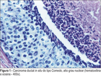
PURPOSE: to evaluate the clinical, radiological therapeutic and anatomo-pathological aspects in a series of patients with breast ductal carcinoma in situ (DCIS), attended in three public hospitals in Belo Horizonte (MG). METHODS: 179 cases of DCIS, that were selected from all the patients who had been diagnosed with breast cancer between 1985 and 2000, were studied retrospectively. After reviewing all the tissue sections, it was possible to collect all the clinical data, mammogram and treatment information of 85 cases. RESULTS: most patients were not symptomatic and the diagnosis had been done by mammogram (68.2%), being the microcalcification the most common radiological alteration. There has been a progressive increase in the diagnosis of DCIS along the years, following the introduction of periodical mammographic screening. The initial histopathological diagnosis and the review agreed in 72.9% of cases. In three cases, the original diagnosis of DCIS was not confirmed, being classified as atypical hyperplasia. Mammogram microcalcifications were confirmed in the pathological analysis in 95.6% of cases. Half of the patients was treated with mastectomy. All lymph nodes from axillary dissection were negative for metastases. CONCLUSIONS: The present study is in agreement with the recent literature, which shows an increase in the diagnosis of DCIS since 1990. There has been a great interobserver variation since the initial pathological diagnosis, which tended to malignancy and the present review. There were a great number of radical treatments, such as mastectomy and axillary dissection, which would probably be replaced by conservative treatment and sentinel lymph node biopsy nowadays, according to recent knowledge.
Summary
Revista Brasileira de Ginecologia e Obstetrícia. 2006;28(12):721-727
DOI 10.1590/S0100-72032006001200006
PURPOSE: to evaluate the clinical, radiological therapeutic and anatomo-pathological aspects in a series of patients with breast ductal carcinoma in situ (DCIS), attended in three public hospitals in Belo Horizonte (MG). METHODS: 179 cases of DCIS, that were selected from all the patients who had been diagnosed with breast cancer between 1985 and 2000, were studied retrospectively. After reviewing all the tissue sections, it was possible to collect all the clinical data, mammogram and treatment information of 85 cases. RESULTS: most patients were not symptomatic and the diagnosis had been done by mammogram (68.2%), being the microcalcification the most common radiological alteration. There has been a progressive increase in the diagnosis of DCIS along the years, following the introduction of periodical mammographic screening. The initial histopathological diagnosis and the review agreed in 72.9% of cases. In three cases, the original diagnosis of DCIS was not confirmed, being classified as atypical hyperplasia. Mammogram microcalcifications were confirmed in the pathological analysis in 95.6% of cases. Half of the patients was treated with mastectomy. All lymph nodes from axillary dissection were negative for metastases. CONCLUSIONS: The present study is in agreement with the recent literature, which shows an increase in the diagnosis of DCIS since 1990. There has been a great interobserver variation since the initial pathological diagnosis, which tended to malignancy and the present review. There were a great number of radical treatments, such as mastectomy and axillary dissection, which would probably be replaced by conservative treatment and sentinel lymph node biopsy nowadays, according to recent knowledge.
Summary
Revista Brasileira de Ginecologia e Obstetrícia. 2018;40(11):722-725
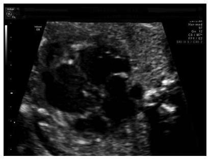
Noncompaction cardiomyopathy (NCCM) and left ventricular noncompaction (LVNC), in their isolated form, are rare cardiomyopathies. They are characterized by a thickened myocardium due to the presence of deep trabeculae recesses, and to thick trabeculae. This condition is associated with a variable clinical phenotype including heart failure, thromboembolism, and sudden death. We report a case of LVNC at 26 weeks and 4 days of gestation revised on the basis of what is currently reported in the literature. A review of the literature was performed to better describe this rare condition. Left ventricular noncompaction is a rare fetal condition and it should be suspected in case of cardiomyopathy.
Summary
Revista Brasileira de Ginecologia e Obstetrícia. 2018;40(11):722-725
Noncompaction cardiomyopathy (NCCM) and left ventricular noncompaction (LVNC), in their isolated form, are rare cardiomyopathies. They are characterized by a thickened myocardium due to the presence of deep trabeculae recesses, and to thick trabeculae. This condition is associated with a variable clinical phenotype including heart failure, thromboembolism, and sudden death. We report a case of LVNC at 26 weeks and 4 days of gestation revised on the basis of what is currently reported in the literature. A review of the literature was performed to better describe this rare condition. Left ventricular noncompaction is a rare fetal condition and it should be suspected in case of cardiomyopathy.
Summary
Revista Brasileira de Ginecologia e Obstetrícia. 2022;44(7):723-736
Summary
Revista Brasileira de Ginecologia e Obstetrícia. 2022;44(7):723-736