Summary
Summary
Summary
Revista Brasileira de Ginecologia e Obstetrícia. 2004;26(1):59-64
DOI 10.1590/S0100-72032004000100009
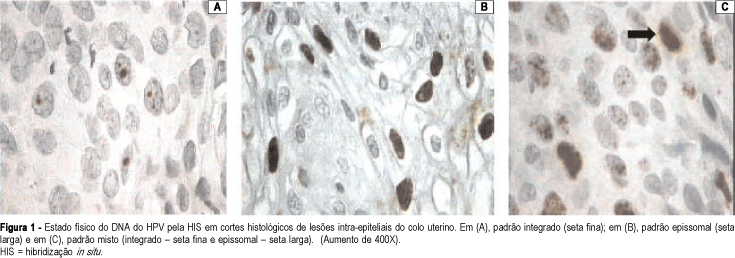
PURPOSE: to carry out a molecular study (in situ hybridization) on patients who present intraepithelial lesions of the uterine cervix, and to assess the frequency and the physical state of the human papillomavirus (HPV). METHODS: histological sections of biopsies of the uterine cervix from 84 patients were evaluated by in situ hybridization, with a broad-spectrum probe, which allows the identification of the HPV types 6, 11, 16, 18, 31, 33, 35, 39, 42, 45, and 56 and with specific probes for HPV types 6, 11, 16, 18, 31, and 33. The physical patterns of HPV DNA found were: episomal, when the entire nucleus stains with biotin (brown); integrated - one or two brown points in the hybridized nucleus, or mixed, associating both patterns. Of the 84 patients evaluated, 31 (36.9%) had low-grade squamous intraepithelial lesions (LSIL), and 53 (63.1%) had high-grade squamous intraepithelial lesions (HSIL) on histological examination. Fisher's exact test was used for the statistical analysis. RESULTS: considering all the cases, 46 (54.7%) were positive for HPV DNA with the broad-spectrum probe. Regarding typing, HPV-16 was the most frequent in HSIL (12 cases - 22.6% - p<0.05). The frequencies of the other HPV types did not show statistically significant differences between the LSIL and HSIL cases. By physical condition assessment of the HPV DNA, the percentage of the episomal (most common in LSIL) and integrated patterns showed no significant differences between the two groups; the mixed HSIL type prevailed when compared to LSIL: 26.4 and 3.2%, respectively (p<0.01). The physical condition of the HPV DNA, integrated in the host cell, was more frequent in the most severe cases. CONCLUSIONS: HPV-16 was the most frequent in HSIL cases. The frequencies of the other HPV types did not show statistically significant differences between the LSIL and HSIL cases. The physical condition of the HPV DNA, integrated in the host cell, was more frequent in the more severe cases.
Summary
Revista Brasileira de Ginecologia e Obstetrícia. 2004;26(1):59-64
DOI 10.1590/S0100-72032004000100009
PURPOSE: to carry out a molecular study (in situ hybridization) on patients who present intraepithelial lesions of the uterine cervix, and to assess the frequency and the physical state of the human papillomavirus (HPV). METHODS: histological sections of biopsies of the uterine cervix from 84 patients were evaluated by in situ hybridization, with a broad-spectrum probe, which allows the identification of the HPV types 6, 11, 16, 18, 31, 33, 35, 39, 42, 45, and 56 and with specific probes for HPV types 6, 11, 16, 18, 31, and 33. The physical patterns of HPV DNA found were: episomal, when the entire nucleus stains with biotin (brown); integrated - one or two brown points in the hybridized nucleus, or mixed, associating both patterns. Of the 84 patients evaluated, 31 (36.9%) had low-grade squamous intraepithelial lesions (LSIL), and 53 (63.1%) had high-grade squamous intraepithelial lesions (HSIL) on histological examination. Fisher's exact test was used for the statistical analysis. RESULTS: considering all the cases, 46 (54.7%) were positive for HPV DNA with the broad-spectrum probe. Regarding typing, HPV-16 was the most frequent in HSIL (12 cases - 22.6% - p<0.05). The frequencies of the other HPV types did not show statistically significant differences between the LSIL and HSIL cases. By physical condition assessment of the HPV DNA, the percentage of the episomal (most common in LSIL) and integrated patterns showed no significant differences between the two groups; the mixed HSIL type prevailed when compared to LSIL: 26.4 and 3.2%, respectively (p<0.01). The physical condition of the HPV DNA, integrated in the host cell, was more frequent in the most severe cases. CONCLUSIONS: HPV-16 was the most frequent in HSIL cases. The frequencies of the other HPV types did not show statistically significant differences between the LSIL and HSIL cases. The physical condition of the HPV DNA, integrated in the host cell, was more frequent in the more severe cases.
Summary
Revista Brasileira de Ginecologia e Obstetrícia. 2019;41(1):59-61
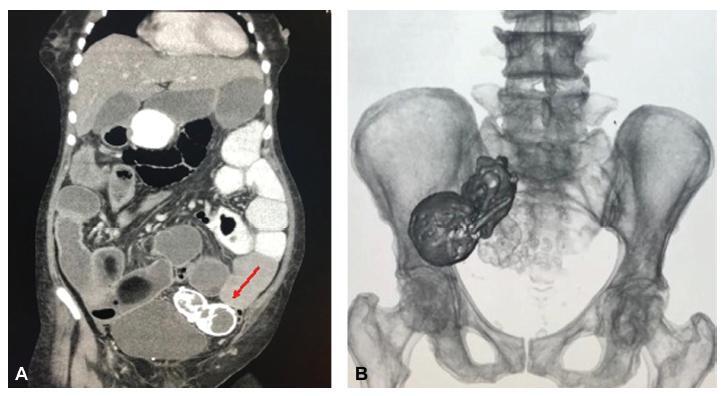
Lithopedion (lithos = rock and paidion = child) is a rare condition that only occurs in 1.5 to 1.8% of extrauterine pregnancies and in 0.00045% of all pregnancies. It consists of an ectopic pregnancy in which the fetus dies but cannot be reabsorbed by the mother’s body, which then coats it in a calcium-rich substance.We present the case of a 77-year-old woman with an incidental diagnosis of a lithopedion, which had been retained in her left pelvis for presumably 40 years.
Summary
Revista Brasileira de Ginecologia e Obstetrícia. 2019;41(1):59-61
Lithopedion (lithos = rock and paidion = child) is a rare condition that only occurs in 1.5 to 1.8% of extrauterine pregnancies and in 0.00045% of all pregnancies. It consists of an ectopic pregnancy in which the fetus dies but cannot be reabsorbed by the mother’s body, which then coats it in a calcium-rich substance.We present the case of a 77-year-old woman with an incidental diagnosis of a lithopedion, which had been retained in her left pelvis for presumably 40 years.
Summary
Revista Brasileira de Ginecologia e Obstetrícia. 1999;21(1):59-59
DOI 10.1590/S0100-72031999000100011
Summary
Revista Brasileira de Ginecologia e Obstetrícia. 1999;21(1):59-59
DOI 10.1590/S0100-72031999000100011
Summary
Revista Brasileira de Ginecologia e Obstetrícia. 1999;21(1):59-59
DOI 10.1590/S0100-72031999000100010
Summary
Revista Brasileira de Ginecologia e Obstetrícia. 1999;21(1):59-59
DOI 10.1590/S0100-72031999000100010
Summary
Revista Brasileira de Ginecologia e Obstetrícia. 2002;24(1):59-65
DOI 10.1590/S0100-72032002000100009
Introduction: the Resolution 196/96 of the Conselho Nacional de Saúde (National Council of Health/Ministry of Health) presents the main Brazilian guidelines on research involving human subjects, including the content of written informed consent. Purpose: to present the knowledge and opinion of Brazilian researchers on the contents of Resolution 196/96, specifically related to the informed consent form. Subjects and methods: forty-six doctors responsible for the area of gynecology at Brazilian universities, four directors of research centers and 31 researchers who participated in a study related to fertility regulation during the 12 months preceding September, 2000. Subjects completed a self-reporting questionnaire. Data were analyzed by the chi² test. Results: most subjects declared that they knew the Resolution 196/96 and considered it adequate, although difficult to comply with; they considered that all studies should have an informed consent form, and knew that its content should guarantee confidentiality. More researchers than those responsible for gynecology department/directors knew that the informed consent form should be prepared by the principal investigator. Significantly more responsible for gynecology department/directors than researchers declared that subjects must always sign (or put their thumb print if they do not know how to write) on the informed consent form. Subjects declared that payment of expenses resulting from participation in a study must always be explained in the informed consent form. Conclusion: despite the wide dissemination of the Resolution 196/96, it was not known by all the researchers nor by all those responsible for gynecology departament/directors. The majority agreed with the contents required by the Resolution for the informed consent form.
Summary
Revista Brasileira de Ginecologia e Obstetrícia. 2015;37(2):59-63
DOI 10.1590/SO100-720320140005180
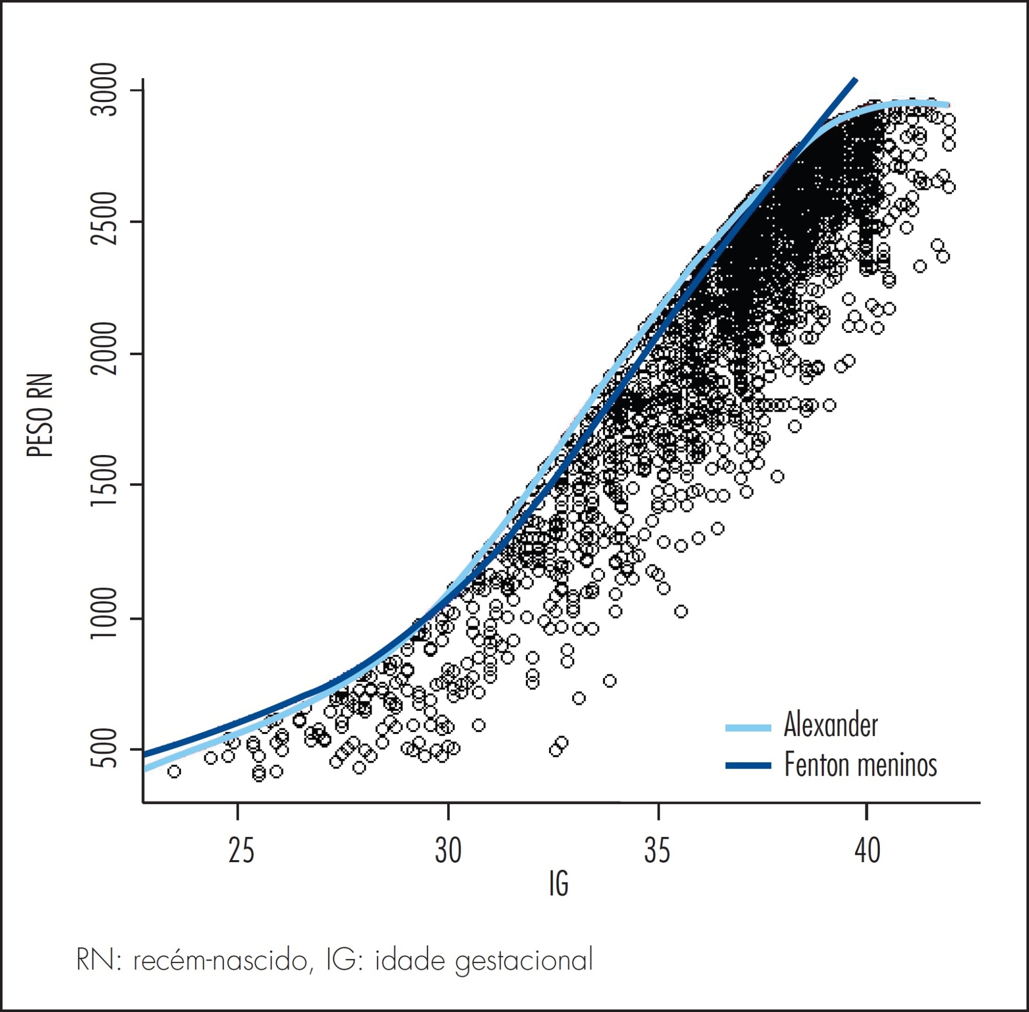
It was to compare the use of two growth curves for the diagnosis of small-for-gestational-age (SGA) infants, having the 10thpercentile as reference.
In a retrospective study, data of 20,567 singleton live births from January 2003 to June 2014 were analyzed, and divided according to gestational age: (a) 23 to 26, (b) 26 to 29, (c) 29 to 32, (d) 32 to 35, (e) 35 to 38, (f) 38 to 41 and (g) >41 weeks. Data were paired and analyzed using the McNemar test, with the level of significance set at 0.05.
The curve designed by Alexander indicated a higher percentage of diagnosis of SGA than the curve constructed by Fenton for every category of gestational age up to 41 weeks, more markedly in the 32-35 week group (18.5%). Between 37 and 40 weeks of gestational age, Alexander's curve exceeded Fenton's curve in 9.1% of the cases in the diagnosis of SGA.
The Fenton curve provides a more accurate evaluation of an infant's growth since it is gender-specific and allows measurement of three parameters. It has also been constructed with newer data and more sophisticated statistical tools.
Summary
Revista Brasileira de Ginecologia e Obstetrícia. 2015;37(2):59-63
DOI 10.1590/SO100-720320140005180
It was to compare the use of two growth curves for the diagnosis of small-for-gestational-age (SGA) infants, having the 10thpercentile as reference.
In a retrospective study, data of 20,567 singleton live births from January 2003 to June 2014 were analyzed, and divided according to gestational age: (a) 23 to 26, (b) 26 to 29, (c) 29 to 32, (d) 32 to 35, (e) 35 to 38, (f) 38 to 41 and (g) >41 weeks. Data were paired and analyzed using the McNemar test, with the level of significance set at 0.05.
The curve designed by Alexander indicated a higher percentage of diagnosis of SGA than the curve constructed by Fenton for every category of gestational age up to 41 weeks, more markedly in the 32-35 week group (18.5%). Between 37 and 40 weeks of gestational age, Alexander's curve exceeded Fenton's curve in 9.1% of the cases in the diagnosis of SGA.
The Fenton curve provides a more accurate evaluation of an infant's growth since it is gender-specific and allows measurement of three parameters. It has also been constructed with newer data and more sophisticated statistical tools.
Summary
Revista Brasileira de Ginecologia e Obstetrícia. 2006;28(10):590-595
DOI 10.1590/S0100-72032006001000004
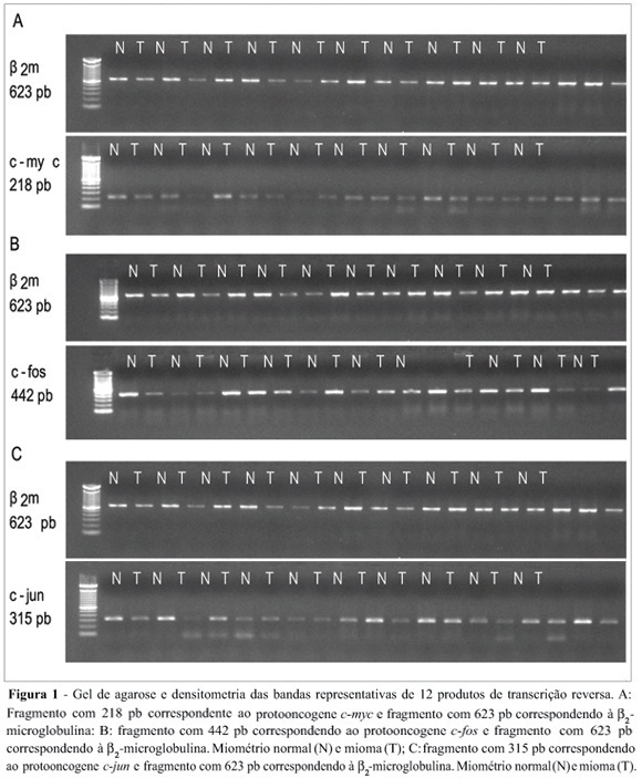
Uterine myomas are common benign tumors of the female genital tract. The expression of growth factor signal transduction cascade components including the protooncogenes c-myc, c-fos, and c-jun seem to be involved in the development of myomas. PURPOSE: To compare the gene (mRNA) and protein expression of the protooncogenes c-fos, c-myc, and c-jun in human normal myometrium and leiomyoma. METHOD: A case-control study was performed. Samples were collected from 12 patients submitted to hysterectomy at the Hospital de Clínicas at Porto Alegre. The expression of the specific mRNA for c-myc, c-fos, c-jun, and beta-microglobulin was assessed through the RT-PCR technique, using specific primers to each gene. The protein expression of these protooncogenes was evaluated through the Western blot technique with specific antibodies. RESULTS: No statistically significant difference was observed in the gene expression for these protooncogenes between normal myometrium and leiomyoma (c-myc: 0,87 ± 0,08 vs 0,87 ± 0,08, p = 0,952; c-fos: 1,10 ± 0,17 vs 1,01 ± 0,11, p = 0,21; c-jun: 1,03 ± 0,12 vs 0,96 ± 0,09, p = 0,168, respectively). No statiscally significant difference was observed for the protein expression of these protooncogenes between normal myometrium and leiomyoma (c-myc: 1,36 ± 0,48 vs 1,53 ± 0,29, p = 0,569; c-fos: 8,85 ± 5,5 vs 6,56 ± 4,22, p = 0,434; e c-jun: 6,47 ± 3,04 vs 5,42 ± 2,03, p = 0,266, respectively). CONCLUSION: No difference was observed in the gene expression (transcription) nor in the protein expression (translation) of the protooncogenes c-myc, c-fos, and c-jun between leiomyoma and myometrium.
Summary
Revista Brasileira de Ginecologia e Obstetrícia. 2006;28(10):590-595
DOI 10.1590/S0100-72032006001000004
Uterine myomas are common benign tumors of the female genital tract. The expression of growth factor signal transduction cascade components including the protooncogenes c-myc, c-fos, and c-jun seem to be involved in the development of myomas. PURPOSE: To compare the gene (mRNA) and protein expression of the protooncogenes c-fos, c-myc, and c-jun in human normal myometrium and leiomyoma. METHOD: A case-control study was performed. Samples were collected from 12 patients submitted to hysterectomy at the Hospital de Clínicas at Porto Alegre. The expression of the specific mRNA for c-myc, c-fos, c-jun, and beta-microglobulin was assessed through the RT-PCR technique, using specific primers to each gene. The protein expression of these protooncogenes was evaluated through the Western blot technique with specific antibodies. RESULTS: No statistically significant difference was observed in the gene expression for these protooncogenes between normal myometrium and leiomyoma (c-myc: 0,87 ± 0,08 vs 0,87 ± 0,08, p = 0,952; c-fos: 1,10 ± 0,17 vs 1,01 ± 0,11, p = 0,21; c-jun: 1,03 ± 0,12 vs 0,96 ± 0,09, p = 0,168, respectively). No statiscally significant difference was observed for the protein expression of these protooncogenes between normal myometrium and leiomyoma (c-myc: 1,36 ± 0,48 vs 1,53 ± 0,29, p = 0,569; c-fos: 8,85 ± 5,5 vs 6,56 ± 4,22, p = 0,434; e c-jun: 6,47 ± 3,04 vs 5,42 ± 2,03, p = 0,266, respectively). CONCLUSION: No difference was observed in the gene expression (transcription) nor in the protein expression (translation) of the protooncogenes c-myc, c-fos, and c-jun between leiomyoma and myometrium.