You searched for:"Marcelo Zugaib"
We found (76) results for your search.Summary
Rev Bras Ginecol Obstet. 2001;23(4):235-241
DOI 10.1590/S0100-72032001000400006
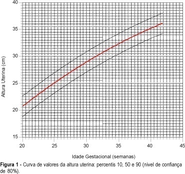
Purpose: to create a uterine height growth curve, according to gestational age, to verify differences among the existing curves and to evaluate the influence of color, parity and maternal weight on the variation of uterine height. Methods: during the period from July 1997 to July 1999, 100 normal pregnant women were submitted to uterine height measurements between the 20th and 42nd week of gestation. All the pregnant women had ultrasonically confirmed gestational age. A total of 726 measurements of uterine height were carried out by the same examiner, using a metric tape from the upper border of the symphysis pubis to the fundus uteri. Results: curves and tables of uterine height according to gestational age were obtained. The average uterine height growth was 0.7 cm/week. The study revealed different average uterine height values in relation to other uterine height growth curves. No statistically significant variations were found between the distributions of uterine heights according to color, parity and weight. Conclusion: the construction of a methodologically accepted uterine height growth curve aimed to detect, as a clinical method, the fetal growth disturbances. This should be analyzed in a posterior study.
Summary
Rev Bras Ginecol Obstet. 2001;23(4):235-241
DOI 10.1590/S0100-72032001000400006
Purpose: to create a uterine height growth curve, according to gestational age, to verify differences among the existing curves and to evaluate the influence of color, parity and maternal weight on the variation of uterine height. Methods: during the period from July 1997 to July 1999, 100 normal pregnant women were submitted to uterine height measurements between the 20th and 42nd week of gestation. All the pregnant women had ultrasonically confirmed gestational age. A total of 726 measurements of uterine height were carried out by the same examiner, using a metric tape from the upper border of the symphysis pubis to the fundus uteri. Results: curves and tables of uterine height according to gestational age were obtained. The average uterine height growth was 0.7 cm/week. The study revealed different average uterine height values in relation to other uterine height growth curves. No statistically significant variations were found between the distributions of uterine heights according to color, parity and weight. Conclusion: the construction of a methodologically accepted uterine height growth curve aimed to detect, as a clinical method, the fetal growth disturbances. This should be analyzed in a posterior study.
Summary
Rev Bras Ginecol Obstet. 2011;33(9):240-245
DOI 10.1590/S0100-72032011000900004
PURPOSE: To determine the accuracy of ultrasound in fetal weight estimation and to evaluate maternal and/or fetal factors that could interfere in the result. METHODS: This was a transverse prospective study, involving 106 patients, with 212 fetal weight evaluations, by two observers, within 24 h to delivery. The following parameters were measured: biparietal diameter, head circumference, abdominal circumference, and femoral length. Fetal weight was estimated using the Hadlock formula and the results were compared to birth weight. The maternal factors examined were: weight, BMI, and skin to uterus distance measured by ultrasound, and the fetal factors were: presentation, position, placental localization and thickness, fetal weight, and amniotic fluid index (AFI). RESULTS: There was good correlation between estimated fetal weight and birth weight (R=0.97). In 79.2% and in 92.4% of cases the estimated fetal weight was within 10% and 15% of birth weight, respectively. The only maternal factor that presented a positive correlation with percent error in the estimate of fetal weight was the skin to uterus distance (R³0.56). Fetal weight showed negative correlation with percent error (R>-0.36; p<0.001), with a significant tendency to overestimate fetal weight in the group of very low weight - <1000 g (p<0.05). The AFI showed a low negative correlation with percent error (R=-0.21; p<0.001) with no difference between AFI groups (p=0.516). CONCLUSION: Ultrasound presented good accuracy in the estimation of fetal weight. The error of weight estimate was directly proportional to the skin to uterus distance and inversely proportional to fetal weight. AFI did not interfere significantly in the ultrasound prediction of fetal weight.
Summary
Rev Bras Ginecol Obstet. 2002;24(4):247-251
DOI 10.1590/S0100-72032002000400006
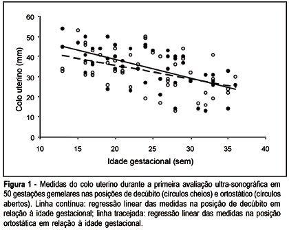
Purpose: to compare cervical length measurements in twin pregnancies obtained by transvaginal ultrasound examination in the recumbent and standing positions. Methods: fifty twin pregnancies underwent transvaginal ultrasound examinations to measure the cervical length with the women in recumbent and standing positions. The study was carried out between May 1999 and December 2000. The scans were repeated every 4 weeks and the total number of evaluations was 136. Two groups were analyzed: one included only the first ultrasound examinations carried out in each woman and the second group included all evaluations. Results: in the first group, cervical length measurements in the standing and recumbent positions correlated inversely with the gestational age (recumbent: r=-0.60; p<0.001; standing: r=-0.46; p=0.008). The mean measure in the recumbent position was 35.2 mm (SD=9.9 mm) and 33.4 mm (SD=9.5 mm) in the standing position. When the difference between the measure obtained in the standing and recumbent positions was expressed as percentage of the measure in the recumbent position, there was no significant association with gestational age (p=0.07). When all evaluations were considered, there was a significant association between cervical length in the recumbent and standing positions (r=0.79; p<0.001). The measures in recumbent and standing positions were inversely correlated with gestational age (recumbent: p<0.0001; standing: p<0.0001). The mean cervical length in the recumbent position was 33.5 mm (SD=10.8 mm) and 31.8 mm (SD=9.6 mm) in the standing position. There was no significant association between cervical length difference expressed as percentage of the measure in the recumbent position and gestation. Conclusion: cervical length measure obtained with the patients in the recumbent and standing positions provided similar information.
Summary
Rev Bras Ginecol Obstet. 2002;24(4):247-251
DOI 10.1590/S0100-72032002000400006
Purpose: to compare cervical length measurements in twin pregnancies obtained by transvaginal ultrasound examination in the recumbent and standing positions. Methods: fifty twin pregnancies underwent transvaginal ultrasound examinations to measure the cervical length with the women in recumbent and standing positions. The study was carried out between May 1999 and December 2000. The scans were repeated every 4 weeks and the total number of evaluations was 136. Two groups were analyzed: one included only the first ultrasound examinations carried out in each woman and the second group included all evaluations. Results: in the first group, cervical length measurements in the standing and recumbent positions correlated inversely with the gestational age (recumbent: r=-0.60; p<0.001; standing: r=-0.46; p=0.008). The mean measure in the recumbent position was 35.2 mm (SD=9.9 mm) and 33.4 mm (SD=9.5 mm) in the standing position. When the difference between the measure obtained in the standing and recumbent positions was expressed as percentage of the measure in the recumbent position, there was no significant association with gestational age (p=0.07). When all evaluations were considered, there was a significant association between cervical length in the recumbent and standing positions (r=0.79; p<0.001). The measures in recumbent and standing positions were inversely correlated with gestational age (recumbent: p<0.0001; standing: p<0.0001). The mean cervical length in the recumbent position was 33.5 mm (SD=10.8 mm) and 31.8 mm (SD=9.6 mm) in the standing position. There was no significant association between cervical length difference expressed as percentage of the measure in the recumbent position and gestation. Conclusion: cervical length measure obtained with the patients in the recumbent and standing positions provided similar information.
Summary
Rev Bras Ginecol Obstet. 2001;23(4):247-251
DOI 10.1590/S0100-72032001000400008
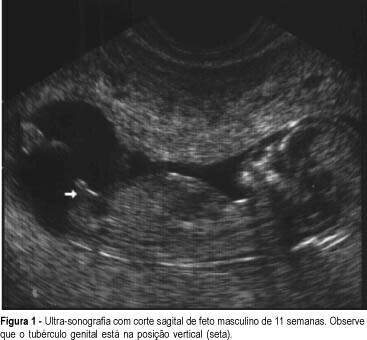
Purpose: to determine the feasibility of early ultrasonographic identification of fetal gender. Methods: a prospective study in a University Department of Obstetrics and Gynecology. A total of 592 women underwent ultrasonography at 11-14 weeks of gestation. Fetal gender was identified according to genital tubercle position (vertical or horizontal) at sagittal plane and confirmed at birth or by karyotype analysis. Results: the overall accuracy of correctly assigning fetal gender was 84%. The success of identification increased with gestational age, being 72%, 85% and 89% at 11, 12 and 13 weeks, respectively. The accuracy of correctly identifying fetal sex significantly changed with operator training, being 83.5% at the beginning and 93.6% at the end of the study. Conclusion: ultrasound determination of fetal gender is feasible, with good accuracy and may be of potential use to avoid invasive testing in family histories of X-linked disorders.
Summary
Rev Bras Ginecol Obstet. 2001;23(4):247-251
DOI 10.1590/S0100-72032001000400008
Purpose: to determine the feasibility of early ultrasonographic identification of fetal gender. Methods: a prospective study in a University Department of Obstetrics and Gynecology. A total of 592 women underwent ultrasonography at 11-14 weeks of gestation. Fetal gender was identified according to genital tubercle position (vertical or horizontal) at sagittal plane and confirmed at birth or by karyotype analysis. Results: the overall accuracy of correctly assigning fetal gender was 84%. The success of identification increased with gestational age, being 72%, 85% and 89% at 11, 12 and 13 weeks, respectively. The accuracy of correctly identifying fetal sex significantly changed with operator training, being 83.5% at the beginning and 93.6% at the end of the study. Conclusion: ultrasound determination of fetal gender is feasible, with good accuracy and may be of potential use to avoid invasive testing in family histories of X-linked disorders.
Summary
Rev Bras Ginecol Obstet. 2002;24(4):261-269
DOI 10.1590/S0100-72032002000400008
Purpose: to develop a mathematical model based on the fetal heart rate (FHR) monitoring to predict metabolic acidosis at birth (base excess < -10 mEq/L), in pregnancies with absent or reversed end-diastolic velocity (AREDV) in the umbilical arteries. Methods: the last FHR tracing of 127 AREDV cases was studied by visual analysis. The analyzed parameters included: gestational age, interval between AREDV diagnosis and delivery, FHR variability, FHR accelerations, decelerations, and sinusoidal- like pattern. Multivariate logistic regression was applied to find the best mathematical model to predict acidosis. Results: metabolic acidosis at birth occurred in 51 cases (40.2%). The model included the parameters: interval between AREDV diagnosis and delivery (X1), gestational age in weeks (X2), FHR variability <5 bpm (X3), and FHR variability 5-9 bpm (X4). To each variability parameter was assigned the value of 1 when present or 0 when absent. The z value is: z = 2.2348 + (-0.0117 X1) + (-0.09 X2) + (1.9552 X3)+ (-0.4474 X4). By applying the expression p=e z/(1 + e z), the probability is estimated. Conclusion: the mathematical model allowed us to estimate the probability of metabolic acidosis at birth, in pregnancies with AREDV, studying FHR-monitoring parameters.
Summary
Rev Bras Ginecol Obstet. 2014;36(6):264-268
DOI 10.1590/S0100-720320140004935
The aim of this study was to analize and describe some characteristics related to a false diagnosis of intrauterine growth restriction (IUGR).
We retrospectively included 48 pregnant women referred to our service with a suspected diagnosis of IUGR that was not confirmed after birth and we compared them to another group with confirmed IUGR. We then analyzed the characteristics of the false-positive results. The results of the study were divided into continuous and categorical variables for analysis. The χ2test or Fisher exact test was applied to compare proportions. The level of significance was set at p<0.05 for all tests.
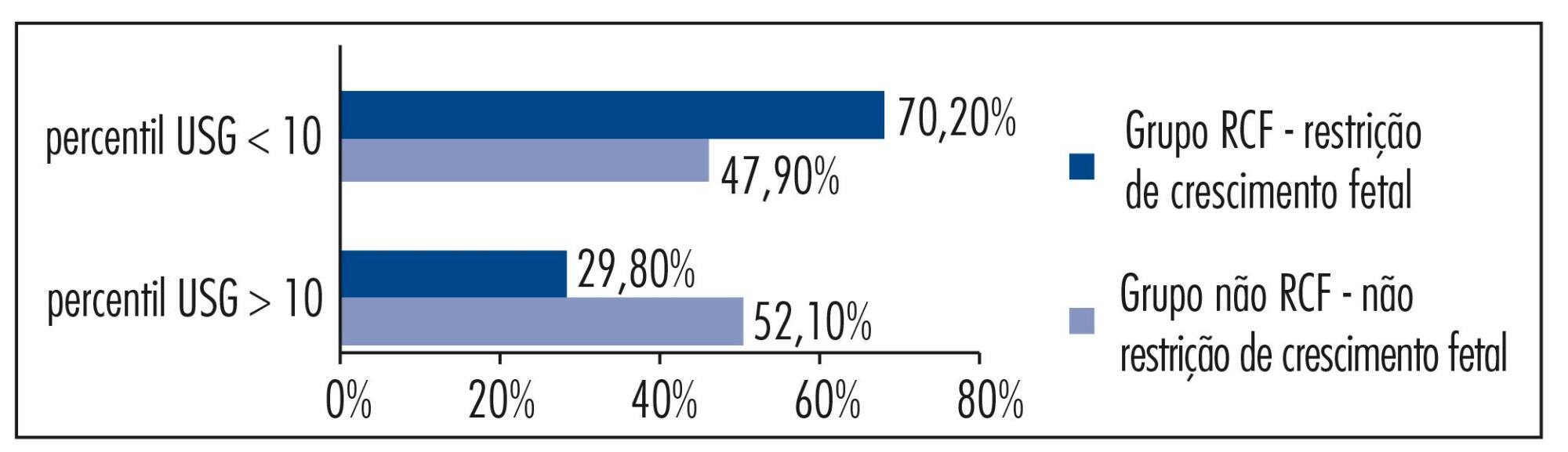
In our sample, pregnant women with a false diagnosis of IUGR had the following characteristics: they were referred earlier (mean gestational age of 32.8 weeks); were submitted to 2 to 6 ultrasound examinations before been registered in our service; in 25% of cases ultrasound examination was performed before 12 weeks; in 66.7% of cases the symphysis-fundal height measurement was normal; in 52.1% of cases they had at least 1 sonographic exam above the 10th percentile; on average, the last ultrasound examination (performed on average at 36 weeks) was above the 18th percentile; the women were submitted to a mean number of 5 ultrasound examinations and to a mean number of 4.6 vitality exams.
The false diagnosis of IUGR involves high hospital costs and higher demand for specialists. The symphysis-fundal height measurement must be valued, and the diagnosis of IUGR must be confirmed with ultrasonography in the last weeks of pregnancy before any obstetric management is taken.
Summary
Rev Bras Ginecol Obstet. 2014;36(6):264-268
DOI 10.1590/S0100-720320140004935
The aim of this study was to analize and describe some characteristics related to a false diagnosis of intrauterine growth restriction (IUGR).
We retrospectively included 48 pregnant women referred to our service with a suspected diagnosis of IUGR that was not confirmed after birth and we compared them to another group with confirmed IUGR. We then analyzed the characteristics of the false-positive results. The results of the study were divided into continuous and categorical variables for analysis. The χ2test or Fisher exact test was applied to compare proportions. The level of significance was set at p<0.05 for all tests.
In our sample, pregnant women with a false diagnosis of IUGR had the following characteristics: they were referred earlier (mean gestational age of 32.8 weeks); were submitted to 2 to 6 ultrasound examinations before been registered in our service; in 25% of cases ultrasound examination was performed before 12 weeks; in 66.7% of cases the symphysis-fundal height measurement was normal; in 52.1% of cases they had at least 1 sonographic exam above the 10th percentile; on average, the last ultrasound examination (performed on average at 36 weeks) was above the 18th percentile; the women were submitted to a mean number of 5 ultrasound examinations and to a mean number of 4.6 vitality exams.
The false diagnosis of IUGR involves high hospital costs and higher demand for specialists. The symphysis-fundal height measurement must be valued, and the diagnosis of IUGR must be confirmed with ultrasonography in the last weeks of pregnancy before any obstetric management is taken.
Summary
Rev Bras Ginecol Obstet. 2013;35(1):27-32
DOI 10.1590/S0100-72032013000100006
PURPOSE: To compare and analyze socioeconomic aspects and the emotional experience of women with spontaneous or induced abortion and in women living in the outskirts of São Paulo. METHODS: A prospective case-control study carried out from July 2008 to March 2010, involving semi-structured interviews with women who presented a previous diagnosis of abortion and who had been admitted to two public hospitals in the outskirts of São Paulo. The study included 100 women with diagnosis of abortion and were hospitalized for curettage. Eleven women who reported induced abortion (11%) represented the case group. The control group (n=22) was selected at a 2:1 ratio according to the following procedure: for every case of induced abortion, the next two cases of spontaneous abortion at the same hospital. A semistructured interview was conducted with questions regarding emotional aspects and family, social and economic context. RESULTS: The women with induced abortion compared to the group with spontaneous abortion had lower educational level, with more frequent elementary level (82 versus 36%, p=0.04), lower income (median, R$ 1,000.00 versus R$ 1,400.00, p=0.04), lower personal income (median, R$ 200.00 versus R$ 333.00, p=0.04), higher frequency of negative feelings upon suspicion (82 versus 22%, p=0.004) and confirmation (72 versus 22%, p=0.03) of pregnancy. CONCLUSION: Among women looking for health care in hospitals in the outskirts of São Paulo, induced abortion is related to unfavorable socioeconomic conditions, which affects the emotional experiences of suspicion and confirmation of pregnancy.
Summary
Rev Bras Ginecol Obstet. 2000;22(5):275-279
DOI 10.1590/S0100-72032000000500004
Objective: to evaluate ultrasound findings in pregnant women with threatened abortion in the first trimester of pregnancy. Methods: transabdominal and transvaginal ultrasound scans were performed in patients with vaginal bleeding with previous positive pregnancy test. Patients with 6-14-week gestation (by the last menstrual period or ultrasound scan), with closed cervix on clinical evaluation were included. Multiple pregnancies and those patients who have tried abortion by using abortive drugs or manipulation were excluded. Results: in 132 of 247 (53.4%) the pregnancy was viable and in 46.6% (115/247) the pregnancy was nonviable. Incomplete miscarriage was found in 19% (47/247), complete miscarriage in 8.5% (21/247), missed abortion in 7.7% (19/247), anembryonic pregnancy in 6.1% (15/247), ectopic pregnancy in 4.5% (11/247) and hydatidiform mole in 0.8% (2/247). Conclusion: almost half (46.6%) of the pregnancies with threatened abortion in the first trimester were diagnosed as a nonviable pregnancy. The ultrasound scan can help to define this condition and the management of the pregnancy.