You searched for:"Antonio Carlos Vieira Cabral"
We found (23) results for your search.Summary
Revista Brasileira de Ginecologia e Obstetrícia. 2001;23(10):653-657
DOI 10.1590/S0100-72032001001000007
Purpose: to verify if there is an association between the mean blood velocity in the descending thoracic aorta and fetal anemia diagnosis. Methods: this is a prospective, cross-sectional study in which the mean blood velocities in the fetal aorta, in 66 fetuses at risk for severe anemia due to severe Rh immunization, and cord blood hemoglobin levels were analyzed comparatively. The hemoglobin level was obtained by cordocentesis if an intravascular transfusion was performed for severe anemia, however, if the fetus received an intrauterine transfusion by the intraperitoneal route or if the fetus did not receive a transfusion at all, hemoglobin level was measured at the time of pregnancy termination by umbilical cord puncture. The authors made a statistical association between the mean blood velocity in fetal descending thoracic aorta and the diagnosis of fetal anemia. The c² test was used for statistical analysis and a p value <0,05 was used to indicate significance. Results: there was a significant and indirect association between the mean blood velocity in the descending thoracic aorta and the detection of fetal anemia. The mean blood velocity in fetal thoracic aorta had a sensitivity of 47.4% for the diagnosis of moderate fetal anemia (Hg<10.0 g/dL), with a p value <0.01 by the Fisher exact test, and a sensitivity of 54.5% for severe Rh isoimmunization (Hg<7.0 g/dL), with a p value =0.01. Conclusion: this study revealed a significant indirect correlation between mean blood velocity in the descending thoracic aorta and the detection of fetal anemia due to Rh isoimmunization.
Summary
Revista Brasileira de Ginecologia e Obstetrícia. 2001;23(4):243-246
DOI 10.1590/S0100-72032001000400007
Purpose: to evaluate the possibility and accuracy of fetal karyotyping in pleural effusions. Methods: we studied fifteen fetuses with unilateral or bilateral pleural effusions. All of these fetuses underwent intrauterine thoracocentesis guided by ultrasound examinations. The gestational age varied from 19 to 34 weeks. A morphogenetic ultrasound examination was performed in each case by the authors in order to identify associated structural anomalies. When the cellular cultures of pleural effusion samples were negative, an alternative karyotype was obtained by cordocentesis. A fetal lymphocyte culture was made of pleural effusion samples for karyotype in a similar technique as for fetal blood. Results: the fetal karyotype was successful in 12 cases. There were 4 abnormal results, all of them were Down syndromes, and in the other 8 cases the chromosomal analyses were normal. The fetal karyotype was confirmed and compared by newborn blood chromosomal analysis, genetic evaluation or necropsy. There were no maternal or fetal side effects related to the procedure. Conclusions: the fetal karyotyping performed in pleural effusions obtained by intrauterine thoracocentesis proved to be highly efficient and safe. It must be the method of choice for rapid karyotyping in fetuses with pleural edema.
Summary
Revista Brasileira de Ginecologia e Obstetrícia. 2001;23(5):277-282
DOI 10.1590/S0100-72032001000500002
Objective: to test the effectiveness of the polymerase chain reaction (PCR) in the amniotic fluid for the detection of fetal contamination due to Toxoplasma gondii in pregnant women with acute infection and to correlate it with the inoculation technique and the histology of the placenta. Methods: thirty-seven patients were prospectively studied and the diagnosis was based on the identification of maternal acute infection followed by amniocentesis guided by ultrasound to obtain amniotic fluid for PCR and mice inoculation. The mothers were treated with spiramycin throughout pregnancy; when fetal infection was demonstrated, pyrimethamine and sulfadiazine were added to the regimen. The placentas were processed for histologic examination. The infants were followed for a period that varied from three to 23 months for the confirmation or exclusion of congenital toxoplasmosis. Results: association measures such as sensitivity, specificity and predictive values were calculated for PCR in the amniotic fluid, detection of the parasite through mice inoculation and placental histology and showed the following results: PCR values of sensitivity = 66.7% and specificity = 87.1%; the respective values for mice inoculation were 50 and 100% and for the placental histology were 80 and 66.7%. Conclusion: although PCR should not be used alone for the prenatal diagnosis of congenital toxoplasmosis, it is a promising method and deserves more studies to improve its efficacy.
Summary
Revista Brasileira de Ginecologia e Obstetrícia. 2001;23(5):299-303
DOI 10.1590/S0100-72032001000500005
Purpose: to evaluate the intrauterine treatment of anemic fetuses that underwent intrauterine transfusions due to rhesus isoimmunization. Methods: the authors studied sixty-one fetuses undergoing intrauterine transfusions by the intravascular, intraperitoneal or both routes. The hydropic fetuses (19.7%) received only intravascular intrauterine transfusions. There was an overall number of 163 intrauterine transfusions with a mean of 2.7 procedures for each case. The indications for intrauterine transfusions were high values of bilirubin in amniotic fluid analyses by the Liley method or a hemoglobin concentration of cord blood below 10.0 g/mL. Results: the overall perinatal survival rate was 46% for hydropic fetuses and 84% for the nonhydropic ones. There were no maternal side effects related to the procedures. Half of the intrauterine transfusions were performed by the intravascular route. The mean gestational age at the delivery was 34.8 weeks. Conclusions: despite better perinatal results with intrauterine transfusions guided by ultrasound, especially using intravascular procedures, rhesus isoimmunization remains as an important cause of high rates of perinatal morbidity and mortality.
Summary
Revista Brasileira de Ginecologia e Obstetrícia. 2001;23(7):431-438
DOI 10.1590/S0100-72032001000700004
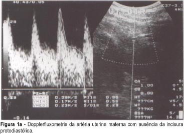
Purpose: to evaluate the association between the presence of diastolic notch in the maternal uterine arteries, and the histopathological changes of the uteroplacental vessels. Methods: transversal study of 144 women with single pregnancy interrupted by cesarean section between 27 and 41 weeks. In this sample, 84 had pregnancies complicated by preeclampsia and the other 60 were normal. In this group, Doppler study of both uterine arteries and placental bed biopsy was performed. Results: of the total of 144 patients, 88 patients (61%) had a biopsy fragment that was considered representative of the placental bed. The diastolic notch was present in 40 patients (70%) of the total of cases with inadequate physiologic alterations and absent in 28 patients (90%) of the total of cases with physiologic alterations (p=0.0000). The Doppler study showed 70% sensitivity, 90% specificity, 44% positive predictive value and 97% negative predictive value. The association between bilateral diastolic notch of uterine arteries and acute atherosis in the placental bed was also significant (24 out of 25 cases -- p=0.000). The Doppler study showed 96% sensitivity, 70% specificity, 26% positive predictive value and 99% negative predictive value, while for arteriolosclerosis its results were 80% sensitivity, 55% specificity, 17% positive predictive value and 96% negative predictive value. Conclusions: the diastolic notch in the maternal uterine is a safe indicator of pathological vessel alteration in the placental bed. The adequate trophoblast migration into the myometrium, revealed by physiologic changes, results in the absence of bilateral diastolic notch of the maternal uterine arteries.

Summary
Revista Brasileira de Ginecologia e Obstetrícia. 2002;24(10):663-668
DOI 10.1590/S0100-72032002001000005
PURPOSE: to evaluate the effect of intravascular transfusion on ductus venosus and inferior vena cava Doppler ultrasound indexes (SV/CA) and to relate it to hemoglobin levels before transfusion. METHODS: this is a transversal prospective study. A total of 62 intravascular transfusions were performed in 27 fetuses from pregnancies with red blood cell isoimmunization. The 62 cases were divided into two groups: (1) fetuses with hemoglobin levels before transfusion £10 g/dL and (2) fetuses with hemoglobin levels before transfusion >10 g/dL. The SV/CA and CA/SV indexes were measured using color Doppler ultrasound 6 h before and 12 h after intravascular transfusion. The index values before and after transfusion in all 62 cases were compared. Thereafter we compared these indexes before and after transfusion regarding each group. The Wilcoxon test was used and the results were considered statiscally significant when p<0.05. RESULTS: when we studied the whole group (62 cases) no significant difference was observed between the CA/SV index before and after transfusion (p=0.775). On the other hand, a significant increase in the SV/CA index was observed after transfusion (p=0.004). No significant differences were observed in both the SV/CA and CA/SV indexes before and after transfusion in the group of fetuses with hemoglobin levels before transfusion £10 g/dL (p=0.061 and p=0.345, respectively). There was a significant increase in the CA/SV index after transfusion in fetuses with hemoglobin levels before transfusion >10 g/dL (p=0.049), but the SV/CA index did not change in this group (p=0.086). CONCLUSION: venous Doppler study may be useful to understand fetal hemodynamic adjustment after intravascular transfusion. An increase in SV/CA without change in CA/SV after transfusion in anemic fetuses may be an important compensatory mechanism to increase intravascular volume. The increase in CA/SV index in fetuses with hemoglobin levels before transfusion <10 g/dL suggests a state of fetal hypervolemia.
Summary
Revista Brasileira de Ginecologia e Obstetrícia. 2002;24(1):09-13
DOI 10.1590/S0100-72032002000100002
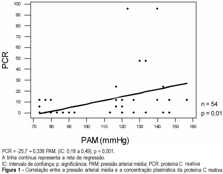
Purpose: to investigate the association between serum C-reactive protein concentration and preeclampsia occurrence, as well as its relation to the disease severity. Patients and Methods: twenty-seven preeclamptic pregnant women and 27 pregnant women with no clinical intercurrences, in the third trimester of pregnancy, were evaluated in a transversal case-control study. Serum C-reactive protein dosage, besides clinical examination and laboratory tests for the diagnosis of the disease, were performed in the antenatal period. The association between C-reactive protein and the presence of preeclampsia, and the correlation between plasma protein values and blood pressure values were investigated. The chi² significance test and regression analysis through the square minimum technique were used, and the results were considered to be statistically significant when p<0.05. Results: the preeclamptic pregnant women presented mean blood pressure levels higher than their controls (129.9±12.1 and 87.2±6.5 mmHg, respectively) and significantly higher C-reactive protein mean values than the normotensive women (18.9±4.9 and 1.56±0.8 mg/L, respectively). There was a significant association between the C-reactive protein concentration increase and preeclampsia occurrence (p<0.0001, odds ratio: 20.1). It was also observed that the mean arterial pressure and proteinuria presented a direct correlation with the circulating C-reactive protein in maternal blood (p=0.001 and p<0.001, respectively). Conclusion: C-reactive protein is an effective marker of preeclampsia occurrence and significantly correlates with the disease severity. The use of this test for the differential diagnosis of pregnant women in several hypertensive situations and its utilization as a marker of preeclampsia prognosis deserve further studies.
