Summary
Revista Brasileira de Ginecologia e Obstetrícia. 2003;25(7):533-533
DOI 10.1590/S0100-72032003000700013
Summary
Revista Brasileira de Ginecologia e Obstetrícia. 2003;25(7):533-533
DOI 10.1590/S0100-72032003000700013
Summary
Revista Brasileira de Ginecologia e Obstetrícia. 2003;25(7):533-533
DOI 10.1590/S0100-72032003000700012
Summary
Revista Brasileira de Ginecologia e Obstetrícia. 2003;25(7):533-533
DOI 10.1590/S0100-72032003000700012
Summary
Revista Brasileira de Ginecologia e Obstetrícia. 1999;21(9):533-538
DOI 10.1590/S0100-72031999000900006
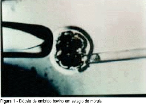
Purpose: to develop an animal model for the study of, and training in, bovine biopsies. Methods: cow ovaries were obtained from a slaughterhouse and transported to the laboratory where the oocytes were aspirated, maturated and submitted to in vitro fertilization. On the 5th day after fertilization, the embryos were biopsied, with the zona pellucida being opened with a cutting blade fitted to the light microscope. One or two blastomeres were removed from the embryos and left in coculture for three additional days. After this time, embryo development was evaluated in comparison to a control group by morphological study and cell counts using specific staining for nuclei. Results: forty of the 57 biopsied embryos reached the blastocyst stage (70.2%) and hatching was observed in 11 (27.5%). Forty-two blastocysts were obtained in the control group (73.7%) and 11 of them hatched (26.2%). Cell counts showed no significant differences between groups. Conclusions: we conclude that the proposed protocol is technically feasible and supplies a good number of embryos because of the easy technique for obtaining bovine oocytes, thus representing a method that could be adopted for training.
Summary
Revista Brasileira de Ginecologia e Obstetrícia. 1999;21(9):533-538
DOI 10.1590/S0100-72031999000900006
Purpose: to develop an animal model for the study of, and training in, bovine biopsies. Methods: cow ovaries were obtained from a slaughterhouse and transported to the laboratory where the oocytes were aspirated, maturated and submitted to in vitro fertilization. On the 5th day after fertilization, the embryos were biopsied, with the zona pellucida being opened with a cutting blade fitted to the light microscope. One or two blastomeres were removed from the embryos and left in coculture for three additional days. After this time, embryo development was evaluated in comparison to a control group by morphological study and cell counts using specific staining for nuclei. Results: forty of the 57 biopsied embryos reached the blastocyst stage (70.2%) and hatching was observed in 11 (27.5%). Forty-two blastocysts were obtained in the control group (73.7%) and 11 of them hatched (26.2%). Cell counts showed no significant differences between groups. Conclusions: we conclude that the proposed protocol is technically feasible and supplies a good number of embryos because of the easy technique for obtaining bovine oocytes, thus representing a method that could be adopted for training.
Summary
Revista Brasileira de Ginecologia e Obstetrícia. 2015;37(11):533-546
DOI 10.1590/SO100-720320150005330
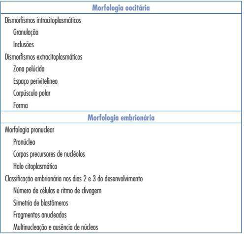
In order to increase the success rate of in vitro fertilization cycles, several studies have focused on the identification of the embryo with higher implantation potential. Despite recent advances in the reproductive medicine, based on the OMICs technology, routinely applicable methodologies are still needed. Thus, in most fertilization centers embryo selection for transfer is still based on morphological parameters evaluated under light microscopy. Several morphological parameters may be evaluated, ranging from the pronuclear to blastocyst stage. In general, despite the day of transfer, some criteria are suggested to present a predictive value for embryo viability when analyzed independently or combined. However, the subjectivity of morphological evaluation, as well as the wide diversity of embryo classification systems used by different fertilization centers shows contrasting results, making the implementation of a consensus regarding different morphological criteria and their predictive value a difficult task. The optimization of embryo selection represents a large potential to increase treatment success rates, allowing the transfer of a reduced number of embryos and inimizing the risks of multiple pregnancy.
Summary
Revista Brasileira de Ginecologia e Obstetrícia. 2015;37(11):533-546
DOI 10.1590/SO100-720320150005330
In order to increase the success rate of in vitro fertilization cycles, several studies have focused on the identification of the embryo with higher implantation potential. Despite recent advances in the reproductive medicine, based on the OMICs technology, routinely applicable methodologies are still needed. Thus, in most fertilization centers embryo selection for transfer is still based on morphological parameters evaluated under light microscopy. Several morphological parameters may be evaluated, ranging from the pronuclear to blastocyst stage. In general, despite the day of transfer, some criteria are suggested to present a predictive value for embryo viability when analyzed independently or combined. However, the subjectivity of morphological evaluation, as well as the wide diversity of embryo classification systems used by different fertilization centers shows contrasting results, making the implementation of a consensus regarding different morphological criteria and their predictive value a difficult task. The optimization of embryo selection represents a large potential to increase treatment success rates, allowing the transfer of a reduced number of embryos and inimizing the risks of multiple pregnancy.
Summary
Revista Brasileira de Ginecologia e Obstetrícia. 2013;35(12):533-535
Summary
Revista Brasileira de Ginecologia e Obstetrícia. 2013;35(12):533-535
Summary
Revista Brasileira de Ginecologia e Obstetrícia. 1998;20(9):533-536
DOI 10.1590/S0100-72031998000900007
Purpose: to evaluate the effects of tamoxifen (TAM) on plasma levels of estradiol, progesterone, prolactin, luteinizing hormone (LH), follicle-stimulating hormone (FSH) and steroid hormone-binding globulin (SHBG) when given to premenopausal women in the doses of 10 and 20 mg/day for 22 days. Patients and Methods: a randomized double-blind study was performed with 43 premenopausal eumenorrheic women. The patients were divided into three groups: A (N = 15, placebo); B (N = 15, TAM 10 mg/day) and C (N = 13, 20 mg/day). They started taking an oral dose of TAM or placebo on the very first day of the menstrual cycle. Two hormone determinations were performed, both on the 22nd day of the menstrual cycle: the first in the cycle that preceded the use of the drug and the second, in the following cycle, after 22 days of using the medication. We used the Levine and Student tests in order to evaluate the homogeneity of the sample and the variation of the hormone determinations respectively. Results:serum levels of estradiol, progesterone and SHBG increased significantly in groups B and C. In group C, we also observed increase in serum level of FSH (p < 0.0045) and a fall in prolactin level (p < 0.0055). Conclusions: TAM promoted a significant increase in serum concentrations of estradiol, progesterone and SHBG either in the doses of 10 or 20 mg/day. However, significant increase in FSH and decrease in prolactin were obtained only with the dose of 20 mg/day.
Summary
Revista Brasileira de Ginecologia e Obstetrícia. 2003;25(7):534-534
DOI 10.1590/S0100-72032003000700015
Summary
Revista Brasileira de Ginecologia e Obstetrícia. 2003;25(7):534-534
DOI 10.1590/S0100-72032003000700015
Summary
Revista Brasileira de Ginecologia e Obstetrícia. 2009;31(11):534-539
DOI 10.1590/S0100-72032009001100002
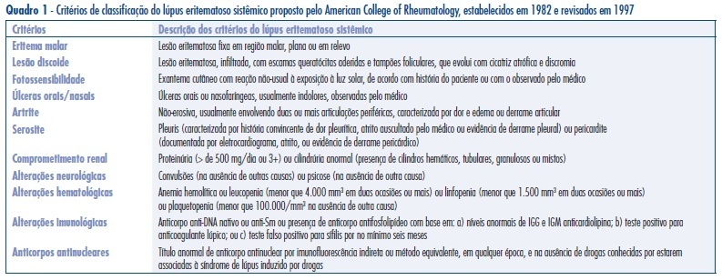
PURPOSE: to analyze the ophthalmic artery functioning in pregnant women with systemic lupus erythematosus (PL) without active renal disease as compared to non-pregnant women with lupus (NPL) without active renal disease, and to normal pregnant women (PN). METHODS: observational study that analyzed ophthalmic artery dopplervelocimetric variables of 20 PN, 10 PL and 17 NPL women. The variables analyzed were: pulsatility index (PI), final diastolic velocity (FDV) and velocity peak ratio (VPR). Mean and standard deviation of these indexes were calculated. For group mean comparison, analysis of variance (ANOVA) and the post-hoc Tukey test have been used, with confidence interval of 95% (p<0.05). RESULTS: the PN group showed the following means and standard deviations of ophthalmic artery parameters: PI=2,4±0,3; VPR=0,5±0,1 e FDV=5,1±2,1 cm/s. The PL and NPL groups showed the following values, respectively: PI=2,0±0,4 and 1,9±0,4; VPR=0,6±0,1 and 0,6±0,1; FDV=9,7±3,9 cm/s and 8,1±4,3 cm/s. There was not significant mean difference between the PL and NPL groups for PI, VPR or FDV. However, statistically significant mean differences were observed between PN and PL for PI, VPR and FDV, with higher values of FDV and VPR in the PL group. CONCLUSIONS: there was a reduction of ophthalmic artery vascular impedance with orbital hyperfusion in the two groups of women with lupus erythematosus as compared to normal pregnant women. These results may help to improve the understanding on pathophysiology of systemic lupus erythematosus. In addition, the present method may be applied in future studies as a complementary procedure for the differential diagnosis between pre-eclampsia and renal failure due to lupus.
Summary
Revista Brasileira de Ginecologia e Obstetrícia. 2009;31(11):534-539
DOI 10.1590/S0100-72032009001100002
PURPOSE: to analyze the ophthalmic artery functioning in pregnant women with systemic lupus erythematosus (PL) without active renal disease as compared to non-pregnant women with lupus (NPL) without active renal disease, and to normal pregnant women (PN). METHODS: observational study that analyzed ophthalmic artery dopplervelocimetric variables of 20 PN, 10 PL and 17 NPL women. The variables analyzed were: pulsatility index (PI), final diastolic velocity (FDV) and velocity peak ratio (VPR). Mean and standard deviation of these indexes were calculated. For group mean comparison, analysis of variance (ANOVA) and the post-hoc Tukey test have been used, with confidence interval of 95% (p<0.05). RESULTS: the PN group showed the following means and standard deviations of ophthalmic artery parameters: PI=2,4±0,3; VPR=0,5±0,1 e FDV=5,1±2,1 cm/s. The PL and NPL groups showed the following values, respectively: PI=2,0±0,4 and 1,9±0,4; VPR=0,6±0,1 and 0,6±0,1; FDV=9,7±3,9 cm/s and 8,1±4,3 cm/s. There was not significant mean difference between the PL and NPL groups for PI, VPR or FDV. However, statistically significant mean differences were observed between PN and PL for PI, VPR and FDV, with higher values of FDV and VPR in the PL group. CONCLUSIONS: there was a reduction of ophthalmic artery vascular impedance with orbital hyperfusion in the two groups of women with lupus erythematosus as compared to normal pregnant women. These results may help to improve the understanding on pathophysiology of systemic lupus erythematosus. In addition, the present method may be applied in future studies as a complementary procedure for the differential diagnosis between pre-eclampsia and renal failure due to lupus.