Summary
Revista Brasileira de Ginecologia e Obstetrícia. 2015;37(11):505-511
DOI 10.1590/SO100-720320150005400
To evaluate the factors associated with anemia among pregnant women receiving public health care in a capital city in Northeastern Brazil.
This was a cross-sectional study conducted on a sample of 428 patients obtained on the basis of the estimated prevalence of anemia during pregnancy (50%), a 95% confidence interval (95%CI), an error of 5% and a sample loss of 20%. Pregnant women who lived in the city and were served by the municipal public health network were considered to be eligible for the study. Socioeconomic, lifestyle, clinical and anthropometric data and dietary iron intake were obtained, and capillary hemoglobin was determined. Anemia was identified as a hemoglobin level <11 g/dL, and its association with risk factors was tested using multivariate Poisson regression analysis, with the results expressed as the Prevalence Ratio (PR) and 95%CI.
The prevalence of anemia was 28.3% and was higher among women with more members in the household (PR=1.49; 95%CI 1.01-2.22; p=0.046) and those living with food insecurity (PR=1.43; 95%CI 1.00-2.04; p=0.047).
The prevalence of anemia among pregnant women receiving care from the public health system of the city is a moderate public health problem, requiring the planning of effective measures for its control.
Summary
Revista Brasileira de Ginecologia e Obstetrícia. 2012;34(11):505-510
DOI 10.1590/S0100-72032012001100005
PURPOSE: To investigate the effect of adding biofeedback (BF) to the training of pelvic floor muscles (PFMT) for the treatment of stress urinary incontinence (SUI). METHODS: A prospective pilot study, randomized and controlled with women with SUI without sphincter deficiency, detected by urodynamic study and who performed the correct PFM contraction. Women with neuromuscular disorders and grade III and IV genital prolapse were excluded. Forty women were randomized into a Control Group and BF Group. The PFMT protocol with BF equipment consisted of three sets of ten slow contractions (tonic), with a holding time of six to eight seconds at each contraction followed by a rest period of equal duration. After each sustained contraction, they performed three to four fast contractions (phasic) in the supine and standing position twice a week, for a total of 12 sessions. We evaluated the effect of adding BF to PFMT on quality of life using King's Health Questionnaire (KHQ) regarding urinary symptoms based on a voiding diary and regarding the function of pelvic floor muscles by digital palpation. The evaluation was performed initially and after 12 treatment sessions. Data are reported as mean and standard deviation. The Mann-Whitney test was used for the analysis of homogeneity and to determine differences between groups, and the Wilcoxon test was used to determine possible differences between the times of observation, with the level of significance set at 0.05. RESULTS: A significant decrease in the scores of the domains assessed by the KHQ was observed in the comparison between groups, except for the general health domain (BF Group: 32.8±26.9 versus Control Group: 48.4±29.5, p<0.13). Accordingly, there was improvement in PFM function after treatment in the BF Group, regarding power (4.3±0.8, p= 0.001), endurance (6.0±2.2, p<0.001) and fast (9.3±1.9, p=0.001). When comparing the groups, the BF Group showed a positive result regarding power (BF Group 4.3±0.8 versus Control Group 2.5±0.9, p<0.001), endurance (6.0±2.2 BF Group versus Control Group 2.7±1.9, p<0.001) and fast (BF Group 9.3±1.9 versus Control Group 4.6 ± 3.2, p<0.001). Reduction of nocturnal urinary frequency (1.2±1.2 versus 0.7±0.9, p=0.02) and of effort urine loss (1.5±1.4 versus 0.6±0.8, p=0.001) was observed in the BF Group. CONCLUSION: The addition of BF to the PFMT for the treatment of SUI, applied according to the protocol described, improved PFM function, reduced urinary symptoms, and improved of the quality of life.
Summary
Revista Brasileira de Ginecologia e Obstetrícia. 2007;29(10):506-510
DOI 10.1590/S0100-72032007001000003
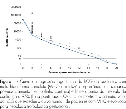
PURPOSE: to evaluate the usefulness of the normal human chorionic gonadotropin (hCG) regression curve in the early diagnosis of post-molar trophoblastic neoplasia (GTN). METHODS: a longitudinal study including 105 patients with complete hydatidiform mole (CHM) followed up at the Botucatu Center of Trophoblastic Diseases from 1998 to 2005. Serial serum hCG titers were measured fortnightly in all patients. Individual curves of the 105 patients were built. Comparison between the normal regression curve established at our center with individual hCG curves was used to screen and diagnose (plateau/rise) GTN. The number of weeks postevacuation when hCG levels exceeded the normal limits was compared with the number of weeks when hCG reached plateau/rise. RESULTS: among the 105 patients with CHM, 80 reached spontaneous remission (SR) and 25 developed GTN. Among the 80 SR patients, 7 (8.7%) initially showed hCG concentrations above normal but eventually achieved remission. All the 25 GTN patients showed deviation from the normal hCG curve at 3.84±2.57 weeks and reached plateau or rise at 8.40±2.94 weeks (p<0.001). CONCLUSIONS: the normal regression curve of post-molar hCG is useful in the early diagnosis of GTN.
Summary
Revista Brasileira de Ginecologia e Obstetrícia. 2007;29(10):506-510
DOI 10.1590/S0100-72032007001000003
PURPOSE: to evaluate the usefulness of the normal human chorionic gonadotropin (hCG) regression curve in the early diagnosis of post-molar trophoblastic neoplasia (GTN). METHODS: a longitudinal study including 105 patients with complete hydatidiform mole (CHM) followed up at the Botucatu Center of Trophoblastic Diseases from 1998 to 2005. Serial serum hCG titers were measured fortnightly in all patients. Individual curves of the 105 patients were built. Comparison between the normal regression curve established at our center with individual hCG curves was used to screen and diagnose (plateau/rise) GTN. The number of weeks postevacuation when hCG levels exceeded the normal limits was compared with the number of weeks when hCG reached plateau/rise. RESULTS: among the 105 patients with CHM, 80 reached spontaneous remission (SR) and 25 developed GTN. Among the 80 SR patients, 7 (8.7%) initially showed hCG concentrations above normal but eventually achieved remission. All the 25 GTN patients showed deviation from the normal hCG curve at 3.84±2.57 weeks and reached plateau or rise at 8.40±2.94 weeks (p<0.001). CONCLUSIONS: the normal regression curve of post-molar hCG is useful in the early diagnosis of GTN.
Summary
Revista Brasileira de Ginecologia e Obstetrícia. 2016;38(10):506-511
To evaluate the accuracy of transvaginal ultrasonography, hysteroscopy and uterine curettage in the diagnosis of endometrial polyp, submucous myoma and endometrial hyperplasia, using as gold standard the histopathological analysis of biopsy samples obtained during hysteroscopy or uterine curettage.
Cross-sectional study performed at the Hospital Universitário de Brasília (HUB). Data were obtained from the charts of patients submitted to hysteroscopy or uterine curettage in the period from July 2007 to July 2012.
One-hundred and ninety-one patients were evaluated, 134 of whom underwent hysteroscopy, and 57, uterine curettage. Hysteroscopy revealed a diagnostic accuracy higher than 90% for all the diseases evaluated, while transvaginal ultrasonography showed an accuracy of 65.9% for polyps, 78.1% for myoma and 63.2% for endometrial hyperplasia. Within the 57 patients submitted to uterine curettage, there was an accuracy of 56% for polyps and 54.6% for endometrial hyperplasia.
Ideally, after initial investigation with transvaginal ultrasonography, guided biopsy of the lesion should be performed by hysteroscopy, whenever necessary, in order to improve the diagnostic accuracy and subsequent clinical management.
Summary
Revista Brasileira de Ginecologia e Obstetrícia. 2018;40(9):507-512
To analyze the use of the measurement of uterine cervix length (MUCL) and the fetal fibronectin (fFN) rapid test as predictors of preterm delivery (PTD) in symptomatic pregnant women assisted at the Santa Casa de Misericórdia de Sobral Maternity Hospital.
This was a prospective and analytic study involving 53 parturients assisted between September of 2015 and July of 2016; the participants were between 24 and 34 weeks of gestational age (GA) and presented complaints related to preterm labor (PTL) prodromes. Vaginal secretion was collected for fFN testing, and the MUCL was obtained via transvaginal ultrasonography.
A total of 58.49% of the subjects showed MUCL < 25 mm, and 41.51% were positive in the fFNrapid test.Atotal of 48 patients were followed-up until their delivery date, and 54.17% resulted in PTL. The relative risk (RR) for PTD in patients with MUCL < 25 mm was 1.83 (p = 0.09, 0.99-3.36, 95% confidence interval [CI]), with a mean time before delivery of 2.98 weeks. Based on fFN positive results, the RR was 3.50 (p = 0.002, 1.39- 8.79, 95%CI) and themean time until delivery was 1.94weeks. The RRwas 2.70 (p = 0.002, 1.08-6.72, 95%CI) when both tests were used. The RR of PTD within 48 hours, and 7 and 14 days were, respectively, 1.30 (p = 0.11, 95% CI 1.02-1.67), 1.43 (p = 0.12, 95% CI % 0.99-2.06), and 2.03 (p = 0.008, 95% CI 1.26-3.27), when based on the MUCL, and 1.75 (p = 0.0006, 95% CI 1.20-2.53), 2.88 (p = 0.0001, 95% CI, 1.57-5.31), and 3.57 (p = 0.0002, 95% CI 1.63-7.81) when based on positive fFN results. The RR at 48 hours and 7 and 14 days considering both tests was 1.74 (p = 0.0001, 95% CI 1.14-2.64), 2.22 (p = 0.0001, 95% CI 1.22-4.04), and 2.76 (p = 0.0002, 95% CI 1.27-5.96), respectively.
In symptomatic pregnant women, we concluded that the MUCL < 25 mm associated with positive fFN rapid test indicate increased the risk for PTD. Further studies with larger sample sizes could contribute in supporting the results presented in the current study.
Summary
Revista Brasileira de Ginecologia e Obstetrícia. 2003;25(7):507-512
DOI 10.1590/S0100-72032003000700007
PURPOSE: to evaluate the prevalence of osteoporosis in climacteric women and analyze the influence of general and reproductive risk factors on bone mineral density. METHODS: a cross-sectional study with the evaluation of the 473 hospital records of climacteric women followed up at the Menopause Outpatient Facility of CAISM/Unicamp, between 03/28/2000 and 04/17/2001. These women were at least 12 months in amenorrhea and presented the results of a bone densitometry study performed at the Nuclear Medicine Department of HC/Unicamp. The following variables were evaluated: age, color, body mass index, level of education, smoking, use of medication, age at menopause, parity, use and length of hormone replacement therapy and its effect on bone mineral density. Statistical analyses were performed using logistic regression ajusted by age and hormone replacement therapy use. RESULTS: the mean age of the studied women was 53.9 years (± 7.1 SD) with mean age at menopause being 45.9 years (± 6.9 SD). Osteoporosis occurred in 14.7% and osteopenia in 38% of the cases in the lumbar vertebrae (L2-L4 interspace) and in 3.8 and 32.7% in the femur, respectively. Logistic regression adjusted to age and hormone therapy showed an association between the following variables: level of education, age at menopause and body mass index. CONCLUSION: there was a high prevalence of osteoporosis and osteopenia in the studied population. Advanced age, lower level of education, late menarche, early menopause and lower body mass index were identified as risk factors for developing decreased bone mass in the studied population.
Summary
Revista Brasileira de Ginecologia e Obstetrícia. 2001;23(8):507-513
DOI 10.1590/S0100-72032001000800005
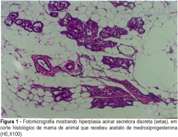
Purpose: to evaluate the effect of hormone replacement therapy on breast cell proliferation and on collagen and elastic fiber formation and to analyze the changes occurring in the breast parenchyma as a whole. Method: a total of 61 adult Wistar rats were divided into 5 groups. The standard group (12 rats) represented the normal hormonal ovarian status. The remaining 49 rats were oophorectomized and, starting on the 96th P.O. day, received the specific drug for 30 days. The CEE group received 50 mg/day conjugated equine estrogens (13 rats); the MPA group, 2.0 mg/day medroxyprogesterone acetate (12 rats); the CEE + MPA group, both drugs (12 rats), and the DW group, distilled water (12 rats). On the 31st day of medication, the animals were sacrificed and the inguinal mammary glands were removed for histological analysis. Cell proliferation was assessed at the ductal and acinar levels using anti-PCNA antibody. Mature collagen (type I) and immature collagen (type III) were quantified by Sirius-Red staining, and elastic fiber formation was quantified by Weigert staining. Anatomopathological analysis was performed by hematoxylin-eosin staining, with the determination of number of acini per terminal duct, number of ducts per field, presence of intraductal secretion, and intensity of intracytoplasmic vacuolization. Results: the CEE + MPA group presented a smaller percentage of proliferating ductal cells (46.1%) (p<0.0001) and a greater proliferation of acinar cells (66.3%), similar to those detected in the MPA group (p=0.075) but differing from those detected in the remaining groups (p<0.004). The CEE group showed the largest amount of immature collagen (33.6%) (p<0.01) and the MPA group showed the highest concentration of elastic fibers (11.7%) (p<0.0001). The CEE + MPA and MPA groups showed secretory acinar hyperplasia that was intense (91.7%) in the CEE + MPA group and mild (41.7%) or moderate (58.3%) in the MPA group, but differering in both cases from the remaining groups (p<0,097). Conclusions: conjugated equine estrogens in combination with medroxyprogesterone inhibit ductal cell proliferation and stimulate acinar cell proliferation causing secretory acinar hyperplasia; conjugated horse estrogens intensify the formation of immature (type III) collagen, and medroxyprogesterone acetate increases the formation of elastic fibers.
Summary
Revista Brasileira de Ginecologia e Obstetrícia. 2001;23(8):507-513
DOI 10.1590/S0100-72032001000800005
Purpose: to evaluate the effect of hormone replacement therapy on breast cell proliferation and on collagen and elastic fiber formation and to analyze the changes occurring in the breast parenchyma as a whole. Method: a total of 61 adult Wistar rats were divided into 5 groups. The standard group (12 rats) represented the normal hormonal ovarian status. The remaining 49 rats were oophorectomized and, starting on the 96th P.O. day, received the specific drug for 30 days. The CEE group received 50 mg/day conjugated equine estrogens (13 rats); the MPA group, 2.0 mg/day medroxyprogesterone acetate (12 rats); the CEE + MPA group, both drugs (12 rats), and the DW group, distilled water (12 rats). On the 31st day of medication, the animals were sacrificed and the inguinal mammary glands were removed for histological analysis. Cell proliferation was assessed at the ductal and acinar levels using anti-PCNA antibody. Mature collagen (type I) and immature collagen (type III) were quantified by Sirius-Red staining, and elastic fiber formation was quantified by Weigert staining. Anatomopathological analysis was performed by hematoxylin-eosin staining, with the determination of number of acini per terminal duct, number of ducts per field, presence of intraductal secretion, and intensity of intracytoplasmic vacuolization. Results: the CEE + MPA group presented a smaller percentage of proliferating ductal cells (46.1%) (p<0.0001) and a greater proliferation of acinar cells (66.3%), similar to those detected in the MPA group (p=0.075) but differing from those detected in the remaining groups (p<0.004). The CEE group showed the largest amount of immature collagen (33.6%) (p<0.01) and the MPA group showed the highest concentration of elastic fibers (11.7%) (p<0.0001). The CEE + MPA and MPA groups showed secretory acinar hyperplasia that was intense (91.7%) in the CEE + MPA group and mild (41.7%) or moderate (58.3%) in the MPA group, but differering in both cases from the remaining groups (p<0,097). Conclusions: conjugated equine estrogens in combination with medroxyprogesterone inhibit ductal cell proliferation and stimulate acinar cell proliferation causing secretory acinar hyperplasia; conjugated horse estrogens intensify the formation of immature (type III) collagen, and medroxyprogesterone acetate increases the formation of elastic fibers.