Summary
Revista Brasileira de Ginecologia e Obstetrícia. 2010;32(8):405-411
DOI 10.1590/S0100-72032010000800008
PURPOSE: the aim of this study was to describe perinatal and maternal outcomes of pregnancies complicated by sickle cell disease (SCD), comparing to pregnancies of women with sickle cell trait (SCT). METHODS: this was a retrospective cohort study, covering the period from March 2001 to April 2008, which included all pregnant women with SCD (n=42) followed up at a university hospital in the Southeast region of Brazil. The maternal and perinatal outcomes were compared to those of pregnant women with SCT (n=56) who were followed up at the same service. RESULTS:SCD-SS was diagnosed in 42 (82.4%) pregnant women and SC in 9 (17.6%). Mean (±SD) maternal age was significantly lower in the SCD group (26.0 years) compared to SCT women (28.7±7.1 years; p=0.018). The following maternal complications were more common among women with SCD in comparison to SCT: urinary tract infection (25.5 versus 8.9%; p=0.04), pneumonia (23.5 versus 1.8%; p=0.002), pulmonary hypertension (15.7 versus 0%; p=0.002), and blood transfusion during delivery or postpartum (33.3 versus 5.4%; p=0.001). Adverse perinatal outcome was more frequent in the SCD group compared to the SCT group: prematurity (49 versus 25%, p=0.01); mean gestational age at delivery (35.2 versus 37.9 weeks, p<0.001); fetal distress (56.9 versus 28.6%, p=0.006); birth weight <2,500 g (62.7 versus 17.9%, p<0.001); mean birth weight (2,183 versus 2,923 g, p<0.001), and small for gestational age infants (29.4 versus 10.7%, p=0.029). Two maternal deaths (3.9%) occurred in the group with SCD. CONCLUSION: Pregnant women with SCD are at greater risk for maternal morbidity and for adverse perinatal outcomes than women with SCT.
Summary
Revista Brasileira de Ginecologia e Obstetrícia. 2016;38(8):405-411
We aimed to evaluate the safety, efficacy and surgical outcomes of combined laparoscopic/vaginal prolapse repair by two surgeons.
A retrospective chart review of all patients (n =135) who underwent apical prolapse repair from February 2009 to December 2012 performed in a collaborative manner by a Minimally Invasive Gynecologic Surgeon and a Urogynecologist. Demographic data (age, body mass index [BMI], race, gravidity, parity) and surgical information (estimated blood loss, operative time, intraoperative complications, readmission and reoperation rates, presence of postoperative infection) were collected.
The majority of patients were postmenopausal (58.91%), multiparous (mean parity =2.49) and overweight (mean BMI =27.71). Nearly 20% had previous prolapse surgery. The most common surgical procedure was laparoscopic supracervical hysterectomy (LSH) with sacrocervicopexy (59.26%), and the most common vaginal repair was of the posterior compartment (78.68%). The median operative time was 149 minutes (82-302), and the estimated blood loss was 100 mL (10-530). Five intraoperative complications, five readmissions and four reoperations were noted. Performance of a concomitant hysterectomy did not affect surgical or anatomical outcomes.
Combination laparoscopic/vaginal prolapse repair by two separate surgeons seems to be an efficient option for operative management.
Summary
Revista Brasileira de Ginecologia e Obstetrícia. 2008;30(8):406-412
DOI 10.1590/S0100-72032008000800006
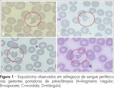
PURPOSE: to evaluate the significance of schizocytes presence in peripheral blood smear of pregnant women with pre-eclampsia, identifying and correlating them with other markers of hemolysis and of the disease severity. METHODS: Seventh six glass slides of peripheral blood smear of pregnant women with pre-eclampsia have been evaluated. After the smear, the slides have been stained with Leishman's dye and stored till they were examined with a Leica, model DLMB microscope, provided with the Qwin Lite 2.5 software that made it possible to record the images of selected fields in CD-ROM. Ten fields with approximately 100 erythrocytes were counted in each glass slide. Schizocytes (irregular fragment or helmet-shaped, bite-shaped or triangular) were considered as present, when their percentage was equal or higher than 0.2%, their presence being correlated with other hemolysis markers (hemoglobin, total bilirubin, lactic desidrogenasis and reticulocytes), pre-eclampsia markers (proteinuria and platelet number). The Statistical Package in Social Science for Windows (SPSS), 10.0 version has been used for statistical analysis, at p<0.05. RESULTS: schizocytes have been present in 31.6% of the pregnant women with pre-eclampsia. In most (75%) of the blood smears there have been three or four schizocytes. There has been no correlation between schizocyte presence and any other hemolysis marker, any pre-eclampsia marker or disease severity. CONCLUSIONS: schizocytes have been identified in a small number and in less than a third of the pregnant women with pre-eclampsia. There has been no correlation with other hemolysis marker parameters or with the disease severity. This way, the presence of schizocytes is not a marker of the clinical evolution of pre-eclampsia.
Summary
Revista Brasileira de Ginecologia e Obstetrícia. 2008;30(8):406-412
DOI 10.1590/S0100-72032008000800006
PURPOSE: to evaluate the significance of schizocytes presence in peripheral blood smear of pregnant women with pre-eclampsia, identifying and correlating them with other markers of hemolysis and of the disease severity. METHODS: Seventh six glass slides of peripheral blood smear of pregnant women with pre-eclampsia have been evaluated. After the smear, the slides have been stained with Leishman's dye and stored till they were examined with a Leica, model DLMB microscope, provided with the Qwin Lite 2.5 software that made it possible to record the images of selected fields in CD-ROM. Ten fields with approximately 100 erythrocytes were counted in each glass slide. Schizocytes (irregular fragment or helmet-shaped, bite-shaped or triangular) were considered as present, when their percentage was equal or higher than 0.2%, their presence being correlated with other hemolysis markers (hemoglobin, total bilirubin, lactic desidrogenasis and reticulocytes), pre-eclampsia markers (proteinuria and platelet number). The Statistical Package in Social Science for Windows (SPSS), 10.0 version has been used for statistical analysis, at p<0.05. RESULTS: schizocytes have been present in 31.6% of the pregnant women with pre-eclampsia. In most (75%) of the blood smears there have been three or four schizocytes. There has been no correlation between schizocyte presence and any other hemolysis marker, any pre-eclampsia marker or disease severity. CONCLUSIONS: schizocytes have been identified in a small number and in less than a third of the pregnant women with pre-eclampsia. There has been no correlation with other hemolysis marker parameters or with the disease severity. This way, the presence of schizocytes is not a marker of the clinical evolution of pre-eclampsia.
Summary
Revista Brasileira de Ginecologia e Obstetrícia. 2002;24(6):407-412
DOI 10.1590/S0100-72032002000600008
Purpose: to evaluate the agreement between the theory about informed consent, represented by Resolution 01/88, and the practice according to the report of researchers and of women who were subjects of their research. Methods: eleven researchers from three centers of excellence in research related to fertility regulation in Brazil and 18 women, subjects of their research. Information was obtained through in-depth interviews and content analysis was carried out. Results: the report of the researchers agreed with the requirements of the Resolution; however, the women's report showed that most of the required items were not referred to when they were invited to participate in the research. Conclusion: a disagreement was observed between theory and practice in obtaining informed consent. This may be explained by difficulties in complying with the requirements of the Resolution in force at the time. On the other hand, it is also possible to imagine difficulties experienced by researchers when approaching the women and/or that the women also forgot the received information. Finally, a bias may have resulted from the researchers and women who had to give consent to participate in this study.
Summary
Revista Brasileira de Ginecologia e Obstetrícia. 2013;35(9):407-412
DOI 10.1590/S0100-72032013000900005
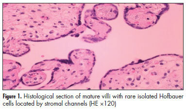
PURPOSE: In placentas from uncomplicated pregnancies, Hofbauer cells either disappear or become scanty after the fourth to fifth month of gestation. Immunohistochemistry though, reveals that a high percentage of stromal cells belong to Hofbauer cells. The aim of this study was to investigate the changes in morphology and density of Hofbauer cells in placentas from normal and pathological pregnancies. METHODS: Seventy placentas were examined: 16 specimens from normal term pregnancies, 10 from first trimester's miscarriages, 26 from cases diagnosed with chromosomal abnormality of the fetus, and placental tissue specimens complicated with intrauterine growth restriction (eight) or gestational diabetes mellitus (10). A histological study of hematoxylin-eosin (HE) sections was performed and immunohistochemical study was performed using the markers: CD 68, Lysozyme, A1 Antichymotrypsine, CK-7, vimentin, and Ki-67. RESULTS: In normal term pregnancies, HE study revealed Hofbauer cells in 37.5% of cases while immunohistochemistry revealed in 87.5% of cases. In first trimester's miscarriages and in cases with prenatal diagnosis of fetal chromosomal abnormalities, both basic and immunohistochemical study were positive for Hofbauer cells. In pregnancies complicated with intrauterine growth restriction or gestational diabetes mellitus, a positive immunoreaction was observed in 100 and 70% of cases, respectively. CONCLUSIONS: Hofbauer cells are present in placental villi during pregnancy, but with progressively reducing density. The most specific marker for their detection seems to be A1 Antichymotrypsine. It is remarkable that no mitotic activity of Hofbauer cells was noticed in our study, as the marker of cellular multiplication Ki-67 was negative in all examined specimens.
Summary
Revista Brasileira de Ginecologia e Obstetrícia. 2013;35(9):407-412
DOI 10.1590/S0100-72032013000900005
PURPOSE: In placentas from uncomplicated pregnancies, Hofbauer cells either disappear or become scanty after the fourth to fifth month of gestation. Immunohistochemistry though, reveals that a high percentage of stromal cells belong to Hofbauer cells. The aim of this study was to investigate the changes in morphology and density of Hofbauer cells in placentas from normal and pathological pregnancies. METHODS: Seventy placentas were examined: 16 specimens from normal term pregnancies, 10 from first trimester's miscarriages, 26 from cases diagnosed with chromosomal abnormality of the fetus, and placental tissue specimens complicated with intrauterine growth restriction (eight) or gestational diabetes mellitus (10). A histological study of hematoxylin-eosin (HE) sections was performed and immunohistochemical study was performed using the markers: CD 68, Lysozyme, A1 Antichymotrypsine, CK-7, vimentin, and Ki-67. RESULTS: In normal term pregnancies, HE study revealed Hofbauer cells in 37.5% of cases while immunohistochemistry revealed in 87.5% of cases. In first trimester's miscarriages and in cases with prenatal diagnosis of fetal chromosomal abnormalities, both basic and immunohistochemical study were positive for Hofbauer cells. In pregnancies complicated with intrauterine growth restriction or gestational diabetes mellitus, a positive immunoreaction was observed in 100 and 70% of cases, respectively. CONCLUSIONS: Hofbauer cells are present in placental villi during pregnancy, but with progressively reducing density. The most specific marker for their detection seems to be A1 Antichymotrypsine. It is remarkable that no mitotic activity of Hofbauer cells was noticed in our study, as the marker of cellular multiplication Ki-67 was negative in all examined specimens.
Summary
Revista Brasileira de Ginecologia e Obstetrícia. 2005;27(7):407-414
DOI 10.1590/S0100-72032005000700007
PURPOSE: to evaluate the prevalence of vulval squamous intraepithelial lesions and associated factors in HIV-infected patients attended at the public health services of Rio de Janeiro city. METHOD: a total of 374 HIV-infected patients were attended at public services in Rio de Janeiro city and submitted to gynecological examination, Pap smear and colposcopic examination of the cervix and vulva. The association of vulval intraepithelial lesion was analyzed according to the results of clinical (age and cervical lesions), laboratorial (CD4 count) and behavioral (number of partners and smoking habit) variables. The study (independent) variables were the epidemiological data, the immunologic status and the results of gynecological propaedeutic. Thus, age, the smoking habit, number of sexual partners, count of T CD4 lymphocites, and cervical intraepithelial lesion were selected. In the beginning, a bivariate analysis was performed, aiming at assessing the association between the presence of vulval intraepithelial lesion (ultimate variable) and the independent variables (age, smoking habits, number of sexual partners, cytology, colposcopy and CD4 count). Thereafter, the results with statistical significance (p<0.05) were submitted to a multiple logistic regression, and the probability ratio with the respective 95% confidence interval was established. RESULTS: the prevalence of vulval intraepithelial lesions was 40%. In the multivariate analysis CD4 count below 500 cells/mm³ OR=2.69 [IC 95%: 1.61-4.52], abnormal colposcopy OR=1.64 [IC 95%: 1.01-2.67] and age under 26 OR=1.98 [IC 95%: 1.18-3.30] were significant. In the vulval and cervical simultaneous lesion subgroup, age under 26 OR=3.30 [IC 95%: 1.65-6.59] and CD4 count below 500 cells/mm³ OR=4.15 [IC 95%: 1.92-8.96], were significant on analysis. CONCLUSIONS: the prevalence of vulval squamous intraepithelial lesions in HIV-infected patients is high. Immunodeficiency, presence of cervical intraepithelial lesions and age under 26 were associated with the presence of vulval intraepithelial lesions.
Summary
Revista Brasileira de Ginecologia e Obstetrícia. 2007;29(8):408-414
DOI 10.1590/S0100-72032007000800005
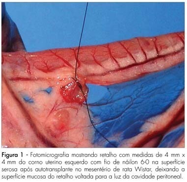
PURPOSE: to analyze the macroscopic and histological changes that occur with the use of sinvastatin in experimental endometriosis in female rats. METHODS: forty Wistar female rats were submitted to the technique of uterine self-transplant in mesenterium. After three weeks, 24 of them developed experimental endometriosis grade III, and were divided in two groups: one group received sinvastatin orally (20 mg/kg/day) and the other (control group) received 0.9% of sodium chloride orally (1 mL/100 g of body weight/day). Both groups received gavage for 14 days, followed by death. The implant volume was calculated [4pi (lenght/2) x (width/2) x (height/2)/3] at the surgical intervention and after the animal’s death. The self-transplants were removed, dyed with hematoxylin-eosin and analyzed by light microscopy. The Mann-Whitney’s test was used in the independent samples and the Wilcoxon’s test for the related samples. The Fisher’s exact test was used for the histological evaluation, with a significance level of 5%. RESULTS: the difference between groups of the initial average volumes of the self-transplants was not significant (p=1.00), but became significant for the final average volumes (p=0.04). There was a significant increase (p=0.01) between the initial and final average volumes in the control group, and a no significant decrease in the sinvastatin group (p=0.95). Histologically, the sinvastatin group (n=9) presented seven cases (77.8%) of moderately preserved and two cases (22.2%) of well preserved epithelial wall, while the control group (n=12) presented seven cases (58.3%) of moderately preserved and five cases (41.7%) of well preserved epithelial wall. CONCLUSIONS: sinvastatin prevented the growth of experimental endometriosis. Studies with sinvastatin for longer periods are promising.
Summary
Revista Brasileira de Ginecologia e Obstetrícia. 2007;29(8):408-414
DOI 10.1590/S0100-72032007000800005
PURPOSE: to analyze the macroscopic and histological changes that occur with the use of sinvastatin in experimental endometriosis in female rats. METHODS: forty Wistar female rats were submitted to the technique of uterine self-transplant in mesenterium. After three weeks, 24 of them developed experimental endometriosis grade III, and were divided in two groups: one group received sinvastatin orally (20 mg/kg/day) and the other (control group) received 0.9% of sodium chloride orally (1 mL/100 g of body weight/day). Both groups received gavage for 14 days, followed by death. The implant volume was calculated [4pi (lenght/2) x (width/2) x (height/2)/3] at the surgical intervention and after the animal’s death. The self-transplants were removed, dyed with hematoxylin-eosin and analyzed by light microscopy. The Mann-Whitney’s test was used in the independent samples and the Wilcoxon’s test for the related samples. The Fisher’s exact test was used for the histological evaluation, with a significance level of 5%. RESULTS: the difference between groups of the initial average volumes of the self-transplants was not significant (p=1.00), but became significant for the final average volumes (p=0.04). There was a significant increase (p=0.01) between the initial and final average volumes in the control group, and a no significant decrease in the sinvastatin group (p=0.95). Histologically, the sinvastatin group (n=9) presented seven cases (77.8%) of moderately preserved and two cases (22.2%) of well preserved epithelial wall, while the control group (n=12) presented seven cases (58.3%) of moderately preserved and five cases (41.7%) of well preserved epithelial wall. CONCLUSIONS: sinvastatin prevented the growth of experimental endometriosis. Studies with sinvastatin for longer periods are promising.
Summary
Revista Brasileira de Ginecologia e Obstetrícia. 2011;33(12):408-413
DOI 10.1590/S0100-72032011001200006
PURPOSE: To evaluate the influence of physical activity on the quality of life of middle-aged women. METHODS: A population-based cross-sectional study was conducted on 370 women aged 40 to 65 years-old recruited from a population-based sample. Enrollment took place in Basic Health Units in each health district of the city (North, South, East, and West) from June to September 2011. According to the Municipal Health Department of the City, 20,801 women were assisted at the Basic Health Units during a one-year period. The sample size calculation was stratified by district and based on a 95% confidence level with a power of 80%, as well as an error estimate of 5% and it was considered proportional to the number of patients classified as having adequate quality of life (indicator >26) in the general population. Data were collected while women waited for their routine appointment at the Health Unit. WHOQOL-Bref was used to evaluate the quality of life, and menopause rating scale (MRS) was used to determine climacteric symptoms. The level of physical activity was assessed by means of the International Physical Activity Questionnaire (IPAQ). To obtain the classification of PA levels, we used three categories: sedentary, moderately active, and very active. Statistical analysis was performed using the Minitab software, version 16. RESULTS: The mean age of the subjects was 49.8 years-old (±8.1) and they were predominantly Caucasian (72.7%), married (61.6%), non-smokers (93.5%), and had High School education (47.8%). Using the WHOQOL, mean scores were found to be significantly different between the groups (low, moderate, and vigorous physical activity), classified according to the domains of quality of life (p<0.01). Concerning physical activity and climacteric symptoms, significant differences were found for all domains: psychological (p<0.01), vegetative-somatic (p<0.01), and urogenital (p<0.01). CONCLUSIONS: Physical activity improves quality of life in middle-aged women.