Summary
Revista Brasileira de Ginecologia e Obstetrícia. 2000;22(6):385-385
DOI 10.1590/S0100-72032000000600010
Summary
Revista Brasileira de Ginecologia e Obstetrícia. 2000;22(6):385-385
DOI 10.1590/S0100-72032000000600010
Summary
Revista Brasileira de Ginecologia e Obstetrícia. 2000;22(6):385-385
DOI 10.1590/S0100-72032000000600011
Summary
Revista Brasileira de Ginecologia e Obstetrícia. 2000;22(6):385-385
DOI 10.1590/S0100-72032000000600011
Summary
Revista Brasileira de Ginecologia e Obstetrícia. 2022;44(4):385-390
To evaluate the role of cervical cytology (Pap smear) in the diagnosis of cervical intraepithelial neoplasia 2 or greater (CIN2+), presented exclusively in the endocervical canal, the clinical-epidemiological characteristics of this lesion, the necessary length of canal to be removed to treat, and the rate of invasive lesion hidden in the endocervical canal.
Cross-sectional study, by database analysis, of patients with abnormal cytology (high-grade squamous intraepithelial lesion [HSIL]), without visible colposcopy lesion, submitted to loop electrosurgical procedure (LEEP) to evaluate the association of cytology results with the histological product of the conization, to identify the epidemiological characteristics of endocervical lesion and clinical evolution, using a pvalue< 0.05 and 95% CI.
In 444 cases, the Pap smear sensitivity for CIN2+ diagnosis was 75% (95% CI: 69.8-79.7), specificity was 40% (95% CI: 30.2-49.5), and the prevalence rate of histological lesion was 73% (95% CI: 70.1-78.7). There was a higher prevalence of CIN2+ in women over 42 years old and invasive cancer in those over 56 years old (p<0.001), and it was necessary to remove 2.6 cm in length of the canal to reduce the chance of recurrence (p<0.006). The rate of invasive cancer was 2.7%.
Cytology was related to a high prevalence to histological lesion (73%) in the diagnosis of CIN2+ in the endocervical disease; older patients presented a higher relationship with histological lesions in the canal disease, and it was necessary to remove an average of 2.6 cm in length of the endocervical canal to avoid the persistence and progression of CIN. The rate of occult neoplasia in the endocervical canal was 2.7%.
Summary
Revista Brasileira de Ginecologia e Obstetrícia. 2009;31(8):385-390
DOI 10.1590/S0100-72032009000800003
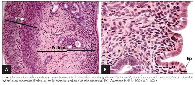
PURPOSE: to evaluate the effect of hyperprolactinemia induced by metoclopramide on the endometrium and myometrium of female mice in the proestrus phase. METHODS: 24 female mice were randomly divided in two groups: CtrG/control and ExpG/treated with metoclopramide (6.7 mg/g daily). After 50 days, the animals were sacrificed in the proestrus phase, and the blood was collected to determine the levels of estradiol, progesterone and prolactin. The uterine horns were removed, fixed in 10% formaldehyde and processed before being included in paraffin. Slices of 4 µm were stained by hematoxylin and eosin (H/E). In the morphological analysis, a Carl Zeiss light microscope, with objectives varying from 4 to 400 X was used for each histological slice characterization. In the morphometrical analysis, the superficial epithelium, the lamina propria and the myometrium thickness were evaluated, with the help of an image analyzer (AxionVision - Carl Zeiss) attached to the light microscope (Carl Zeiss). The statistical analysis was done by ANOVA, followed by the Wilcoxon test. P-value was considered as significant, when <0.05. RESULTS: our findings have shown an increase in the seric levels of prolactin (295.6±38.0 ng/mL) and significant decrease in the progesterone levels (11.3±0.9 ng/mL) in the ExpG, as compared to the CtrG (45.5±5.2 ng/mL and 18.2±1.6 ng/mL, respectively; p<0.001). Concerning the seric level of estradiol, significant differences between the groups were not obtained (ExpG=119.1±12.3 pg/mL and CtrG=122.7±8.4 pg/mL; p=0.418). The morphological study has shown that the uterus from the ExpG presented the endometrium with more developed superficial epithelium and lamina propria, as compared to the CtrG, the same happening with the myometrium. The thickness morphometrical values of the luminal epithelium (8.0±1.1 µm) and endometrium (116.2±21.1x10² µm) from the CtrG were lower than the ones from the ExpG (10.2±0.8 µm and 163.2±23.3x10² µm, respectively) with p<0.05. Nevertheless, data obtained in the myometrium have not shown significant differences between the groups (CtrG=152.2±25.2x10² µm and ExpG=140.8±18.0x10² µm). CONCLUSIONS: data have shown that hyperprolactinemia induced by metoclopramide determines endometrial proliferation and interferes with the ovarian function, mainly in the progesterone production.
Summary
Revista Brasileira de Ginecologia e Obstetrícia. 2009;31(8):385-390
DOI 10.1590/S0100-72032009000800003
PURPOSE: to evaluate the effect of hyperprolactinemia induced by metoclopramide on the endometrium and myometrium of female mice in the proestrus phase. METHODS: 24 female mice were randomly divided in two groups: CtrG/control and ExpG/treated with metoclopramide (6.7 mg/g daily). After 50 days, the animals were sacrificed in the proestrus phase, and the blood was collected to determine the levels of estradiol, progesterone and prolactin. The uterine horns were removed, fixed in 10% formaldehyde and processed before being included in paraffin. Slices of 4 µm were stained by hematoxylin and eosin (H/E). In the morphological analysis, a Carl Zeiss light microscope, with objectives varying from 4 to 400 X was used for each histological slice characterization. In the morphometrical analysis, the superficial epithelium, the lamina propria and the myometrium thickness were evaluated, with the help of an image analyzer (AxionVision - Carl Zeiss) attached to the light microscope (Carl Zeiss). The statistical analysis was done by ANOVA, followed by the Wilcoxon test. P-value was considered as significant, when <0.05. RESULTS: our findings have shown an increase in the seric levels of prolactin (295.6±38.0 ng/mL) and significant decrease in the progesterone levels (11.3±0.9 ng/mL) in the ExpG, as compared to the CtrG (45.5±5.2 ng/mL and 18.2±1.6 ng/mL, respectively; p<0.001). Concerning the seric level of estradiol, significant differences between the groups were not obtained (ExpG=119.1±12.3 pg/mL and CtrG=122.7±8.4 pg/mL; p=0.418). The morphological study has shown that the uterus from the ExpG presented the endometrium with more developed superficial epithelium and lamina propria, as compared to the CtrG, the same happening with the myometrium. The thickness morphometrical values of the luminal epithelium (8.0±1.1 µm) and endometrium (116.2±21.1x10² µm) from the CtrG were lower than the ones from the ExpG (10.2±0.8 µm and 163.2±23.3x10² µm, respectively) with p<0.05. Nevertheless, data obtained in the myometrium have not shown significant differences between the groups (CtrG=152.2±25.2x10² µm and ExpG=140.8±18.0x10² µm). CONCLUSIONS: data have shown that hyperprolactinemia induced by metoclopramide determines endometrial proliferation and interferes with the ovarian function, mainly in the progesterone production.
Summary
Revista Brasileira de Ginecologia e Obstetrícia. 2013;35(9):385-387
Summary
Revista Brasileira de Ginecologia e Obstetrícia. 2013;35(9):385-387
Summary
Revista Brasileira de Ginecologia e Obstetrícia. 2000;22(6):386-386
DOI 10.1590/S0100-72032000000600012
Summary
Revista Brasileira de Ginecologia e Obstetrícia. 2000;22(6):386-386
DOI 10.1590/S0100-72032000000600012
Summary
Revista Brasileira de Ginecologia e Obstetrícia. 2010;32(8):386-392
DOI 10.1590/S0100-72032010000800005
PURPOSE: to evaluate the incidence and direct economic impact of cervical cancer (CC) in Roraima, in 2009, and to analyze the epidemiological profile of patients with this disease. METHODS: the histopathologic reports issued in Roraima in 2009 were reviewed, as were hospital records of female patients under treatment for cancer. Clinical data and medical procedures related to CC were recorded. CC carriers were treated under expenses of the public Brazilian health system (SUS) in Roraima underwent an interview dealing with socio-economic topics. RESULTS: we registered 90 cases of CC and high grade pre-invasive lesions. Roraima has the highest incidence of CC of Brazil (46.21 cases/100,000 women), which is 3 times higher than that of breast cancer, comparable to low-income developing countries. The epidemiological profile shows patients with economic deprivation, social disadvantage, low education, early first intercourse (mean age is 13.8 years), and high parity (medium of 5.5 gestations). Among the patients included in this report, 71.7% had never been submited to a Pap smear, and ignorance about it was the main reported reason (47.4%). As a public health problem, the management of CC generates direct annual expenditures of more than R$ 600,000, with an average cost per patient of R$ 8,711. CONCLUSIONS: CC is the most common cancer among women from Roraima, and represents a serious public health problem in Roraima. Its high economic impact favors the implementation of preventive strategies from the standpoint of cost-effectiveness. The profile of patients reveals the ineffectiveness of preventive services in reaching patients with a socio-economic exclusion profile at high risk for cervical cancer.
Summary
Revista Brasileira de Ginecologia e Obstetrícia. 2012;34(8):386-393
DOI 10.1590/S0100-72032012000800008
PURPOSE: To evaluate sociodemographic, behavioral and reproductive factors and morbidities associated with inadequate weight gain during pregnancy. METHODS: Cohort study conducted from December 2007 to August 2008 with women in the first trimester of pregnancy looking for prenatal care in the Public Health System who lived in the cities of Petrópolis or Queimados, Rio de Janeiro state (Brazil). Women with multiple pregnancy, who had a miscarriage in the index pregnancy or who lacked information for the assessment of pregravid nutritional status or weight gain were excluded. Pregravid nutritional status and weight gain during pregnancy were determined according to the criterion established by the Institute of Medicine (IOM). Statistical analysis was performed using a multinomial logistic regression model. RESULTS: A total of 1,287 women were included in the study; 26.6% of them were overweight or obese while 11% were underweight. Inadequate weight gain during pregnancy was observed in 71.4% of pregnant women; 35.6% of them did not gain enough weight while 35.8% gained more weight than recommended by the IOM. In the multivariate analysis, women with hypertension (OR=2.1; 95%CI 1.4-3.1), pregravid overweight (OR=2.5; 95%CI 1.4-4.5) or obesity (OR=2.7; 95%CI 1.8-3.9) and who had a higher educational level were more likely to gain more weight than recommended, while pregravid underweight (OR=0.6; 95%CI 0.4-0.9) represented a protection against excessive gain. CONCLUSION: Pregravid nutritional diagnosis and weight gain monitoring should be actions effectively instituted in the routine of health professionals.