You searched for:"Rogerio Dias"
We found (13) results for your search.Summary
Rev Bras Ginecol Obstet. 2007;29(10):525-531
DOI 10.1590/S0100-72032007001000006
PURPOSE: to evaluate changes in mammographic breast density in postmenopausal women using raloxifene. METHODS: in this clinical trial, 80 women (mean age=61.1 years) were studied prospectively. Forty patients received 60 mg/day raloxifene, and 40 women comprised the non-treated group (control), paired by age and time of menopause. The treated group was composed of patients with osteoporosis of the lumbar spine. Those with history of breast surgery and users of hormone therapy up to six months prior to the study were excluded. The breast density was assessed qualitatively (subjective) and quantitatively (objective) in two moments, initial and final, after a 6-month follow-up. The 320 mammograms (craniocaudal and oblique) were interpreted qualitatively by the Breast Imaging Reporting and Data System (BI-RADS) classification and quantitatively by digital scanning and computer-assisted segmentation. For statistical analysis t-test, Wilcoxon Mann-Whitney, Spearman correlation and the kappa index were used. RESULTS: on the initial statistical comparison, the groups were considered homogenous for the variables: analyzed age, time of menopause, parity, breast feeding, previous hormonal therapy and body mass index. Baseline breast density, by qualitative and quantitative methods, correlated negatively with the age in both groups (p<0.05). Concerning the other variables, there was no correlation. After six months, no alteration was observed in the mammographic breast density in 38 women of raloxifene group and 38 of the control group, by qualitative method. However, by quantitative method, no alteration was observed in 30 women of the raloxifene group and 27 controls (p>0.05). It was observed a weak agreement rate (kappa=0.25) between the BI-RADS classification and digital scanning/computer-assisted segmentation. CONCLUSIONS: in post-menopausal women with osteoporosis, submitted to raloxifene treatment for six months, no alterations were observed on the mammographic breast density.
Summary
Rev Bras Ginecol Obstet. 2000;22(9):567-572
DOI 10.1590/S0100-72032000000900005
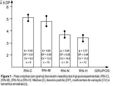
Purpose: to study the repercussion of arterial hypertension regarding body weight gain and body length, as well as liver and brain weight of offspring. Methods: a total of 82 animals in reproductive age were used. They were randomly assigned to 4 different groups (control, handled, nephrectomized and hypertensive). Renal hypertension was produced by a controlled constriction of the main left renal artery and contralateral nephrectomy, according to the technique described by Goldblatt (Goldblatt I: one kidney - one clip hypertension). Afterwards, they were distributed among nonpregnant and pregnant groups. The following newborn groups resulted from the pregnant groups: RN-C (control-newborn group of pregnant rats without surgical treatment), RN-M (manipulation-newborn group of the pregnant rats with surgical manipulation), RN-N (nephrectomized-newborn group of pregnant rats with nephrectomy) and Rn-H (hypertensive-newborn group of pregnant rats with hypertension). Results: the RN-N and RN-H groups showed body weight gain ( = 3,64 ± 0,50; or = 3,37 ± 0,44), body length ( = 3,89 ± 0,36; or = 3,68 ± 0,32) and brain weight ( = 0,16 ± 0,01; or = 0,16 ± 0,05), respectively, smaller than the control group ( = 5,40 ± 0,51; or = 4,95 ± 0,23 and or = 0,22 ± 0,04, respectively). In addition, the RN-H group showed the lowest liver weight ( = 0,22 ± 0,03) compared with the other three groups. Conclusion: after statistical analysis, the results obtained showed that the arterial hypertension determined a reduction in body weight, body length, and liver and brain weight of the offspring.
Summary
Rev Bras Ginecol Obstet. 2000;22(9):567-572
DOI 10.1590/S0100-72032000000900005
Purpose: to study the repercussion of arterial hypertension regarding body weight gain and body length, as well as liver and brain weight of offspring. Methods: a total of 82 animals in reproductive age were used. They were randomly assigned to 4 different groups (control, handled, nephrectomized and hypertensive). Renal hypertension was produced by a controlled constriction of the main left renal artery and contralateral nephrectomy, according to the technique described by Goldblatt (Goldblatt I: one kidney - one clip hypertension). Afterwards, they were distributed among nonpregnant and pregnant groups. The following newborn groups resulted from the pregnant groups: RN-C (control-newborn group of pregnant rats without surgical treatment), RN-M (manipulation-newborn group of the pregnant rats with surgical manipulation), RN-N (nephrectomized-newborn group of pregnant rats with nephrectomy) and Rn-H (hypertensive-newborn group of pregnant rats with hypertension). Results: the RN-N and RN-H groups showed body weight gain ( = 3,64 ± 0,50; or = 3,37 ± 0,44), body length ( = 3,89 ± 0,36; or = 3,68 ± 0,32) and brain weight ( = 0,16 ± 0,01; or = 0,16 ± 0,05), respectively, smaller than the control group ( = 5,40 ± 0,51; or = 4,95 ± 0,23 and or = 0,22 ± 0,04, respectively). In addition, the RN-H group showed the lowest liver weight ( = 0,22 ± 0,03) compared with the other three groups. Conclusion: after statistical analysis, the results obtained showed that the arterial hypertension determined a reduction in body weight, body length, and liver and brain weight of the offspring.
Summary
Rev Bras Ginecol Obstet. 2001;23(2):87-91
DOI 10.1590/S0100-72032001000200005
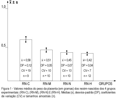
Purpose: to study the repercussion regarding placental weight and placental index determined by induced experimental hypertension in rats. Methods: a total of 82 rats in reproductive age were used. They were randomly assigned to 4 different groups (control, handled, nephrectomy and hypertension) and renal hypertension was produced by a controlled constriction of the main left renal artery, according to the technique described by Goldblatt, and contralateral nephrectomy (Goldblatt I one kidney--one clip model). Furthermore, they were distributed into non-pregnant groups and pregnant (P) groups. From the pregnant groups, the following newborn groups were obtained: RN-C (control -- newborn group from the pregnant rats without surgical treatment), RN-M (manipulation -- newborn group from the pregnant rats with surgical manipulation), RN-N (nephrectomy -- newborn group from the pregnant rats with nephrectomy) and RN-H (hypertension -- newborn group from pregnant rats with hypertension). Results: the RN-C newborn group ( = 0.58 ± 0,12) showed placental weight higher than the other three groups (RN-M: or = 0.51 ± 0.05; RN-N: or = 0.45 ± 0.07 and RN-H: or = 0.42 ± 0.04). On the other hand, it was possible to observe that the placental weight of the RN-M was higher than that of RN-N and RN-H, respectively, but no difference was observed between the RN-N and RN-H groups. The placental index showed no difference between P-C (Md = 0.1085) and P-M (Md = 0.1110), and also between P-N (0.1175) and P-H (0.1211), but it was observed that the placental indexes of P-C and P-M were smaller that those of P-N and P-H. Conclusion: unilateral nephrectomy and hypertension determined a reduction in placental weight and an increase in the placental index, showing a repercussion regarding placental and fetal development.
Summary
Rev Bras Ginecol Obstet. 2001;23(2):87-91
DOI 10.1590/S0100-72032001000200005
Purpose: to study the repercussion regarding placental weight and placental index determined by induced experimental hypertension in rats. Methods: a total of 82 rats in reproductive age were used. They were randomly assigned to 4 different groups (control, handled, nephrectomy and hypertension) and renal hypertension was produced by a controlled constriction of the main left renal artery, according to the technique described by Goldblatt, and contralateral nephrectomy (Goldblatt I one kidney--one clip model). Furthermore, they were distributed into non-pregnant groups and pregnant (P) groups. From the pregnant groups, the following newborn groups were obtained: RN-C (control -- newborn group from the pregnant rats without surgical treatment), RN-M (manipulation -- newborn group from the pregnant rats with surgical manipulation), RN-N (nephrectomy -- newborn group from the pregnant rats with nephrectomy) and RN-H (hypertension -- newborn group from pregnant rats with hypertension). Results: the RN-C newborn group ( = 0.58 ± 0,12) showed placental weight higher than the other three groups (RN-M: or = 0.51 ± 0.05; RN-N: or = 0.45 ± 0.07 and RN-H: or = 0.42 ± 0.04). On the other hand, it was possible to observe that the placental weight of the RN-M was higher than that of RN-N and RN-H, respectively, but no difference was observed between the RN-N and RN-H groups. The placental index showed no difference between P-C (Md = 0.1085) and P-M (Md = 0.1110), and also between P-N (0.1175) and P-H (0.1211), but it was observed that the placental indexes of P-C and P-M were smaller that those of P-N and P-H. Conclusion: unilateral nephrectomy and hypertension determined a reduction in placental weight and an increase in the placental index, showing a repercussion regarding placental and fetal development.
Summary
Rev Bras Ginecol Obstet. 2002;24(2):93-99
DOI 10.1590/S0100-72032002000200004
Purpose: to evaluate the correlation between the laparoscopic aspects and the stromal histologic findings of peritoneal endometriosis in order to understand the evolutive theory of endometriosis. Methods: sixty-seven women were submitted to laparoscopy for pelvic pain, infertility, ovarian tumor and other pathologies. A peritoneal biopsy was taken from the typical (puckered black) and atypical endometriotic implants. The different aspects of endometriosis were classified as follows: red lesions (Group V), black lesions (Group N) and white lesions (Group B). The histological sections were examined according to a standardized protocol. The histologic parameters used were: depth of the lesion, presence of hemosiderin, vascularization of the stroma and fibrotic tissue in stroma. Results: regarding lesion depth, there were significant differences between the groups. Red lesions were located consistently on the surface of the peritoneum (100%) and black lesions were superficial in 55.6%, intermediate in 38.9% and deep in 5.5%. White lesions were superficial in 28%, intermediate in 68% and deep in 4%. The presence of hemosiderin showed equivalent results in the 3 groups. The large stromal vascularization was present in the red lesions (60%), which a statistically significant difference compared to the other groups. Fibrotic tissue was present in 70.6% of the white lesions (group B), a fact that was significantly different when compared to groups V and N. Conclusion: the parameters analyzed in this study confirmed the importance of the evolutive theory of endometriosis.
Summary
Rev Bras Ginecol Obstet. 2000;22(2):95-100
DOI 10.1590/S0100-72032000000200006
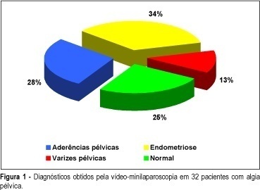
SUMMARY Purpose: to study the usefulness of minilaparoscopy in diagnosing the cause of pelvic pain. Methods: women with pelvic pain were prospectively analyzed and underwent an office video-microlaparoscopy. We analyzed the data regarding procedure time, stay in the recovery room, acceptance of anesthesia, and morbidity. Results: the average procedure time of the office video-microlaparoscopy was 19 min, the average stay for recovery was 43 min, and the technical quality of the image was excellent or good in 100% of the selected patients. The following laparoscopic findings were reported: 34.4% endometriosis, 28.1% pelvic adhesion, 12.5% pelvic varices, and 25% normal. Based on Bordhal et al.'s¹ criteria, a low frequency of pain manifestation during local anesthesia (12.5%) and discomfort on pneumoperitoneum (46.9%) were noticed. It could also be observed that, according to Milki and Tazuke's² criteria, the tolerance to the method was excellent and good (96.9%). Twenty-four hours after the procedure the morbidity rate was in accordance with Chung et al.'s³ criteria, showing a high frequency of pain at the incision area (59.4%) and sleepiness (43.8%). Only 3.1% reported they felt pain during the procedure, which shows the acceptance of the method by the patients. Conclusions: the acceptance of anesthesia and of the surgical procedure and the low morbidity allow the use of minilaparoscopy as a very important method in investigating patients with pelvic pain.
Summary
Rev Bras Ginecol Obstet. 2000;22(2):95-100
DOI 10.1590/S0100-72032000000200006
SUMMARY Purpose: to study the usefulness of minilaparoscopy in diagnosing the cause of pelvic pain. Methods: women with pelvic pain were prospectively analyzed and underwent an office video-microlaparoscopy. We analyzed the data regarding procedure time, stay in the recovery room, acceptance of anesthesia, and morbidity. Results: the average procedure time of the office video-microlaparoscopy was 19 min, the average stay for recovery was 43 min, and the technical quality of the image was excellent or good in 100% of the selected patients. The following laparoscopic findings were reported: 34.4% endometriosis, 28.1% pelvic adhesion, 12.5% pelvic varices, and 25% normal. Based on Bordhal et al.'s¹ criteria, a low frequency of pain manifestation during local anesthesia (12.5%) and discomfort on pneumoperitoneum (46.9%) were noticed. It could also be observed that, according to Milki and Tazuke's² criteria, the tolerance to the method was excellent and good (96.9%). Twenty-four hours after the procedure the morbidity rate was in accordance with Chung et al.'s³ criteria, showing a high frequency of pain at the incision area (59.4%) and sleepiness (43.8%). Only 3.1% reported they felt pain during the procedure, which shows the acceptance of the method by the patients. Conclusions: the acceptance of anesthesia and of the surgical procedure and the low morbidity allow the use of minilaparoscopy as a very important method in investigating patients with pelvic pain.