You searched for:"Ricardo Santos Simões"
We found (16) results for your search.Summary
Revista Brasileira de Ginecologia e Obstetrícia. 2012;34(7):323-328
DOI 10.1590/S0100-72032012000700006
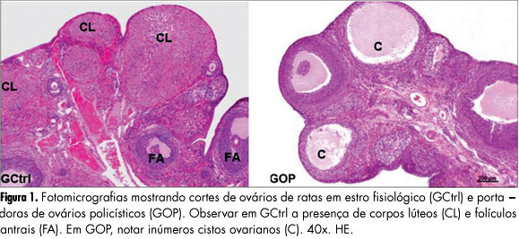
PURPOSES: To evaluate the histomorphometry of ovarian interstitial cells, as well as the blood sex steroid concentrations of female rats with polycystic ovaries induced by continuous light. METHODS: Twenty female rats were divided into two groups: Control Group - in the estrous phase (CtrlG), and a group of rats with polycystic ovaries induced by continuous illumination (POG). CtrlG animals were maintained on a light period from 07:00 a.m. to 07:00 p.m., and POG animals with continuous illumination (400 Lux) for 60 days. After this period all animals were anesthetized and blood was collected for the determination of serum estradiol (E2), progesterone (P4), and testosterone (T), followed by removal of the ovaries that were fixed in 10% formalin and processed for paraffin embedding. Five-µm histological sections were stained with hematoxylin and eosin and used for histomorphometric analysis. Morphological analyses, cyst count, determination of concentration and of the nuclear volume of interstitial cells were performed with the aid of a light microscope adapted to a high resolution camera (AxioCam), whose images were transmitted to and analyzed by the computer using AxioVision Rel 4.8 software (Carl Zeiss). Data were analyzed statistically by the Student's t-test (p<0.05). RESULTS: Morphological analysis showed the presence of ovarian cysts in POG animals and corpora lutea in CtrlG animals, as well as evidence of the origin of interstitial cells from the internal theca of these cysts. POG animals presented increased serum estradiol levels (pg/mL) compared to CtrlG animals (POG=124.9±4.2>CtrlG=73.2±6.5, p<0.05), the same occurring with testosterone levels (pg/mL) (POG=116.9±4.6>CtrlG=80.6±3.9, p<0.05). However, progesterone levels (ng/mL) were higher in CtrlG than in POG animals (CtrlG=16.3±2.0>POG=4.2±1.5, p<0.05). Morphometry showed a significant increase in nuclear volume in POG animals (POG=102.1±5.2>CtrlG=63.6±16.5, p<0.05), as well as in the area occupied (%) by interstitial cells (POG=24.4±6.9>CtrlG=6.9±3.2, p<0.05) compared to CtrlG animals. CONCLUSION: The interstitial cells of the rat polycystic ovary probably originate from ovarian cysts due to the degeneration of granulosa cells and differentiation of the internal theca cells. The elevations of serum testosterone and estradiol were probably due to the significant increase in cell activity and in the area occupied by interstitial cells.
Summary
Revista Brasileira de Ginecologia e Obstetrícia. 2012;34(7):323-328
DOI 10.1590/S0100-72032012000700006
PURPOSES: To evaluate the histomorphometry of ovarian interstitial cells, as well as the blood sex steroid concentrations of female rats with polycystic ovaries induced by continuous light. METHODS: Twenty female rats were divided into two groups: Control Group - in the estrous phase (CtrlG), and a group of rats with polycystic ovaries induced by continuous illumination (POG). CtrlG animals were maintained on a light period from 07:00 a.m. to 07:00 p.m., and POG animals with continuous illumination (400 Lux) for 60 days. After this period all animals were anesthetized and blood was collected for the determination of serum estradiol (E2), progesterone (P4), and testosterone (T), followed by removal of the ovaries that were fixed in 10% formalin and processed for paraffin embedding. Five-µm histological sections were stained with hematoxylin and eosin and used for histomorphometric analysis. Morphological analyses, cyst count, determination of concentration and of the nuclear volume of interstitial cells were performed with the aid of a light microscope adapted to a high resolution camera (AxioCam), whose images were transmitted to and analyzed by the computer using AxioVision Rel 4.8 software (Carl Zeiss). Data were analyzed statistically by the Student's t-test (p<0.05). RESULTS: Morphological analysis showed the presence of ovarian cysts in POG animals and corpora lutea in CtrlG animals, as well as evidence of the origin of interstitial cells from the internal theca of these cysts. POG animals presented increased serum estradiol levels (pg/mL) compared to CtrlG animals (POG=124.9±4.2>CtrlG=73.2±6.5, p<0.05), the same occurring with testosterone levels (pg/mL) (POG=116.9±4.6>CtrlG=80.6±3.9, p<0.05). However, progesterone levels (ng/mL) were higher in CtrlG than in POG animals (CtrlG=16.3±2.0>POG=4.2±1.5, p<0.05). Morphometry showed a significant increase in nuclear volume in POG animals (POG=102.1±5.2>CtrlG=63.6±16.5, p<0.05), as well as in the area occupied (%) by interstitial cells (POG=24.4±6.9>CtrlG=6.9±3.2, p<0.05) compared to CtrlG animals. CONCLUSION: The interstitial cells of the rat polycystic ovary probably originate from ovarian cysts due to the degeneration of granulosa cells and differentiation of the internal theca cells. The elevations of serum testosterone and estradiol were probably due to the significant increase in cell activity and in the area occupied by interstitial cells.
Summary
Revista Brasileira de Ginecologia e Obstetrícia. 2007;29(7):346-351
DOI 10.1590/S0100-72032007000700004
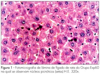
PURPOSE: to evaluate the effect of the chronic administration of three different doses of Ritonavir in the liver and kidneys of pregnant albino rats and their concepts from a morphological standpoint. METHODS: forty pregnant albino EPM-1 Wistar rats were randomly divided into four groups: Contr (vehicle control), and three experimental groups, Exp20, Exp60, Exp180, which received daily 20, 60 or 180 mg/kg of Ritonavir, respectively. The drug and the vehicle (propyleneglycol) were orally administered by gavage, from the first up to the 20th day of pregnancy. At the last experimental day, all the animals were sacrificed under deep anesthesia, and fragments from the maternal and fetal liver and kidneys were taken and prepared for histological analysis by light microscope. RESULTS: no morphological changes were identified in Exp20 and control group. In the Exp60 group, we found hepatocytes with signs of atrophy and apoptosis (eosinophilic cytoplasm and picnotic nuclei) and marked sinusoid capillary vasodilation (congestion). The proximal convoluted tubules of maternal kidneys and liver showed eosinophilic areas and hyperchromatic nuclei, as well as signs of vasodilation. The maternal kidneys and livers of the Exp180 rats presented more prominent morphological changes than the ones of Exp60. Regarding the fetal organs, no histomorphological abnormalities were observed in all the groups. CONCLUSIONS: our results show that the administration of Ritonavir to pregnant rats, in higher than conventional doses causes morphological changes in the maternal liver and kidneys. On the other hand, the lack of abnormalities in the fetal organs may be due to the protective role of glycoprotein P.
Summary
Revista Brasileira de Ginecologia e Obstetrícia. 2007;29(7):346-351
DOI 10.1590/S0100-72032007000700004
PURPOSE: to evaluate the effect of the chronic administration of three different doses of Ritonavir in the liver and kidneys of pregnant albino rats and their concepts from a morphological standpoint. METHODS: forty pregnant albino EPM-1 Wistar rats were randomly divided into four groups: Contr (vehicle control), and three experimental groups, Exp20, Exp60, Exp180, which received daily 20, 60 or 180 mg/kg of Ritonavir, respectively. The drug and the vehicle (propyleneglycol) were orally administered by gavage, from the first up to the 20th day of pregnancy. At the last experimental day, all the animals were sacrificed under deep anesthesia, and fragments from the maternal and fetal liver and kidneys were taken and prepared for histological analysis by light microscope. RESULTS: no morphological changes were identified in Exp20 and control group. In the Exp60 group, we found hepatocytes with signs of atrophy and apoptosis (eosinophilic cytoplasm and picnotic nuclei) and marked sinusoid capillary vasodilation (congestion). The proximal convoluted tubules of maternal kidneys and liver showed eosinophilic areas and hyperchromatic nuclei, as well as signs of vasodilation. The maternal kidneys and livers of the Exp180 rats presented more prominent morphological changes than the ones of Exp60. Regarding the fetal organs, no histomorphological abnormalities were observed in all the groups. CONCLUSIONS: our results show that the administration of Ritonavir to pregnant rats, in higher than conventional doses causes morphological changes in the maternal liver and kidneys. On the other hand, the lack of abnormalities in the fetal organs may be due to the protective role of glycoprotein P.
Summary
Revista Brasileira de Ginecologia e Obstetrícia. 2010;32(8):374-380
DOI 10.1590/S0100-72032010000800003
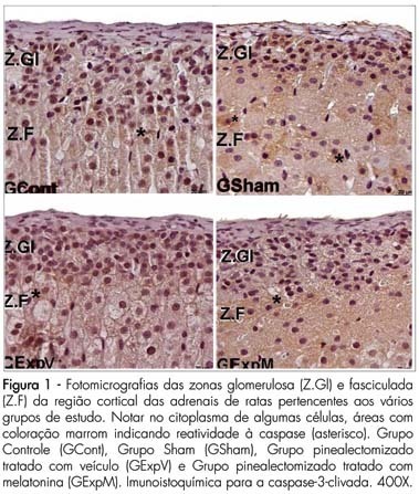
PURPOSE: to evaluate the reactivity of VEGF-A and cleaved caspase-3 in the adrenal gland cortex of female pinealectomized rats treated with melatonin. METHODS: forty adult female rats were divided into 4 groups (G) of 10 animals: GI - no surgical intervention, with vehicle administration; GII - sham pinealectomized with vehicle administration; GIII - pinealectomized with vehicle administration; GIV - pinealectomized with melatonin administration (10 µg/animal) during the night. After 60 days of treatment, all animals were anesthetized, and the adrenal glands were removed and fixed in 10% formaldehyde (phosphate buffered) for histological processing and paraffin embedding. Sections (5 µm thick) were collected on silanized slides and submitted to imunnohistochemical methods for the detection of cleaved caspase-3 (apoptosis) and of vascular endothelial growth factor (VEGF-A) in the adrenal cortex. The data obtained were submitted to analysis of variance (ANOVA) complemented by the Tukey-Kramer test (p<0.05). RESULTS: reactivity to cleaved Caspase-3 was noted in the zona glomerulosa of the adrenal glands in all studied groups. There were no significant differences in the zona glomerulosa; however, the zona fasciculata (15.51±3.12*, p<0.05) and the zona reticularis (8.11±1.90*, p<0.05) presented the smallest percentage of apoptosis in the pinealectomized group (GIII). The reactivity to the VEGF-A was stronger in the zona glomerulosa and weaker in the zona reticularis in all groups. We found a stronger VEGF-A reactivity in the zona fasciculata in the pinealectomized group (GIII). CONCLUSIONS: the pineal gland affects the arrangement of the zona glomerulosa and reticularis of the adrenal glands, which are related to the production of sex hormones.
Summary
Revista Brasileira de Ginecologia e Obstetrícia. 2010;32(8):374-380
DOI 10.1590/S0100-72032010000800003
PURPOSE: to evaluate the reactivity of VEGF-A and cleaved caspase-3 in the adrenal gland cortex of female pinealectomized rats treated with melatonin. METHODS: forty adult female rats were divided into 4 groups (G) of 10 animals: GI - no surgical intervention, with vehicle administration; GII - sham pinealectomized with vehicle administration; GIII - pinealectomized with vehicle administration; GIV - pinealectomized with melatonin administration (10 µg/animal) during the night. After 60 days of treatment, all animals were anesthetized, and the adrenal glands were removed and fixed in 10% formaldehyde (phosphate buffered) for histological processing and paraffin embedding. Sections (5 µm thick) were collected on silanized slides and submitted to imunnohistochemical methods for the detection of cleaved caspase-3 (apoptosis) and of vascular endothelial growth factor (VEGF-A) in the adrenal cortex. The data obtained were submitted to analysis of variance (ANOVA) complemented by the Tukey-Kramer test (p<0.05). RESULTS: reactivity to cleaved Caspase-3 was noted in the zona glomerulosa of the adrenal glands in all studied groups. There were no significant differences in the zona glomerulosa; however, the zona fasciculata (15.51±3.12*, p<0.05) and the zona reticularis (8.11±1.90*, p<0.05) presented the smallest percentage of apoptosis in the pinealectomized group (GIII). The reactivity to the VEGF-A was stronger in the zona glomerulosa and weaker in the zona reticularis in all groups. We found a stronger VEGF-A reactivity in the zona fasciculata in the pinealectomized group (GIII). CONCLUSIONS: the pineal gland affects the arrangement of the zona glomerulosa and reticularis of the adrenal glands, which are related to the production of sex hormones.
Summary
Revista Brasileira de Ginecologia e Obstetrícia. 2009;31(8):385-390
DOI 10.1590/S0100-72032009000800003
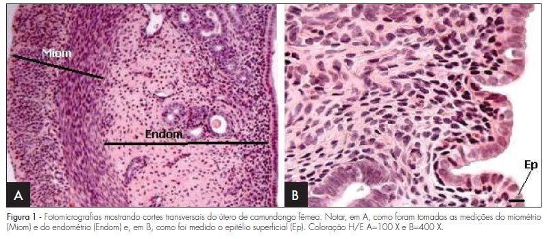
PURPOSE: to evaluate the effect of hyperprolactinemia induced by metoclopramide on the endometrium and myometrium of female mice in the proestrus phase. METHODS: 24 female mice were randomly divided in two groups: CtrG/control and ExpG/treated with metoclopramide (6.7 mg/g daily). After 50 days, the animals were sacrificed in the proestrus phase, and the blood was collected to determine the levels of estradiol, progesterone and prolactin. The uterine horns were removed, fixed in 10% formaldehyde and processed before being included in paraffin. Slices of 4 µm were stained by hematoxylin and eosin (H/E). In the morphological analysis, a Carl Zeiss light microscope, with objectives varying from 4 to 400 X was used for each histological slice characterization. In the morphometrical analysis, the superficial epithelium, the lamina propria and the myometrium thickness were evaluated, with the help of an image analyzer (AxionVision - Carl Zeiss) attached to the light microscope (Carl Zeiss). The statistical analysis was done by ANOVA, followed by the Wilcoxon test. P-value was considered as significant, when <0.05. RESULTS: our findings have shown an increase in the seric levels of prolactin (295.6±38.0 ng/mL) and significant decrease in the progesterone levels (11.3±0.9 ng/mL) in the ExpG, as compared to the CtrG (45.5±5.2 ng/mL and 18.2±1.6 ng/mL, respectively; p<0.001). Concerning the seric level of estradiol, significant differences between the groups were not obtained (ExpG=119.1±12.3 pg/mL and CtrG=122.7±8.4 pg/mL; p=0.418). The morphological study has shown that the uterus from the ExpG presented the endometrium with more developed superficial epithelium and lamina propria, as compared to the CtrG, the same happening with the myometrium. The thickness morphometrical values of the luminal epithelium (8.0±1.1 µm) and endometrium (116.2±21.1x10² µm) from the CtrG were lower than the ones from the ExpG (10.2±0.8 µm and 163.2±23.3x10² µm, respectively) with p<0.05. Nevertheless, data obtained in the myometrium have not shown significant differences between the groups (CtrG=152.2±25.2x10² µm and ExpG=140.8±18.0x10² µm). CONCLUSIONS: data have shown that hyperprolactinemia induced by metoclopramide determines endometrial proliferation and interferes with the ovarian function, mainly in the progesterone production.
Summary
Revista Brasileira de Ginecologia e Obstetrícia. 2009;31(8):385-390
DOI 10.1590/S0100-72032009000800003
PURPOSE: to evaluate the effect of hyperprolactinemia induced by metoclopramide on the endometrium and myometrium of female mice in the proestrus phase. METHODS: 24 female mice were randomly divided in two groups: CtrG/control and ExpG/treated with metoclopramide (6.7 mg/g daily). After 50 days, the animals were sacrificed in the proestrus phase, and the blood was collected to determine the levels of estradiol, progesterone and prolactin. The uterine horns were removed, fixed in 10% formaldehyde and processed before being included in paraffin. Slices of 4 µm were stained by hematoxylin and eosin (H/E). In the morphological analysis, a Carl Zeiss light microscope, with objectives varying from 4 to 400 X was used for each histological slice characterization. In the morphometrical analysis, the superficial epithelium, the lamina propria and the myometrium thickness were evaluated, with the help of an image analyzer (AxionVision - Carl Zeiss) attached to the light microscope (Carl Zeiss). The statistical analysis was done by ANOVA, followed by the Wilcoxon test. P-value was considered as significant, when <0.05. RESULTS: our findings have shown an increase in the seric levels of prolactin (295.6±38.0 ng/mL) and significant decrease in the progesterone levels (11.3±0.9 ng/mL) in the ExpG, as compared to the CtrG (45.5±5.2 ng/mL and 18.2±1.6 ng/mL, respectively; p<0.001). Concerning the seric level of estradiol, significant differences between the groups were not obtained (ExpG=119.1±12.3 pg/mL and CtrG=122.7±8.4 pg/mL; p=0.418). The morphological study has shown that the uterus from the ExpG presented the endometrium with more developed superficial epithelium and lamina propria, as compared to the CtrG, the same happening with the myometrium. The thickness morphometrical values of the luminal epithelium (8.0±1.1 µm) and endometrium (116.2±21.1x10² µm) from the CtrG were lower than the ones from the ExpG (10.2±0.8 µm and 163.2±23.3x10² µm, respectively) with p<0.05. Nevertheless, data obtained in the myometrium have not shown significant differences between the groups (CtrG=152.2±25.2x10² µm and ExpG=140.8±18.0x10² µm). CONCLUSIONS: data have shown that hyperprolactinemia induced by metoclopramide determines endometrial proliferation and interferes with the ovarian function, mainly in the progesterone production.
Summary
Revista Brasileira de Ginecologia e Obstetrícia. 2012;34(10):447-452
DOI 10.1590/S0100-72032012001000003
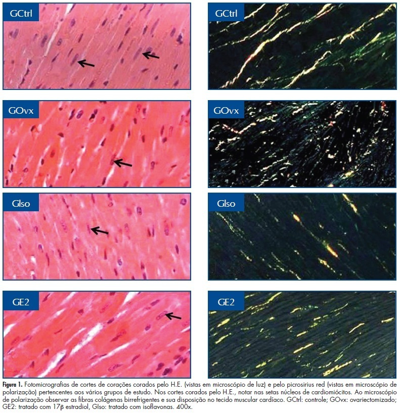
PURPOSES: To evaluate the histomorphometry of cardiomyocytes and collagen present in the myocardium of rats treated with a concentrated extract of soy or 17β-estradiol (E2). METHODS: Twenty-eight rats were divided into four groups: GCtrl - estrus phase; GOvx - ovariectomized (Ovx) and receiving vehicle; GIso - Ovx and treated with soy extract (150 mg/kg per day); GE2 - Ovx and treated with E2 (10 µg/kg per day). The drugs and vehicle (0.2 mL propylene glycol) were administered for 30 consecutive days after ovariectomy. On the last day the animals were anesthetized, the hearts removed, submerged in 10% formaldehyde and fragments of the ventricles underwent histological procedures, and the sections were stained with hematoxylin and eosin or picrosirius-red. Histomorphometric analysis (number and volume of nuclei and quantification of collagen) was performed under a light microscope with AxioVision Rel. 4.2 software, and collagen fibers were quantified using IMAGELAB-2000 software. Data were submitted to ANOVA followed by the Tukey test (p<0.05). RESULTS: We observed a higher number of cardiomyocyte nuclei in animals of the Ovx and Iso groups than in GE2 and GCtrl animals (GOvx=121.7±20.2=GIso=92.8±15.4>GE2=70.5±14,8=GCtrl=66.3±9.6; p <0.05), while the nuclear volume was greater in the Ctrl and E2 groups (GE2=35.7±4.8 GCtrl=29.9±3.6=>GIso=26.5±4.5=GOvx=22.4±2.9; p <0.05). Collagen concentration was higher in the Ovx group (GOvx=5.4±0.1>GCtrl=4.0±0.1=GIso=4.4±0.08=GE2=4.3±0.5; p <0.05). CONCLUSIONS: Estrogen may prevent the reduction of the nuclear volume of cardiomyocytes and collagen deposition between heart muscle fibers, while the administration of isoflavones only prevents the deposition of collagen, which can preserve the mechanical properties of cardiac fibers.
Summary
Revista Brasileira de Ginecologia e Obstetrícia. 2012;34(10):447-452
DOI 10.1590/S0100-72032012001000003
PURPOSES: To evaluate the histomorphometry of cardiomyocytes and collagen present in the myocardium of rats treated with a concentrated extract of soy or 17β-estradiol (E2). METHODS: Twenty-eight rats were divided into four groups: GCtrl - estrus phase; GOvx - ovariectomized (Ovx) and receiving vehicle; GIso - Ovx and treated with soy extract (150 mg/kg per day); GE2 - Ovx and treated with E2 (10 µg/kg per day). The drugs and vehicle (0.2 mL propylene glycol) were administered for 30 consecutive days after ovariectomy. On the last day the animals were anesthetized, the hearts removed, submerged in 10% formaldehyde and fragments of the ventricles underwent histological procedures, and the sections were stained with hematoxylin and eosin or picrosirius-red. Histomorphometric analysis (number and volume of nuclei and quantification of collagen) was performed under a light microscope with AxioVision Rel. 4.2 software, and collagen fibers were quantified using IMAGELAB-2000 software. Data were submitted to ANOVA followed by the Tukey test (p<0.05). RESULTS: We observed a higher number of cardiomyocyte nuclei in animals of the Ovx and Iso groups than in GE2 and GCtrl animals (GOvx=121.7±20.2=GIso=92.8±15.4>GE2=70.5±14,8=GCtrl=66.3±9.6; p <0.05), while the nuclear volume was greater in the Ctrl and E2 groups (GE2=35.7±4.8 GCtrl=29.9±3.6=>GIso=26.5±4.5=GOvx=22.4±2.9; p <0.05). Collagen concentration was higher in the Ovx group (GOvx=5.4±0.1>GCtrl=4.0±0.1=GIso=4.4±0.08=GE2=4.3±0.5; p <0.05). CONCLUSIONS: Estrogen may prevent the reduction of the nuclear volume of cardiomyocytes and collagen deposition between heart muscle fibers, while the administration of isoflavones only prevents the deposition of collagen, which can preserve the mechanical properties of cardiac fibers.
Summary
Revista Brasileira de Ginecologia e Obstetrícia. 2005;27(9):524-528
DOI 10.1590/S0100-72032005000900004
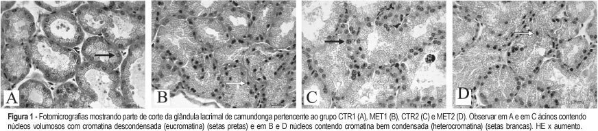
PURPOSE: to evaluate the morphological changes in murine lacrimal glands by metoclopramide-induced hyperprolactinemia during the proestrus phase or pregnancy. METHODS: forty adult mice were divided into two groups: CTR1 (control) and MET1 (treated with metoclopramide). After fifty days, half of the mice were sacrificed. The remaining animals were mated, and then labeled as pregnant controls (CTR2). Part of these animals were treated with metoclopramide and constituted the metoclopramide-treated pregnant (MET2) group. The CTR2 and MET2 groups were sacrificed on the 6th day of pregnancy. The blood was collected for determination of the hormonal levels of estradiol and progesterone by a chemoluminescent method. The lacrimal glands were then removed, fixed in 10% formaldehyde and stained with HE. The morphometric analysis was performed using the Axion Vision program (Carl Zeiss) to measure acinar nuclear and cellular volumes. RESULTS: the nuclear and cellular volumes of the lacrimal glands in the MET1-(152.2±8.7; 6.3±1.6 µm³) and MET2-(278.3±7.9; 27.5±0.9 µm³) treated groups were lower than those in CTR1 (204.2±7.4; 21.9±1.3 µm³) and CTR2 (329.4±2.2; 35.5±2.0 µm³), respectively. There was a significant hormonal level reduction in the animals that received metoclopramide compared to controls (CTR1: estradiol = 156.6±42.2 pg/ml; progesterone = 39.4±5.1 ng/ml; MET1: estradiol = 108.0±33.1 pg/ml; progesterone = 28.0±6.4 ng/ml; CTR2: estradiol = 354.0±56.0 pg/ml; progesterone = 251.0±56.0 ng/ml; MET2: estradiol = 293.0±43.0 pg/ml, progesterone = 184.0±33.0 ng/ml). CONCLUSION: metoclopramide-induced hyperprolactinemia produced morphological signs of reduction of cellular activity in lacrimal glands during the proestrus phase and pregnancy. It is hypothesized that this effect might be related to the hyperprolactinemia-induced decrease in the hormonal production of estrogen and progesterone.
Summary
Revista Brasileira de Ginecologia e Obstetrícia. 2005;27(9):524-528
DOI 10.1590/S0100-72032005000900004
PURPOSE: to evaluate the morphological changes in murine lacrimal glands by metoclopramide-induced hyperprolactinemia during the proestrus phase or pregnancy. METHODS: forty adult mice were divided into two groups: CTR1 (control) and MET1 (treated with metoclopramide). After fifty days, half of the mice were sacrificed. The remaining animals were mated, and then labeled as pregnant controls (CTR2). Part of these animals were treated with metoclopramide and constituted the metoclopramide-treated pregnant (MET2) group. The CTR2 and MET2 groups were sacrificed on the 6th day of pregnancy. The blood was collected for determination of the hormonal levels of estradiol and progesterone by a chemoluminescent method. The lacrimal glands were then removed, fixed in 10% formaldehyde and stained with HE. The morphometric analysis was performed using the Axion Vision program (Carl Zeiss) to measure acinar nuclear and cellular volumes. RESULTS: the nuclear and cellular volumes of the lacrimal glands in the MET1-(152.2±8.7; 6.3±1.6 µm³) and MET2-(278.3±7.9; 27.5±0.9 µm³) treated groups were lower than those in CTR1 (204.2±7.4; 21.9±1.3 µm³) and CTR2 (329.4±2.2; 35.5±2.0 µm³), respectively. There was a significant hormonal level reduction in the animals that received metoclopramide compared to controls (CTR1: estradiol = 156.6±42.2 pg/ml; progesterone = 39.4±5.1 ng/ml; MET1: estradiol = 108.0±33.1 pg/ml; progesterone = 28.0±6.4 ng/ml; CTR2: estradiol = 354.0±56.0 pg/ml; progesterone = 251.0±56.0 ng/ml; MET2: estradiol = 293.0±43.0 pg/ml, progesterone = 184.0±33.0 ng/ml). CONCLUSION: metoclopramide-induced hyperprolactinemia produced morphological signs of reduction of cellular activity in lacrimal glands during the proestrus phase and pregnancy. It is hypothesized that this effect might be related to the hyperprolactinemia-induced decrease in the hormonal production of estrogen and progesterone.
Summary
Revista Brasileira de Ginecologia e Obstetrícia. 2006;28(9):545-550
DOI 10.1590/S0100-72032006000900007
PURPOSE: to evaluate muscular strength of the pelvic floor and the periurethral vessels of postmenopausal women before and after six months of soybean extract treatment. METHODS: the study was conducted on 30 postmenopausal women before and after six consecutive months of soyabean extract (100 mg/day) administration. Urinary loss and muscular strength of the pelvic floor were investigated through digital perineometer and functional evaluation. Digital color Doppler in the periurethral region was used to count the number of vessels. For statistical analysis, the paired Student t test was applied to compare the results before and after the treatment. RESULTS: twenty women reported urinary incontinence before the treatment period. The amelioration of this symptom was observed in 15 (75%) women after the treatment. Vaginal pressure (muscular strength of the pelvic floor) was 12.95±1.73 and 15.86±1.86 Sauers, before and after the treatment, respectively (p<0.001). Twenty-two women (73.3%) presented an increase in the pressure at the end of this study. In relation to the function evaluation, 18 (60%) had improvement in muscular strength and 12 women did not present any change. On ultrasonography (Doppler), the number of vessels was 2.20±0.15 blood vessels/field in the beginning of this study and 3.46±0.25 blood vessels/field at the end of the treatment (p<0.001). An increase in the number of periurethral vessels was detected in 21 women (70%). CONCLUSION: it is important to emphasize that these are preliminary results. A double blind randomized and placebo-controlled clinical trial with a high number of participants is necessary. However, the treatment with concentrated soybean extract (100 mg per day) for six consecutive months may determine an improvement in pelvic floor muscular strength and an increase in the number of periurethral vessels in postmenopausal women.
Summary
Revista Brasileira de Ginecologia e Obstetrícia. 2010;32(11):556-562
DOI 10.1590/S0100-72032010001100007
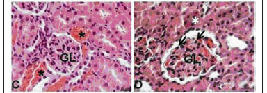
PURPOSE: to evaluate the effect of administration of three different doses of the zidovudine/lamivudine/ritonavir combination on the liver and kidneys of pregnant rats and their concepts from a morphological and physiological standpoint. METHODS: 40 pregnant EPM-1 Wistar rats were randomly divided into 4 groups: 1 control (Ctrl: drug vehicle control, n=10) and 3 experimental groups: Exp1x, Exp3x and Exp9x. An oral solution of the zidovudine/lamivudine/ritonavir combination was administered to the experimental groups from the day 0 to day 20 of pregnancy: Exp1x=10/5/20 mg/kg; Exp3x=30/15/60 mg/kg; Exp9x=90/45/180 mg/kg. On the 20th pregnancy day the rats were anesthetized and blood was taken directly from the ventricular chambers for further biochemical determinations: aspartate-(AST) and alanine-(ALT) aminotransferases (Calorimetric method), urea nitrogen (BUN) by an enzymatic-kinetic method, and creatinine by a kinetic-calorimetric method. Maternal and fetal liver and kidney samples were taken, fixed in 10% formaldehyde and processed histologically for paraffin embedding. Five µm-thick fragments of maternal and fetal livers and kidneys were stained with hematoxilyn-eosin, being analyzed by light microscopy. To interpret the results, the well-known pattern of normality for livers and kidneys was considered on the basis of the following structures: hepatocytes, portal structure, hepatic veins, renal corpuscles, renal tubules and loop of Henle. Regarding the fetal livers, we also considered the erythrocytes in their different stages of development as well as the megacariocytes. If there was a change in the established staining pattern for liver and kidney structures, changes in nuclear morphology, rupture of some cytoplasmic organelles, and presence of vascular congestion, this was considered to be due to the drug doses. Results were submitted to analysis of variance (ANOVA) and to the Tukey-Kramer multiple comparisons test (p<0.05). RESULTS: no morphological changes were observed in the maternal livers of the Ctrl, Exp1x and Exp3x groups. In the maternal liver of the Exp9x group, hepatocytes showed signs of atrophy and apoptosis (eosinophilic cytoplasm and pycnotic nuclei) and marked sinusoid capillary vasodilation (congestion) was observed. The maternal kidneys of the Ctrl and Exp1x groups were normal, with renal corpuscles, convoluted tubules and typical loops of Henle. In contrast, the Exp3x and Exp9x groups showed vascular congestion and small glomeruli rich in cells containing hyperchromatic nuclei which were more intense in Exp9x. Regarding the fetal organs, no morphological or physiological changes were observed. A significant increase of AST (305.70±55.80, p<0.05) and creatinine (0.50±0.09, p<0.05) was observed in group Exp9x. CONCLUSIONS: our results show that the administration of the zidovudine, lamivudine and ritonavir combination to pregnant rats at high doses caused morphological and physiological changes in the maternal liver and kidneys. On the other hand, there were no changes in fetal organs.
Summary
Revista Brasileira de Ginecologia e Obstetrícia. 2010;32(11):556-562
DOI 10.1590/S0100-72032010001100007
PURPOSE: to evaluate the effect of administration of three different doses of the zidovudine/lamivudine/ritonavir combination on the liver and kidneys of pregnant rats and their concepts from a morphological and physiological standpoint. METHODS: 40 pregnant EPM-1 Wistar rats were randomly divided into 4 groups: 1 control (Ctrl: drug vehicle control, n=10) and 3 experimental groups: Exp1x, Exp3x and Exp9x. An oral solution of the zidovudine/lamivudine/ritonavir combination was administered to the experimental groups from the day 0 to day 20 of pregnancy: Exp1x=10/5/20 mg/kg; Exp3x=30/15/60 mg/kg; Exp9x=90/45/180 mg/kg. On the 20th pregnancy day the rats were anesthetized and blood was taken directly from the ventricular chambers for further biochemical determinations: aspartate-(AST) and alanine-(ALT) aminotransferases (Calorimetric method), urea nitrogen (BUN) by an enzymatic-kinetic method, and creatinine by a kinetic-calorimetric method. Maternal and fetal liver and kidney samples were taken, fixed in 10% formaldehyde and processed histologically for paraffin embedding. Five µm-thick fragments of maternal and fetal livers and kidneys were stained with hematoxilyn-eosin, being analyzed by light microscopy. To interpret the results, the well-known pattern of normality for livers and kidneys was considered on the basis of the following structures: hepatocytes, portal structure, hepatic veins, renal corpuscles, renal tubules and loop of Henle. Regarding the fetal livers, we also considered the erythrocytes in their different stages of development as well as the megacariocytes. If there was a change in the established staining pattern for liver and kidney structures, changes in nuclear morphology, rupture of some cytoplasmic organelles, and presence of vascular congestion, this was considered to be due to the drug doses. Results were submitted to analysis of variance (ANOVA) and to the Tukey-Kramer multiple comparisons test (p<0.05). RESULTS: no morphological changes were observed in the maternal livers of the Ctrl, Exp1x and Exp3x groups. In the maternal liver of the Exp9x group, hepatocytes showed signs of atrophy and apoptosis (eosinophilic cytoplasm and pycnotic nuclei) and marked sinusoid capillary vasodilation (congestion) was observed. The maternal kidneys of the Ctrl and Exp1x groups were normal, with renal corpuscles, convoluted tubules and typical loops of Henle. In contrast, the Exp3x and Exp9x groups showed vascular congestion and small glomeruli rich in cells containing hyperchromatic nuclei which were more intense in Exp9x. Regarding the fetal organs, no morphological or physiological changes were observed. A significant increase of AST (305.70±55.80, p<0.05) and creatinine (0.50±0.09, p<0.05) was observed in group Exp9x. CONCLUSIONS: our results show that the administration of the zidovudine, lamivudine and ritonavir combination to pregnant rats at high doses caused morphological and physiological changes in the maternal liver and kidneys. On the other hand, there were no changes in fetal organs.