You searched for:"Patrícia Spara"
We found (13) results for your search.Summary
Rev Bras Ginecol Obstet. 2002;24(7):485-489
DOI 10.1590/S0100-72032002000700009
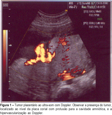
The most frequently nontrophoblastic tumor of the placenta found is chorioangioma, with an incidence of about 1%. When they are small, they do not significantly affect the fetus, but the large ones can cause intrauterine growth restriction, polyhydramnios, premature delivery, congestive heart failure and fetal death. The authors report a case of chorioangioma in a 28-year-old woman, second gestation, whose diagnosis was established at the 32nd week by ultrasound and confirmed by the anatomopathological examination. Ultrasonography evaluations showed chronic fetal distress and the delivery was performed at 36 weeks. The newborn results were satisfactory with Apgar 9-10 and fetal weight 2.460 g.
Summary
Rev Bras Ginecol Obstet. 2002;24(7):485-489
DOI 10.1590/S0100-72032002000700009
The most frequently nontrophoblastic tumor of the placenta found is chorioangioma, with an incidence of about 1%. When they are small, they do not significantly affect the fetus, but the large ones can cause intrauterine growth restriction, polyhydramnios, premature delivery, congestive heart failure and fetal death. The authors report a case of chorioangioma in a 28-year-old woman, second gestation, whose diagnosis was established at the 32nd week by ultrasound and confirmed by the anatomopathological examination. Ultrasonography evaluations showed chronic fetal distress and the delivery was performed at 36 weeks. The newborn results were satisfactory with Apgar 9-10 and fetal weight 2.460 g.
Summary
Rev Bras Ginecol Obstet. 2006;28(10):575-580
DOI 10.1590/S0100-72032006001000002
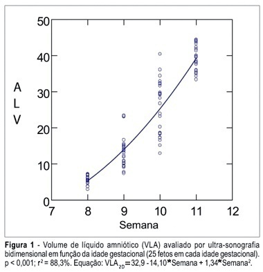
PURPOSE: To determine the values of amniotic fluid in normal fetuses during the first trimester of pregnancy by three- and bi-dimensional ultrasonography. METHODS: In a prospective longitudinal study, 25 normal fetuses were evaluated from the 8th to the 11th week of gestation. Amniotic fluid volume was measured by endovaginal ultrasonography with the three- and two-dimensional modes. The two-dimensional study consisted of volumetric determination by mathematical calculation based on an ellipsoidal shape (constant 0.52) to obtain the amniotic sac and embryo volumes. In the three-dimensional study, the amniotic fluid volume was determined by the VOCAL technique using 6, 9, 15, and 30 degrees of rotation. The amniotic fluid volume obtained by 6-degree rotations was considered to be the final result. In both modes, amniotic fluid volume was obtained by subtracting the volume of the embryo from the volume of the amniotic sac. Data were analyzed statistically for variance (ANOVA), correlation and regression analysis. The level of significance was set at p < 0.05. RESULTS: The amniotic fluid volume as measured by two-dimensional ultrasonography increased from 5.45 to 39.52 cm³ in the range from the 8th to the 11th week (ANOVA - p < 0.05). There was a correlation between gestational age and amniotic fluid volume (p < 0.001, r² = 88.3%). In the three-dimensional study, the amniotic fluid volume increased from 5.7 to 42.9 cm³ in the range from the 8th to the 11th week (ANOVA - p < 0.05), and again a correlation between gestational age and amniotic fluid volume (p < 0.001, r² = 98.1%) was observed. CONCLUSION: an increase in amniotic fluid volume occurs during the first trimester of pregnancy, as determined by the two- and three-dimensional modes. In addition, we have demonstrated that the higher the gestational age, the larger the amniotic fluid volume.
Summary
Rev Bras Ginecol Obstet. 2006;28(10):575-580
DOI 10.1590/S0100-72032006001000002
PURPOSE: To determine the values of amniotic fluid in normal fetuses during the first trimester of pregnancy by three- and bi-dimensional ultrasonography. METHODS: In a prospective longitudinal study, 25 normal fetuses were evaluated from the 8th to the 11th week of gestation. Amniotic fluid volume was measured by endovaginal ultrasonography with the three- and two-dimensional modes. The two-dimensional study consisted of volumetric determination by mathematical calculation based on an ellipsoidal shape (constant 0.52) to obtain the amniotic sac and embryo volumes. In the three-dimensional study, the amniotic fluid volume was determined by the VOCAL technique using 6, 9, 15, and 30 degrees of rotation. The amniotic fluid volume obtained by 6-degree rotations was considered to be the final result. In both modes, amniotic fluid volume was obtained by subtracting the volume of the embryo from the volume of the amniotic sac. Data were analyzed statistically for variance (ANOVA), correlation and regression analysis. The level of significance was set at p < 0.05. RESULTS: The amniotic fluid volume as measured by two-dimensional ultrasonography increased from 5.45 to 39.52 cm³ in the range from the 8th to the 11th week (ANOVA - p < 0.05). There was a correlation between gestational age and amniotic fluid volume (p < 0.001, r² = 88.3%). In the three-dimensional study, the amniotic fluid volume increased from 5.7 to 42.9 cm³ in the range from the 8th to the 11th week (ANOVA - p < 0.05), and again a correlation between gestational age and amniotic fluid volume (p < 0.001, r² = 98.1%) was observed. CONCLUSION: an increase in amniotic fluid volume occurs during the first trimester of pregnancy, as determined by the two- and three-dimensional modes. In addition, we have demonstrated that the higher the gestational age, the larger the amniotic fluid volume.
Summary
Rev Bras Ginecol Obstet. 2003;25(9):673-678
DOI 10.1590/S0100-72032003000900009
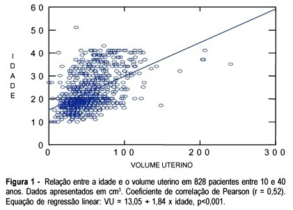
PURPOSE: to evaluate the uterine volume in women between 10 and 40 years in order to observe if the uterine volume in adolescents is smaller than the uterine volume in women between 20 and 40 years. We intend to emphasize the differences between the uterine volume of adolescents and that of adult women and to correlate with the immaturity of the genital tract of adolescents regarding gestation and delivery. METHOD: a cross-sectional study, which included 828 patients between 10 and 40 years old divided into two groups and examined using abdominal ultrasound to obtain the uterine volume measure. The first group consisted of 477 (57.7%) adolescents, and the second group of 351 (42.3%) adult women between 20 and 40 years old. In the adolescent group, ultrasound examination was performed by a single observer and in the group of adult women ultrasound examination was performed by a group of observers who used the same methodology as that of group 1. Image Point HX (Hewlett Packard) and Hitachi 525 ultrasound equipment were used with a multiple frequency probe. For the calculation of the uterine volume we used the longitudinal diameter (LD), anteroposterior diameter (APD) and transverse diameter (TD) with the (LD x APD x TD) x 0.45 formula. RESULTS: adolescents aged 10 to 17 years had a smaller uterine volume than women aged 20 to 40 years (p<0.05). Adolescents who delivered twice had a uterine volume similar to that of the patients between 20 and 40 years old with respective mean values of 62.6 ± 20.6 and 69.0±22.9 (p>0.05). CONCLUSION: adolescents less than 18 years old or primiparous have a smaller uterine volume than women between 20 to 40 years old. However, adolescents aged 18 years or older, or secundipara, have a uterine volume similar to that of women aged 20 to 40 years.
Summary
Rev Bras Ginecol Obstet. 2003;25(9):673-678
DOI 10.1590/S0100-72032003000900009
PURPOSE: to evaluate the uterine volume in women between 10 and 40 years in order to observe if the uterine volume in adolescents is smaller than the uterine volume in women between 20 and 40 years. We intend to emphasize the differences between the uterine volume of adolescents and that of adult women and to correlate with the immaturity of the genital tract of adolescents regarding gestation and delivery. METHOD: a cross-sectional study, which included 828 patients between 10 and 40 years old divided into two groups and examined using abdominal ultrasound to obtain the uterine volume measure. The first group consisted of 477 (57.7%) adolescents, and the second group of 351 (42.3%) adult women between 20 and 40 years old. In the adolescent group, ultrasound examination was performed by a single observer and in the group of adult women ultrasound examination was performed by a group of observers who used the same methodology as that of group 1. Image Point HX (Hewlett Packard) and Hitachi 525 ultrasound equipment were used with a multiple frequency probe. For the calculation of the uterine volume we used the longitudinal diameter (LD), anteroposterior diameter (APD) and transverse diameter (TD) with the (LD x APD x TD) x 0.45 formula. RESULTS: adolescents aged 10 to 17 years had a smaller uterine volume than women aged 20 to 40 years (p<0.05). Adolescents who delivered twice had a uterine volume similar to that of the patients between 20 and 40 years old with respective mean values of 62.6 ± 20.6 and 69.0±22.9 (p>0.05). CONCLUSION: adolescents less than 18 years old or primiparous have a smaller uterine volume than women between 20 to 40 years old. However, adolescents aged 18 years or older, or secundipara, have a uterine volume similar to that of women aged 20 to 40 years.
Summary
Rev Bras Ginecol Obstet. 2005;27(11):707-707
Summary
Rev Bras Ginecol Obstet. 2005;27(11):707-707
Summary
Rev Bras Ginecol Obstet. 1998;20(2):77-81
DOI 10.1590/S0100-72031998000200004
Electronic fetal heart rate monitoring (EFM) is the most widely used method of direct fetal surveillance especially during labor. In an attempt to elucidate the effect of EFM on cesarean section (CS) rates, a retrospective study was performed at the University Hospital of Santa Maria (HUSM). We studied two groups of patients, consisting of 2114 pregnant women: EFM group (n=517) and intermittent auscultation (IA) group (n=1597). In the EFM group we observed 38.0% of CS vs. 27.2% in the IA group. For all patients, the CS rate was 29.9%. Fetal distress was the most common indication for CS in the EFM group (40.6%), while previous CS was the third cause (10.1%). On the IA group, fetal distress was the third cause in CS (14.3%), while previous CS was the most common cause (32.4%). On the basis of this study, we believe that EFM has no effect in itself on cesarean section rates considering overall deliveries at HUSM. With proper education of the clinician and correct interpretation of the findings, EFM would not increase cesarean section rates, but rather should allow for a more accurate description of intrapartum fetal well-being.