You searched for:"Patrícia El Beitune"
We found (20) results for your search.Summary
Rev Bras Ginecol Obstet. 2003;25(7):465-471
DOI 10.1590/S0100-72032003000700002
PURPOSE: to investigate the effect of antiretroviral drugs on carbohydrate metabolism in HIV-infected pregnant women and on fetal and neonatal prognosis. METHODS: a prospective study was conducted on 57 pregnant women. The women were divided into three groups: ZDV group, taking zidovudine (n=20), TT group, taking zidovudine + lamivudine + nelfinavir (n=25), and control group (n=12). Blood samples were obtained for the determination of the area under the curve (AUC) after a 75-g oral glucose test at four periods during pregnancy (1st=14-20 weeks, 2nd= 21-26 weeks, 3rd=27-32 weeks and 4th=33-38 weeks). Perinatal prognosis was based on prematurity rates, intrauterine growth restriction (IUGR), low birth weight, perinatal mortality, and vertical HIV-1 transmission. Data were analyzed statistically using the nonparametric c² test, Friedman test and Kruskal-Wallis test. RESULTS: the median values of the AUC were 11.685 mg/dL for the control group, 13.477 mg/dL for the ZDV Group, and 13.650 mg/dL for the TT group (p=0.049). The antiretroviral agents had no deleterious effects on prematurity, low birth weight, IUGR rates or on Apgar score. There was no case of vertical transmission of HIV-1. CONCLUSIONS: an association was detected between the use of triple therapy and the development of carbohydrate intolerance during pregnancy. This association was not shown with ZDV alone. The antiretroviral agents had no deleterious effects on perinatal prognosis.
Summary
Rev Bras Ginecol Obstet. 2023;45(1):49-54
Summary
Rev Bras Ginecol Obstet. 2023;45(1):49-54
Summary
Rev Bras Ginecol Obstet. 2004;26(6):497-498
Summary
Rev Bras Ginecol Obstet. 2004;26(6):497-498
Summary
Rev Bras Ginecol Obstet. 2006;28(10):575-580
DOI 10.1590/S0100-72032006001000002
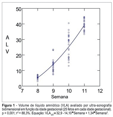
PURPOSE: To determine the values of amniotic fluid in normal fetuses during the first trimester of pregnancy by three- and bi-dimensional ultrasonography. METHODS: In a prospective longitudinal study, 25 normal fetuses were evaluated from the 8th to the 11th week of gestation. Amniotic fluid volume was measured by endovaginal ultrasonography with the three- and two-dimensional modes. The two-dimensional study consisted of volumetric determination by mathematical calculation based on an ellipsoidal shape (constant 0.52) to obtain the amniotic sac and embryo volumes. In the three-dimensional study, the amniotic fluid volume was determined by the VOCAL technique using 6, 9, 15, and 30 degrees of rotation. The amniotic fluid volume obtained by 6-degree rotations was considered to be the final result. In both modes, amniotic fluid volume was obtained by subtracting the volume of the embryo from the volume of the amniotic sac. Data were analyzed statistically for variance (ANOVA), correlation and regression analysis. The level of significance was set at p < 0.05. RESULTS: The amniotic fluid volume as measured by two-dimensional ultrasonography increased from 5.45 to 39.52 cm³ in the range from the 8th to the 11th week (ANOVA - p < 0.05). There was a correlation between gestational age and amniotic fluid volume (p < 0.001, r² = 88.3%). In the three-dimensional study, the amniotic fluid volume increased from 5.7 to 42.9 cm³ in the range from the 8th to the 11th week (ANOVA - p < 0.05), and again a correlation between gestational age and amniotic fluid volume (p < 0.001, r² = 98.1%) was observed. CONCLUSION: an increase in amniotic fluid volume occurs during the first trimester of pregnancy, as determined by the two- and three-dimensional modes. In addition, we have demonstrated that the higher the gestational age, the larger the amniotic fluid volume.
Summary
Rev Bras Ginecol Obstet. 2006;28(10):575-580
DOI 10.1590/S0100-72032006001000002
PURPOSE: To determine the values of amniotic fluid in normal fetuses during the first trimester of pregnancy by three- and bi-dimensional ultrasonography. METHODS: In a prospective longitudinal study, 25 normal fetuses were evaluated from the 8th to the 11th week of gestation. Amniotic fluid volume was measured by endovaginal ultrasonography with the three- and two-dimensional modes. The two-dimensional study consisted of volumetric determination by mathematical calculation based on an ellipsoidal shape (constant 0.52) to obtain the amniotic sac and embryo volumes. In the three-dimensional study, the amniotic fluid volume was determined by the VOCAL technique using 6, 9, 15, and 30 degrees of rotation. The amniotic fluid volume obtained by 6-degree rotations was considered to be the final result. In both modes, amniotic fluid volume was obtained by subtracting the volume of the embryo from the volume of the amniotic sac. Data were analyzed statistically for variance (ANOVA), correlation and regression analysis. The level of significance was set at p < 0.05. RESULTS: The amniotic fluid volume as measured by two-dimensional ultrasonography increased from 5.45 to 39.52 cm³ in the range from the 8th to the 11th week (ANOVA - p < 0.05). There was a correlation between gestational age and amniotic fluid volume (p < 0.001, r² = 88.3%). In the three-dimensional study, the amniotic fluid volume increased from 5.7 to 42.9 cm³ in the range from the 8th to the 11th week (ANOVA - p < 0.05), and again a correlation between gestational age and amniotic fluid volume (p < 0.001, r² = 98.1%) was observed. CONCLUSION: an increase in amniotic fluid volume occurs during the first trimester of pregnancy, as determined by the two- and three-dimensional modes. In addition, we have demonstrated that the higher the gestational age, the larger the amniotic fluid volume.
Summary
Rev Bras Ginecol Obstet. 2004;26(7):583-583
Summary
Rev Bras Ginecol Obstet. 2004;26(7):583-583
Summary
Rev Bras Ginecol Obstet. 2003;25(8):593-598
DOI 10.1590/S0100-72032003000800008
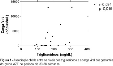
PURPOSE: to investigate the effect of antiretroviral drugs on the lipid metabolism in HIV-infected pregnant women. METHODS: a prospective study was conducted on 57 pregnant women. The women were divided into three groups: ZDV group, consisting of 20 HIV-infected women taking ZDV; TT group, consisting of 25 HIV-1-infected women on triple antiretroviral treatment (ZDV + 3TC + NFV), and control group, consisting of 12 pregnant women considered to be normal from a clinical and laboratory viewpoint. Demographic and anthropometric data were homogeneous. Patients with a personal and family history of hyperlipidemia were excluded. Blood samples were obtained for the determination of fasting lipids (total cholesterol, LDL and HDL, and triglycerides) at four periods during pregnancy (1st = 14-20 weeks; 2nd = 21-26 weeks; 3rd = 27-32 weeks and 4th = 33-38 weeks). Data were analyzed statistically using the nonparametric chi², Friedman and Kruskal-Wallis tests . RESULTS: the use of antiretroviral drugs during pregnancy induced no difference in total or HDL cholesterol but caused an increase from 76.5 and 84 mg/dL to 96 and 105 mg/dL in the concentration of the LDL fraction along gestation in ZDV and TT groups, respectively (p<0.01). A positive significant association was observed between triglycerides and viral burden in the ZDV group (r: 0.534; p=0.015). CONCLUSION: Antiretroviral agents during pregnancy increase serum LDL-colesterol levels. The risk of pregnancy regarding potentiation of long-term antiretroviral effects on lipid metabolism, remains to be established.
Summary
Rev Bras Ginecol Obstet. 2003;25(8):593-598
DOI 10.1590/S0100-72032003000800008
PURPOSE: to investigate the effect of antiretroviral drugs on the lipid metabolism in HIV-infected pregnant women. METHODS: a prospective study was conducted on 57 pregnant women. The women were divided into three groups: ZDV group, consisting of 20 HIV-infected women taking ZDV; TT group, consisting of 25 HIV-1-infected women on triple antiretroviral treatment (ZDV + 3TC + NFV), and control group, consisting of 12 pregnant women considered to be normal from a clinical and laboratory viewpoint. Demographic and anthropometric data were homogeneous. Patients with a personal and family history of hyperlipidemia were excluded. Blood samples were obtained for the determination of fasting lipids (total cholesterol, LDL and HDL, and triglycerides) at four periods during pregnancy (1st = 14-20 weeks; 2nd = 21-26 weeks; 3rd = 27-32 weeks and 4th = 33-38 weeks). Data were analyzed statistically using the nonparametric chi², Friedman and Kruskal-Wallis tests . RESULTS: the use of antiretroviral drugs during pregnancy induced no difference in total or HDL cholesterol but caused an increase from 76.5 and 84 mg/dL to 96 and 105 mg/dL in the concentration of the LDL fraction along gestation in ZDV and TT groups, respectively (p<0.01). A positive significant association was observed between triglycerides and viral burden in the ZDV group (r: 0.534; p=0.015). CONCLUSION: Antiretroviral agents during pregnancy increase serum LDL-colesterol levels. The risk of pregnancy regarding potentiation of long-term antiretroviral effects on lipid metabolism, remains to be established.
Summary
Rev Bras Ginecol Obstet. 2002;24(10):647-652
DOI 10.1590/S0100-72032002001000003
PURPOSE: to evaluate experimentally the effects of antiretroviral drugs used alone and in association upon the fertility of pregnant Wistar rats and the perinatal effects on the offspring. METHODS: adult female pregnant Wistar rats weighing 200-230 g were used. The antiretroviral drugs zidovudine (AZT), lamivudine (3TC) and nelfinavir (NFV) were used alone and in association at daily doses of ten times the dose normally used in pregnant women, proportionally to the animal's body weight. Seven groups were studied, including the control one. The experiment started on day 0 and the pregnant animals were sacrificed on day 21. The alive and dead fetuses, the total implantation sites and the total numbers of corporea lutea were used to calculate the fertility values. The statistical analysis was performed by Student's t test and by the Mann-Whitney test. RESULTS: there were no significant statistical differences regarding preimplantation loss and implantation efficiency values of the rats treated with isolated and associated antiretroviral drugs. There was a significant increase in the postimplantation loss values (control group: 7.6%; drug groups variation: 20.2-26.7%), a decrease in the fetal viability values (control group: 92.4%, drug groups variation: 73.3-79.8%), and a decreasing number of fetuses per animal (control group: 14.7; drug groups variation: 11.1-12.7). There was a significant weight reduction of the female rats and of the offspring of animals treated with 3TC, AZT + 3TC and AZT + 3TC + NFV. CONCLUSION: with the administration of high antiretroviral doses, important fertility effects could be observed, which showed that less histotoxic antiretroviral drugs must be studied in order to warrant the safety of using these medicines in pregnant HIV-1 - infected women.
Summary
Rev Bras Ginecol Obstet. 2020;42(11):697-704
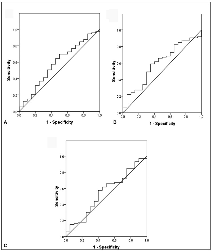
Recent observations support the hypothesis that an imbalance between angiogenic factors has a fundamental role in the pathogenesis of pre-eclampsia and is responsible for the clinical manifestations of the disease. The goal of the present study was to evaluate the sensitivity, specificity, and the best accuracy level of Soluble fms-like tyrosine kinase-1 (sFlt-1), placental growth factor (PlGF), and sFlt-1/PlGF ratio in maternal serum and protein/creatinine ratio in urine sample to define the best cutoff point of these tests to discriminate between the patients with gestational hypertension and the patients with pre-eclampsia, to evaluate the possibility of using them as diagnostic methods.
A prospective longitudinal study was performed, and blood samples were collected from 95 pregnant patients with hypertension to measure serum concentrations of biomarkers sFlt-1 and PlGF. Urine samples were collected for protein screening. Significance was set as p < 0.05.
The sFlt-1/PlGF ratio demonstrated a sensitivity of 57.5% and a specificity of 60% using 50.4 as a cutoff point. The test that showed the best accuracy in the diagnosis of pre-eclampsia was protein/creatinine ratio, with a sensitivity of 78.9% and a specificity of 70% using 0.4 as a cutoff point and showing an area under the receiver operating characteristic curve of 0.80 (p < 0.001).
No studied laboratory test proved to be fairly accurate for the diagnosis of pre-eclampsia, except for the protein/creatinine ratio. The evidence is insufficient to recommend biomarkers sFlt-1 and PlGF to be used for the diagnosis of pre-eclampsia.
Summary
Rev Bras Ginecol Obstet. 2020;42(11):697-704
Recent observations support the hypothesis that an imbalance between angiogenic factors has a fundamental role in the pathogenesis of pre-eclampsia and is responsible for the clinical manifestations of the disease. The goal of the present study was to evaluate the sensitivity, specificity, and the best accuracy level of Soluble fms-like tyrosine kinase-1 (sFlt-1), placental growth factor (PlGF), and sFlt-1/PlGF ratio in maternal serum and protein/creatinine ratio in urine sample to define the best cutoff point of these tests to discriminate between the patients with gestational hypertension and the patients with pre-eclampsia, to evaluate the possibility of using them as diagnostic methods.
A prospective longitudinal study was performed, and blood samples were collected from 95 pregnant patients with hypertension to measure serum concentrations of biomarkers sFlt-1 and PlGF. Urine samples were collected for protein screening. Significance was set as p < 0.05.
The sFlt-1/PlGF ratio demonstrated a sensitivity of 57.5% and a specificity of 60% using 50.4 as a cutoff point. The test that showed the best accuracy in the diagnosis of pre-eclampsia was protein/creatinine ratio, with a sensitivity of 78.9% and a specificity of 70% using 0.4 as a cutoff point and showing an area under the receiver operating characteristic curve of 0.80 (p < 0.001).
No studied laboratory test proved to be fairly accurate for the diagnosis of pre-eclampsia, except for the protein/creatinine ratio. The evidence is insufficient to recommend biomarkers sFlt-1 and PlGF to be used for the diagnosis of pre-eclampsia.