You searched for:"Luiz Henrique Gebrim"
We found (13) results for your search.Summary
Revista Brasileira de Ginecologia e Obstetrícia. 2002;24(6):420-420
DOI 10.1590/S0100-72032002000600014
Summary
Revista Brasileira de Ginecologia e Obstetrícia. 2002;24(6):420-420
DOI 10.1590/S0100-72032002000600014
Summary
Revista Brasileira de Ginecologia e Obstetrícia. 2000;22(7):429-433
DOI 10.1590/S0100-72032000000700005
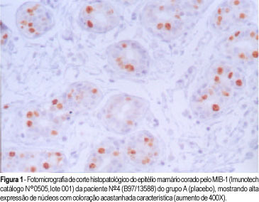
Purpose: to study the monoclonal antibody MIB-1 in the normal breast epithelium adjacent to a fibroadenoma in women in the luteal phase of the menstrual cycle treated with tamoxifen. Patients and methods: the proliferative activity of the mammary epithelium adjacent to the fibroadenoma was studied by immunohistochemistry based on immunoexpression of the monoclonal antibody MIB-1. The study was randomized and double blind and was conducted on 44 women with fibroadenomas, divided into 3 groups: A (n = 16; placebo), B (n = 15; tamoxifen, 10 mg), and C (n = 13; tamoxifen, 20 mg). Tamoxifen was administered for 22 days starting on the 2nd day of the menstrual cycle and a biopsy was taken on the 23rd day. Results: the mean percentage of stained nuclei per 1000 cells was 9.2 in group A, 4.5 in group B, and 3.2 in group C. Fisher's test revealed that tamoxifen significantly reduced the immunoexpression of MIB-1 at the doses of 10 and 20 mg compared to the placebo group (p<0.0001), with no significant differences between doses in terms of proliferative activity (p = 0.21). Conclusion: we conclude that tamoxifen significantly reduced the proliferative activity of the mammary epithelium at the doses of 10 and 20 mg/day.
Summary
Revista Brasileira de Ginecologia e Obstetrícia. 2000;22(7):429-433
DOI 10.1590/S0100-72032000000700005
Purpose: to study the monoclonal antibody MIB-1 in the normal breast epithelium adjacent to a fibroadenoma in women in the luteal phase of the menstrual cycle treated with tamoxifen. Patients and methods: the proliferative activity of the mammary epithelium adjacent to the fibroadenoma was studied by immunohistochemistry based on immunoexpression of the monoclonal antibody MIB-1. The study was randomized and double blind and was conducted on 44 women with fibroadenomas, divided into 3 groups: A (n = 16; placebo), B (n = 15; tamoxifen, 10 mg), and C (n = 13; tamoxifen, 20 mg). Tamoxifen was administered for 22 days starting on the 2nd day of the menstrual cycle and a biopsy was taken on the 23rd day. Results: the mean percentage of stained nuclei per 1000 cells was 9.2 in group A, 4.5 in group B, and 3.2 in group C. Fisher's test revealed that tamoxifen significantly reduced the immunoexpression of MIB-1 at the doses of 10 and 20 mg compared to the placebo group (p<0.0001), with no significant differences between doses in terms of proliferative activity (p = 0.21). Conclusion: we conclude that tamoxifen significantly reduced the proliferative activity of the mammary epithelium at the doses of 10 and 20 mg/day.
Summary
Revista Brasileira de Ginecologia e Obstetrícia. 2000;22(7):455-458
DOI 10.1590/S0100-72032000000700009
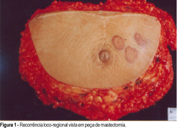
Primary angiosarcoma of the breast is a rare tumor, which appears between 14 and 82 years, with an average of 35 years of age. Its predominant clinical aspect is a painful mass with diffuse increase in the breast and violet or blackened color. Equally to other cases of sarcoma, the medium size of the lesion is approximately 5 cm at the diagnosis. Histologically, it is characterized by the proliferation of endothelial cells that form vascular channels linked to each other infiltrating glandular structures and fatty tissue. Its histological diagnosis is difficult and not always the right diagnosis is immediately established, mainly in the cases of a low malignancy degree, due to limited biopsy material. Because of the difficult diagnosis and aggressivity, it is a neoplasia with ominous prognosis, due to frequent metastasis. In our service, a 18-year-old patient presented with a painful lump which grew quickly. It was biopsied and a hemangioma was diagnosed, a wide excision being indicated. Three months later, she suffered a tumoral relapse, that was biopsied again and mastectomy was indicated, because it was an angiosarcoma with low degree of malignancy. After other relapses, chemotherapy was indicated and later, radiotherapy. During radiotherapy she developed new metastases, and died of pulmonary metastasis.
Summary
Revista Brasileira de Ginecologia e Obstetrícia. 2000;22(7):455-458
DOI 10.1590/S0100-72032000000700009
Primary angiosarcoma of the breast is a rare tumor, which appears between 14 and 82 years, with an average of 35 years of age. Its predominant clinical aspect is a painful mass with diffuse increase in the breast and violet or blackened color. Equally to other cases of sarcoma, the medium size of the lesion is approximately 5 cm at the diagnosis. Histologically, it is characterized by the proliferation of endothelial cells that form vascular channels linked to each other infiltrating glandular structures and fatty tissue. Its histological diagnosis is difficult and not always the right diagnosis is immediately established, mainly in the cases of a low malignancy degree, due to limited biopsy material. Because of the difficult diagnosis and aggressivity, it is a neoplasia with ominous prognosis, due to frequent metastasis. In our service, a 18-year-old patient presented with a painful lump which grew quickly. It was biopsied and a hemangioma was diagnosed, a wide excision being indicated. Three months later, she suffered a tumoral relapse, that was biopsied again and mastectomy was indicated, because it was an angiosarcoma with low degree of malignancy. After other relapses, chemotherapy was indicated and later, radiotherapy. During radiotherapy she developed new metastases, and died of pulmonary metastasis.
Summary
Revista Brasileira de Ginecologia e Obstetrícia. 1998;20(8):475-479
DOI 10.1590/S0100-72031998000800008
Purpose: to analyze the frequency of preoperative bilateral synchronic cancer and occult metastases in 454 operable breast cancer patients, at Instituto Nacional de Câncer (Brazil). Methods: the preoperative evaluation consisted of mammography, bone scan with X-ray if necessary, and chest X-ray. 260 (57.3 %) of 454 patients underwent liver echography. We calculated the cost X effectiveness ratio considering only the direct costs (monetary value) and the effectiveness was analyzed based on the number of metastases identifid by the screening tests. Results: we did not find any case of bilateral synchronic cancer, and the frequency of patients with metastasis was 2% (9/454). The diagnosis of bone metastasis was 1.5 % (7/454). The percentage of lung (2/454) and liver (1/260) metastasis was the same, 0.4 %. Most of the patients with metastases were in stage IIIb (44.5 %). The results of the screening tests showed the alteration of the initial clinical stage in 9 patients only (2%). The total cost of the screening tests for the diagnosis of systemic disease in 9 patients, was US$ 131,020.00. The cost of each diagnosed metastasise, for a total of 10 (two were found in one of the patients), was US$ 29,221.85 and the cost/effectiveness ratio was 22.3%. Conclusious: the results showed that screening for metastases in the preoperative clinical staging of breast cancer should be limited to patients symptomatic for systemic disease or in clinical stage III and that the cost/effectiveness ratio of the tests demonstrated a reduced benefit in the preoperative evaluation.
Summary
Revista Brasileira de Ginecologia e Obstetrícia. 1998;20(9):533-536
DOI 10.1590/S0100-72031998000900007
Purpose: to evaluate the effects of tamoxifen (TAM) on plasma levels of estradiol, progesterone, prolactin, luteinizing hormone (LH), follicle-stimulating hormone (FSH) and steroid hormone-binding globulin (SHBG) when given to premenopausal women in the doses of 10 and 20 mg/day for 22 days. Patients and Methods: a randomized double-blind study was performed with 43 premenopausal eumenorrheic women. The patients were divided into three groups: A (N = 15, placebo); B (N = 15, TAM 10 mg/day) and C (N = 13, 20 mg/day). They started taking an oral dose of TAM or placebo on the very first day of the menstrual cycle. Two hormone determinations were performed, both on the 22nd day of the menstrual cycle: the first in the cycle that preceded the use of the drug and the second, in the following cycle, after 22 days of using the medication. We used the Levine and Student tests in order to evaluate the homogeneity of the sample and the variation of the hormone determinations respectively. Results:serum levels of estradiol, progesterone and SHBG increased significantly in groups B and C. In group C, we also observed increase in serum level of FSH (p < 0.0045) and a fall in prolactin level (p < 0.0055). Conclusions: TAM promoted a significant increase in serum concentrations of estradiol, progesterone and SHBG either in the doses of 10 or 20 mg/day. However, significant increase in FSH and decrease in prolactin were obtained only with the dose of 20 mg/day.