You searched for:"Julio Cesar Rosa-e-Silva"
We found (13) results for your search.Summary
Rev Bras Ginecol Obstet. 2018;40(10):606-613
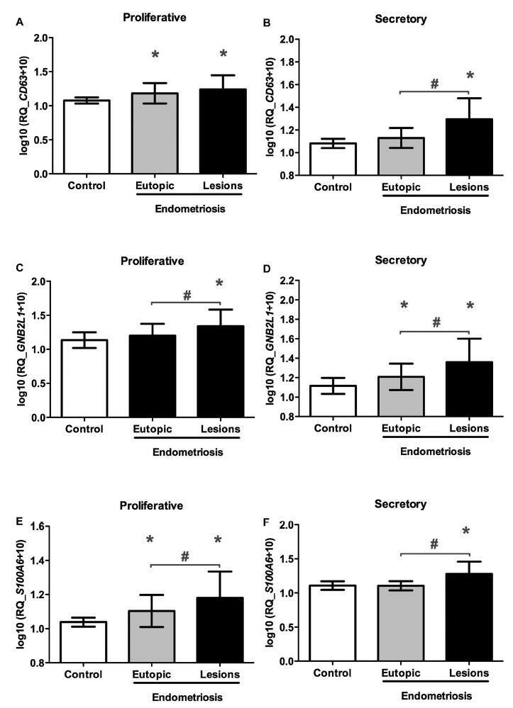
The aim of the present study was to analyze the expression of the CD63, S100A6, and GNB2L1genes, which participate in mechanisms related to the complex pathophysiology of endometriosis.
A case-control study was conducted with 40 women who were diagnosed with endometriosis, and 15 fertile and healthy women. Paired samples of eutopic endometrium and endometriotic lesions (peritoneal and ovarian endometriotic implants) were obtained from the women with endometriosis in the proliferative (n = 20) or secretory phases (n = 20) of the menstrual cycle. As controls, paired endometrial biopsy samples were collected from the healthy women in the proliferative (n = 15) and secretory (n = 15) phases of the samemenstrual cycle.We analyzed the expression levels of the CD63, S100A6, and GNB2L1 genes by real-time polymerase chain reaction.
An increase in CD63, S100A6, and GNB2L1 gene transcript levels was observed in the ectopic implants compared with the eutopic endometrium of the women with and without endometriosis, regardless of the phase of the menstrual cycle.
These findings suggest that the CD63, S100A6, and GNB2L1 genesmay be involved in the pathogenesis of endometriosis, since they participate in mechanisms such as inhibition of apoptosis, angiogenesis and cell proliferation, which lead to the loss of cell homeostasis in the ectopic endometrium, thus contributing to the implantation and survival of the tissue in the extrauterine environment.
Summary
Rev Bras Ginecol Obstet. 2018;40(10):606-613
The aim of the present study was to analyze the expression of the CD63, S100A6, and GNB2L1genes, which participate in mechanisms related to the complex pathophysiology of endometriosis.
A case-control study was conducted with 40 women who were diagnosed with endometriosis, and 15 fertile and healthy women. Paired samples of eutopic endometrium and endometriotic lesions (peritoneal and ovarian endometriotic implants) were obtained from the women with endometriosis in the proliferative (n = 20) or secretory phases (n = 20) of the menstrual cycle. As controls, paired endometrial biopsy samples were collected from the healthy women in the proliferative (n = 15) and secretory (n = 15) phases of the samemenstrual cycle.We analyzed the expression levels of the CD63, S100A6, and GNB2L1 genes by real-time polymerase chain reaction.
An increase in CD63, S100A6, and GNB2L1 gene transcript levels was observed in the ectopic implants compared with the eutopic endometrium of the women with and without endometriosis, regardless of the phase of the menstrual cycle.
These findings suggest that the CD63, S100A6, and GNB2L1 genesmay be involved in the pathogenesis of endometriosis, since they participate in mechanisms such as inhibition of apoptosis, angiogenesis and cell proliferation, which lead to the loss of cell homeostasis in the ectopic endometrium, thus contributing to the implantation and survival of the tissue in the extrauterine environment.
Summary
Rev Bras Ginecol Obstet. 2019;41(11):668-672
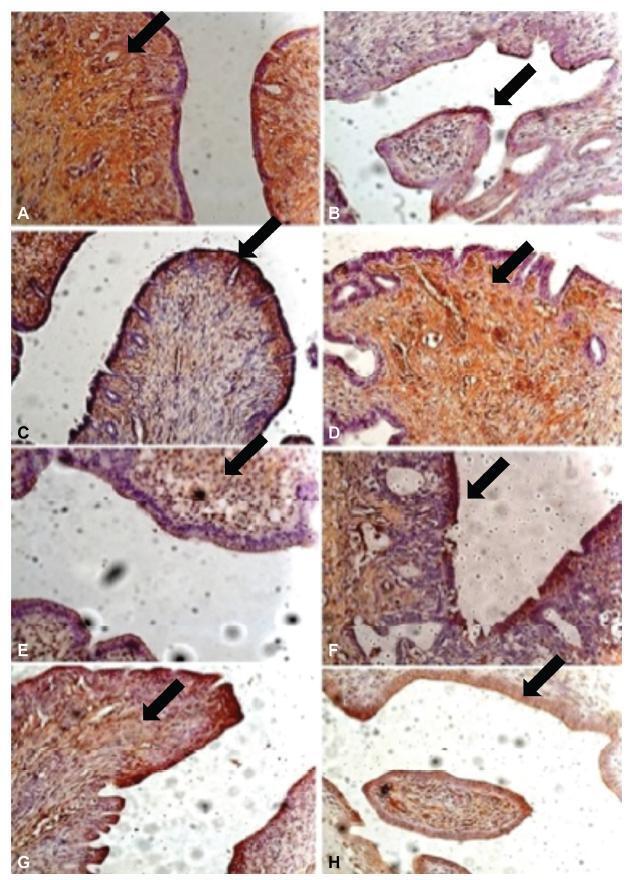
To analyze the effect of thalidomide on the progression of endometriotic lesions experimentally induced in rats and to characterize the pattern of cell proliferation by immunohistochemical Proliferating Cell Nuclear Antigen (PCNA) labeling of eutopic and ectopic endometrium.
Fifteen female Wistar rats underwent laparotomy for endometriosis induction by resection of one uterine horn, isolation of the endometrium and fixation of a tissue segment to the pelvic peritoneum. Four weeks after, the animals were divided into 3 groups: control (I), 10mg/kg/day (II) and 1mg/kg/day (III) intraperitoneal thalidomide for 10 days. The lesion was excised together with the opposite uterine horn for endometrial gland and stroma analysis. Eutopic and ectopic endometrial tissue was submitted to immunohistochemistry for analysis of cell proliferation by PCNA labeling and the cell proliferation index (CPI) was calculated as the number of labeled cells per 1,000 cells.
Group I showed a mean CPI of 0.248 ± 0.0513 in the gland and of 0.178 ± 0.046 in the stroma. In contrast, Groups II and III showed a significantly lower CPI, that is, 0.088 ± 0.009 and 0.080 ± 0.021 for the gland (p < 0.001) and 0.0945 ± 0.0066 and 0.075 ± 0.018 for the stroma (p < 0.001), respectively. Also, the mean lesion area of Group I was 69.2mm2, a significantly higher value compared with Group II (49.4mm2, p = 0.023) and Group III (48.6mm2, p = 0.006). No significant difference was observed between Groups II and III.
Thalidomide proved to be effective in reducing the lesion area and CPI of the experimental endometriosis implants both at the dose of 1mg/kg/day and at the dose of 10 mg/kg/day.
Summary
Rev Bras Ginecol Obstet. 2019;41(11):668-672
To analyze the effect of thalidomide on the progression of endometriotic lesions experimentally induced in rats and to characterize the pattern of cell proliferation by immunohistochemical Proliferating Cell Nuclear Antigen (PCNA) labeling of eutopic and ectopic endometrium.
Fifteen female Wistar rats underwent laparotomy for endometriosis induction by resection of one uterine horn, isolation of the endometrium and fixation of a tissue segment to the pelvic peritoneum. Four weeks after, the animals were divided into 3 groups: control (I), 10mg/kg/day (II) and 1mg/kg/day (III) intraperitoneal thalidomide for 10 days. The lesion was excised together with the opposite uterine horn for endometrial gland and stroma analysis. Eutopic and ectopic endometrial tissue was submitted to immunohistochemistry for analysis of cell proliferation by PCNA labeling and the cell proliferation index (CPI) was calculated as the number of labeled cells per 1,000 cells.
Group I showed a mean CPI of 0.248 ± 0.0513 in the gland and of 0.178 ± 0.046 in the stroma. In contrast, Groups II and III showed a significantly lower CPI, that is, 0.088 ± 0.009 and 0.080 ± 0.021 for the gland (p < 0.001) and 0.0945 ± 0.0066 and 0.075 ± 0.018 for the stroma (p < 0.001), respectively. Also, the mean lesion area of Group I was 69.2mm2, a significantly higher value compared with Group II (49.4mm2, p = 0.023) and Group III (48.6mm2, p = 0.006). No significant difference was observed between Groups II and III.
Thalidomide proved to be effective in reducing the lesion area and CPI of the experimental endometriosis implants both at the dose of 1mg/kg/day and at the dose of 10 mg/kg/day.
Summary
Rev Bras Ginecol Obstet. 2018;40(11):705-712
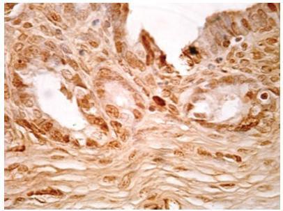
To characterize the patterns of cell differentiation, proliferation, and tissue invasion in eutopic and ectopic endometrium of rabbits with induced endometriotic lesions via a well- known experimental model, 4 and 8 weeks after the endometrial implantation procedure.
Twenty-nine female New Zealand rabbits underwent laparotomy for endometriosis induction through the resection of one uterine horn, isolation of the endometrium, and fixation of tissue segment to the pelvic peritoneum. Two groups of animals (one with 14 animals, and the other with15) were sacrificed 4 and 8 weeks after endometriosis induction. The lesion was excised along with the opposite uterine horn for endometrial gland and stroma determination. Immunohistochemical reactions were performed in eutopic and ectopic endometrial tissues for analysis of the following markers: metalloprotease (MMP-9) and tissue inhibitor of metalloprotease (TIMP-2), which are involved in the invasive capacity of the endometrial tissue; and metallothionein (MT) and p63, which are involved in cell differentiation and proliferation.
The intensity of the immunostaining for MMP9, TIMP-2, MT, and p63 was higher in ectopic endometria than in eutopic endometria. However, when the ectopic lesions were compared at 4 and 8 weeks, no significant difference was observed, with the exception of the marker p63, which was more evident after 8 weeks of evolution of the ectopic endometrial tissue.
Ectopic endometrial lesions seem to express greater power for cell differentiation and tissue invasion, compared with eutopic endometria, demonstrating a potentially invasive, progressive, and heterogeneous presentation of endometriosis.
Summary
Rev Bras Ginecol Obstet. 2018;40(11):705-712
To characterize the patterns of cell differentiation, proliferation, and tissue invasion in eutopic and ectopic endometrium of rabbits with induced endometriotic lesions via a well- known experimental model, 4 and 8 weeks after the endometrial implantation procedure.
Twenty-nine female New Zealand rabbits underwent laparotomy for endometriosis induction through the resection of one uterine horn, isolation of the endometrium, and fixation of tissue segment to the pelvic peritoneum. Two groups of animals (one with 14 animals, and the other with15) were sacrificed 4 and 8 weeks after endometriosis induction. The lesion was excised along with the opposite uterine horn for endometrial gland and stroma determination. Immunohistochemical reactions were performed in eutopic and ectopic endometrial tissues for analysis of the following markers: metalloprotease (MMP-9) and tissue inhibitor of metalloprotease (TIMP-2), which are involved in the invasive capacity of the endometrial tissue; and metallothionein (MT) and p63, which are involved in cell differentiation and proliferation.
The intensity of the immunostaining for MMP9, TIMP-2, MT, and p63 was higher in ectopic endometria than in eutopic endometria. However, when the ectopic lesions were compared at 4 and 8 weeks, no significant difference was observed, with the exception of the marker p63, which was more evident after 8 weeks of evolution of the ectopic endometrial tissue.
Ectopic endometrial lesions seem to express greater power for cell differentiation and tissue invasion, compared with eutopic endometria, demonstrating a potentially invasive, progressive, and heterogeneous presentation of endometriosis.
Summary
Rev Bras Ginecol Obstet. 2022;44(8):737-739
Summary
Rev Bras Ginecol Obstet. 2022;44(8):737-739
Summary
Rev Bras Ginecol Obstet. 2012;34(2):92-96
DOI 10.1590/S0100-72032012000200009
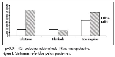
PURPOSE: To characterize patients with indeterminate values of hyperprolactinemia (PEG test for the identification of macroprolactinemias with recovery between 30 and 65%) (PRLi) or macroprolactinemia (PRLm), in relation to clinical characteristics, such as the presence or absence of symptoms, as well as their intensity and variation, and the presence or absence of central nervous system tumors. METHODS: This is a cross-sectional retrospective survey of records of 24 patients with hyperprolactinemia, in reproductive ages, with prolactin >25 ng/dL. Eleven women with PRLm and 13 with PRLi were included. Records from the two groups were extracted for analysis: age, parity, body mass index, presence of galactorrhea, infertility, and central nervous system tumor. Anthropometrics data were expressed as mean and standard deviation. To compare groups regarding the presence of central nervous system tumor, galactorrhea, as well as infertility we used the Student's t-test. RESULTS: Galactorrhea was more prevalent in patients with PRLi (p=0.01). Seventy percent of women with PRLi presented pituitary tumor (microprolactinoma), whereas this finding was evident in 17% of the PRLm Group (p=0.04). Among the patients with and PRLm PRLi, nine were not investigated with the image of the central nervous system because they have low levels of prolactin (five carriers and four PRLm PRLi). There were no significant differences regarding the occurrence of infertility or irregular menstrual cycles between groups. DISCUSSION: Women with intermediate hyperprolactinemia present more galactorrhea symptoms as well as central nervous system tumors than women with macroprolactinemia.
Summary
Rev Bras Ginecol Obstet. 2012;34(2):92-96
DOI 10.1590/S0100-72032012000200009
PURPOSE: To characterize patients with indeterminate values of hyperprolactinemia (PEG test for the identification of macroprolactinemias with recovery between 30 and 65%) (PRLi) or macroprolactinemia (PRLm), in relation to clinical characteristics, such as the presence or absence of symptoms, as well as their intensity and variation, and the presence or absence of central nervous system tumors. METHODS: This is a cross-sectional retrospective survey of records of 24 patients with hyperprolactinemia, in reproductive ages, with prolactin >25 ng/dL. Eleven women with PRLm and 13 with PRLi were included. Records from the two groups were extracted for analysis: age, parity, body mass index, presence of galactorrhea, infertility, and central nervous system tumor. Anthropometrics data were expressed as mean and standard deviation. To compare groups regarding the presence of central nervous system tumor, galactorrhea, as well as infertility we used the Student's t-test. RESULTS: Galactorrhea was more prevalent in patients with PRLi (p=0.01). Seventy percent of women with PRLi presented pituitary tumor (microprolactinoma), whereas this finding was evident in 17% of the PRLm Group (p=0.04). Among the patients with and PRLm PRLi, nine were not investigated with the image of the central nervous system because they have low levels of prolactin (five carriers and four PRLm PRLi). There were no significant differences regarding the occurrence of infertility or irregular menstrual cycles between groups. DISCUSSION: Women with intermediate hyperprolactinemia present more galactorrhea symptoms as well as central nervous system tumors than women with macroprolactinemia.