You searched for:"Francisco Mauad Filho"
We found (15) results for your search.Summary
Revista Brasileira de Ginecologia e Obstetrícia. 1998;20(8):443-448
DOI 10.1590/S0100-72031998000800003
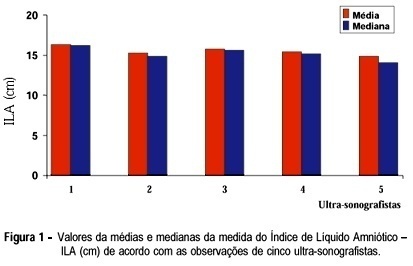
Purpose: to demonstrate the interobserver variation existing in the ultrasonographic measurement of amniotic fluid index (AFI) and in the measurement of pocket area, and to compare these two methods. In addition, an attempt was made to establish the intraobserver variation in the measurement of this index. Methods: values of AFI, described by Phelan et al.18, were studied in a group of 80 pregnant women considered to be clinically normal, seen at the Ultrasonography and Medical Updating School of Ribeirão Preto and in the Department of Gynecology and Obstetrics of the Faculty of Medicine of Ribeirão Preto, University of São Paulo (FMRP-USP). All pregnant women had a gestational age of more than 24 weeks. Fifty of these patients were submitted to AFI evaluation by 5 different ultrasonographists using the same equipment and during the same period of time, in order to determine the interobserver variation of this index. In addition, planimetric measurement of the area was performed by 2 of these 5 ultrasonographists, selected at random, in an attempt to determine interobserver variation in area measurement. Another group of 30 pregnant women was evaluated by the same ultrasonographist in an attempt to evaluate intraobserver variation in terms of AFI measurement. Results: There was a significant interobserver variation in AFI measurement and a significant variation in area measurement. However, the intraobserver variation in AFI measurement was nonsignificant. There was a correlation between AFI and area measurements. Conclusions: we emphasize the obstetrical applicability of this index and the easier execution of this method compared to area measurement, despite the importance of both procedures.
Summary
Revista Brasileira de Ginecologia e Obstetrícia. 1998;20(8):443-448
DOI 10.1590/S0100-72031998000800003
Purpose: to demonstrate the interobserver variation existing in the ultrasonographic measurement of amniotic fluid index (AFI) and in the measurement of pocket area, and to compare these two methods. In addition, an attempt was made to establish the intraobserver variation in the measurement of this index. Methods: values of AFI, described by Phelan et al.18, were studied in a group of 80 pregnant women considered to be clinically normal, seen at the Ultrasonography and Medical Updating School of Ribeirão Preto and in the Department of Gynecology and Obstetrics of the Faculty of Medicine of Ribeirão Preto, University of São Paulo (FMRP-USP). All pregnant women had a gestational age of more than 24 weeks. Fifty of these patients were submitted to AFI evaluation by 5 different ultrasonographists using the same equipment and during the same period of time, in order to determine the interobserver variation of this index. In addition, planimetric measurement of the area was performed by 2 of these 5 ultrasonographists, selected at random, in an attempt to determine interobserver variation in area measurement. Another group of 30 pregnant women was evaluated by the same ultrasonographist in an attempt to evaluate intraobserver variation in terms of AFI measurement. Results: There was a significant interobserver variation in AFI measurement and a significant variation in area measurement. However, the intraobserver variation in AFI measurement was nonsignificant. There was a correlation between AFI and area measurements. Conclusions: we emphasize the obstetrical applicability of this index and the easier execution of this method compared to area measurement, despite the importance of both procedures.
Summary
Revista Brasileira de Ginecologia e Obstetrícia. 2002;24(7):485-489
DOI 10.1590/S0100-72032002000700009
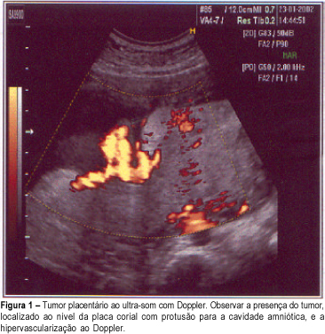
The most frequently nontrophoblastic tumor of the placenta found is chorioangioma, with an incidence of about 1%. When they are small, they do not significantly affect the fetus, but the large ones can cause intrauterine growth restriction, polyhydramnios, premature delivery, congestive heart failure and fetal death. The authors report a case of chorioangioma in a 28-year-old woman, second gestation, whose diagnosis was established at the 32nd week by ultrasound and confirmed by the anatomopathological examination. Ultrasonography evaluations showed chronic fetal distress and the delivery was performed at 36 weeks. The newborn results were satisfactory with Apgar 9-10 and fetal weight 2.460 g.
Summary
Revista Brasileira de Ginecologia e Obstetrícia. 2002;24(7):485-489
DOI 10.1590/S0100-72032002000700009
The most frequently nontrophoblastic tumor of the placenta found is chorioangioma, with an incidence of about 1%. When they are small, they do not significantly affect the fetus, but the large ones can cause intrauterine growth restriction, polyhydramnios, premature delivery, congestive heart failure and fetal death. The authors report a case of chorioangioma in a 28-year-old woman, second gestation, whose diagnosis was established at the 32nd week by ultrasound and confirmed by the anatomopathological examination. Ultrasonography evaluations showed chronic fetal distress and the delivery was performed at 36 weeks. The newborn results were satisfactory with Apgar 9-10 and fetal weight 2.460 g.
Summary
Revista Brasileira de Ginecologia e Obstetrícia. 2008;30(10):494-498
DOI 10.1590/S0100-72032008001000003
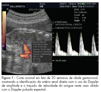
PURPOSE: to describe values found for the resistance index (RI), pulsatility index (PI) and the systole/diastole (S/D) ratio of fetal renal arteries in non-complicated gestations between the 22nd and the 38th week, and to evaluate whether those values vary along that period. METHODS: observational study, where 45 fetuses from non-complicated gestations have been evaluated in the 22nd, 26th, 30th and 38th weeks of gestational age. Doppler ultrasonography has been performed by the same observer, using a device with 4 to 7 MHz transducer. For the acquisition of the renal arteries velocity record, a 1 mm to 2 mm probe has been placed in the mean third of the renal artery for the evaluation through pulsed Doppler ultrasonography. The measurement of RI, PI and S/D ratio from three consecutive waves was performed with the automatic mode. To detect significant differences in the indexes' values along gestation, we have compared values obtained at the different gestational ages, through repeated measures ANOVA, followed by Tukey's post-hoc test. RESULTS: There were no significant differences between the right and left renal arteries, when the RI, IP and S/D ratio were compared. Nevertheless, a change in the values of these parameters has been observed between the 22nd week (RI=0.9 ± 0.02; PI=2.4 ± 0.02; S/D ratio=11.6 ± 2.2; mean ± standard deviation of the combined mean values of the right and left renal artery) and the 38th week (RI=0.8 ± 0.03; PI=2.1 ± 0.2; S/D ratio=8.7 ± 2.3) of gestation. CONCLUSIONS: the parameters evaluated (RI, PI and S/D ratio) have presented decreasing values between the 22nd and 38th, with no difference between the fetus's right and left sides.
Summary
Revista Brasileira de Ginecologia e Obstetrícia. 2008;30(10):494-498
DOI 10.1590/S0100-72032008001000003
PURPOSE: to describe values found for the resistance index (RI), pulsatility index (PI) and the systole/diastole (S/D) ratio of fetal renal arteries in non-complicated gestations between the 22nd and the 38th week, and to evaluate whether those values vary along that period. METHODS: observational study, where 45 fetuses from non-complicated gestations have been evaluated in the 22nd, 26th, 30th and 38th weeks of gestational age. Doppler ultrasonography has been performed by the same observer, using a device with 4 to 7 MHz transducer. For the acquisition of the renal arteries velocity record, a 1 mm to 2 mm probe has been placed in the mean third of the renal artery for the evaluation through pulsed Doppler ultrasonography. The measurement of RI, PI and S/D ratio from three consecutive waves was performed with the automatic mode. To detect significant differences in the indexes' values along gestation, we have compared values obtained at the different gestational ages, through repeated measures ANOVA, followed by Tukey's post-hoc test. RESULTS: There were no significant differences between the right and left renal arteries, when the RI, IP and S/D ratio were compared. Nevertheless, a change in the values of these parameters has been observed between the 22nd week (RI=0.9 ± 0.02; PI=2.4 ± 0.02; S/D ratio=11.6 ± 2.2; mean ± standard deviation of the combined mean values of the right and left renal artery) and the 38th week (RI=0.8 ± 0.03; PI=2.1 ± 0.2; S/D ratio=8.7 ± 2.3) of gestation. CONCLUSIONS: the parameters evaluated (RI, PI and S/D ratio) have presented decreasing values between the 22nd and 38th, with no difference between the fetus's right and left sides.
Summary
Revista Brasileira de Ginecologia e Obstetrícia. 2002;24(8):521-526
DOI 10.1590/S0100-72032002000800004
Purpose: to assess the evolution of epileptic seizures during pregnancy and the occurrence of malformations in neonates born to epileptic mothers who used anticonvulsant drugs during pregnancy, as well as the perinatal characteristics of the newborns. Methods: a total of 126 medical records of epileptic patients seen at the high-risk pregnancy outpatient clinic were analyzed retrospectively in terms of the following variables: age, parity, diagnosis of the type of epileptic seizure, anticonvulsant drug used during the prenatal period, evolution of epileptic seizures during the prenatal period, type of delivery, gestational age at resolution, and perinatal characteristics of the newborns. Results: the incidence of pregnant women with epilepsy was 0.2% in relation to prenatal patients, with simple partial epilepsy being the most frequent type (40% of cases). Monotherapy was applied to 75% of the patients and carbamazepine was the most frequently used drug. Among the 111 patients evaluated in terms of course of the disease during pregnancy, 53% showed no change, 31% became worse and 16% improved. Normal delivery was performed in 62.5% of cases, with a satisfactory perinatal result in terms of Apgar score, and with a rate of low birth weight neonates above the values for low-risk populations. No fetal malformations were observed. Conclusion: epilepsy showed a favorable course during pregnancy and was not aggravated by the latter, with cases of worsening of signs and symptoms being associated with epilepsy of difficult control before pregnancy. Evaluation of the perinatal characteristics of the neonates showed satisfactory Apgar scores and evolution, indicating that epilepsy and anticonvulsant drugs do not cause severe impairment of intrapartum vitality. No cases of malformations or hemorrhagic complications were detected in the present study.
Summary
Revista Brasileira de Ginecologia e Obstetrícia. 2001;23(8):523-527
DOI 10.1590/S0100-72032001000800007
Purpose: to study perinatal outcome in asthmatic pregnant patients who required hospital admission to control acute exacerbations. Patients and Method: retrospective study of 12 pregnant asthmatic patients admitted at the Hospital das Clínicas, Department of Obstetrics and Gynecology, FMRP-USP, during the period between 1992 and 1996. The analyzed data included: maternal age, prenatal care, length of hospitalization, gestational age at delivery, type of delivery, neonatal weight and Apgar score. Results: among the 12 asthmatic pregnant patients 7 did not have prenatal care for acute exacerbation treatment before hospitalization. Three of 12 developed preeclampsia (one with premature rupture of membranes and infection of the amniotic cavity and one with premature separation of the placenta); 2 of 12 were diagnosed with premature placental aging (one with premature labor and twin-to-twin transfusion syndrome and one with oligohydramnios); 1 of 12 was diagnosed with oligohydramnios and fetal death and had pneumonia, and 1 of 12 was diagnosed with polyhydramnios. Among the infants, 3 were small for gestational age. Conclusions: perinatal complications were more frequent in asthmatic pregnant patients who required hospital care for the acute exacerbations. Chronic asthmatic patients in reproductive age should be advised before pregnancy about the prophylactic measures to reduce the incidence of acute crises and exacerbations during pregnancy should be treated promptly.
Summary
Revista Brasileira de Ginecologia e Obstetrícia. 2006;28(10):575-580
DOI 10.1590/S0100-72032006001000002
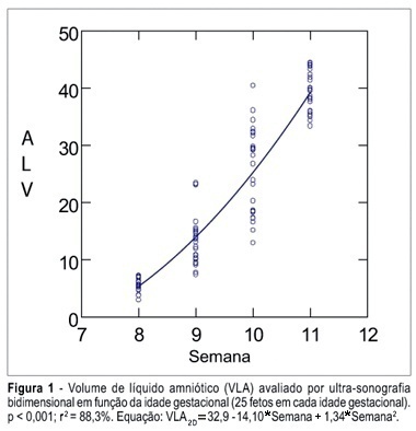
PURPOSE: To determine the values of amniotic fluid in normal fetuses during the first trimester of pregnancy by three- and bi-dimensional ultrasonography. METHODS: In a prospective longitudinal study, 25 normal fetuses were evaluated from the 8th to the 11th week of gestation. Amniotic fluid volume was measured by endovaginal ultrasonography with the three- and two-dimensional modes. The two-dimensional study consisted of volumetric determination by mathematical calculation based on an ellipsoidal shape (constant 0.52) to obtain the amniotic sac and embryo volumes. In the three-dimensional study, the amniotic fluid volume was determined by the VOCAL technique using 6, 9, 15, and 30 degrees of rotation. The amniotic fluid volume obtained by 6-degree rotations was considered to be the final result. In both modes, amniotic fluid volume was obtained by subtracting the volume of the embryo from the volume of the amniotic sac. Data were analyzed statistically for variance (ANOVA), correlation and regression analysis. The level of significance was set at p < 0.05. RESULTS: The amniotic fluid volume as measured by two-dimensional ultrasonography increased from 5.45 to 39.52 cm³ in the range from the 8th to the 11th week (ANOVA - p < 0.05). There was a correlation between gestational age and amniotic fluid volume (p < 0.001, r² = 88.3%). In the three-dimensional study, the amniotic fluid volume increased from 5.7 to 42.9 cm³ in the range from the 8th to the 11th week (ANOVA - p < 0.05), and again a correlation between gestational age and amniotic fluid volume (p < 0.001, r² = 98.1%) was observed. CONCLUSION: an increase in amniotic fluid volume occurs during the first trimester of pregnancy, as determined by the two- and three-dimensional modes. In addition, we have demonstrated that the higher the gestational age, the larger the amniotic fluid volume.
Summary
Revista Brasileira de Ginecologia e Obstetrícia. 2006;28(10):575-580
DOI 10.1590/S0100-72032006001000002
PURPOSE: To determine the values of amniotic fluid in normal fetuses during the first trimester of pregnancy by three- and bi-dimensional ultrasonography. METHODS: In a prospective longitudinal study, 25 normal fetuses were evaluated from the 8th to the 11th week of gestation. Amniotic fluid volume was measured by endovaginal ultrasonography with the three- and two-dimensional modes. The two-dimensional study consisted of volumetric determination by mathematical calculation based on an ellipsoidal shape (constant 0.52) to obtain the amniotic sac and embryo volumes. In the three-dimensional study, the amniotic fluid volume was determined by the VOCAL technique using 6, 9, 15, and 30 degrees of rotation. The amniotic fluid volume obtained by 6-degree rotations was considered to be the final result. In both modes, amniotic fluid volume was obtained by subtracting the volume of the embryo from the volume of the amniotic sac. Data were analyzed statistically for variance (ANOVA), correlation and regression analysis. The level of significance was set at p < 0.05. RESULTS: The amniotic fluid volume as measured by two-dimensional ultrasonography increased from 5.45 to 39.52 cm³ in the range from the 8th to the 11th week (ANOVA - p < 0.05). There was a correlation between gestational age and amniotic fluid volume (p < 0.001, r² = 88.3%). In the three-dimensional study, the amniotic fluid volume increased from 5.7 to 42.9 cm³ in the range from the 8th to the 11th week (ANOVA - p < 0.05), and again a correlation between gestational age and amniotic fluid volume (p < 0.001, r² = 98.1%) was observed. CONCLUSION: an increase in amniotic fluid volume occurs during the first trimester of pregnancy, as determined by the two- and three-dimensional modes. In addition, we have demonstrated that the higher the gestational age, the larger the amniotic fluid volume.
Summary
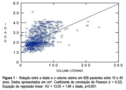
Revista Brasileira de Ginecologia e Obstetrícia. 2003;25(9):673-678
DOI 10.1590/S0100-72032003000900009
PURPOSE: to evaluate the uterine volume in women between 10 and 40 years in order to observe if the uterine volume in adolescents is smaller than the uterine volume in women between 20 and 40 years. We intend to emphasize the differences between the uterine volume of adolescents and that of adult women and to correlate with the immaturity of the genital tract of adolescents regarding gestation and delivery. METHOD: a cross-sectional study, which included 828 patients between 10 and 40 years old divided into two groups and examined using abdominal ultrasound to obtain the uterine volume measure. The first group consisted of 477 (57.7%) adolescents, and the second group of 351 (42.3%) adult women between 20 and 40 years old. In the adolescent group, ultrasound examination was performed by a single observer and in the group of adult women ultrasound examination was performed by a group of observers who used the same methodology as that of group 1. Image Point HX (Hewlett Packard) and Hitachi 525 ultrasound equipment were used with a multiple frequency probe. For the calculation of the uterine volume we used the longitudinal diameter (LD), anteroposterior diameter (APD) and transverse diameter (TD) with the (LD x APD x TD) x 0.45 formula. RESULTS: adolescents aged 10 to 17 years had a smaller uterine volume than women aged 20 to 40 years (p<0.05). Adolescents who delivered twice had a uterine volume similar to that of the patients between 20 and 40 years old with respective mean values of 62.6 ± 20.6 and 69.0±22.9 (p>0.05). CONCLUSION: adolescents less than 18 years old or primiparous have a smaller uterine volume than women between 20 to 40 years old. However, adolescents aged 18 years or older, or secundipara, have a uterine volume similar to that of women aged 20 to 40 years.
Summary
Revista Brasileira de Ginecologia e Obstetrícia. 2003;25(9):673-678
DOI 10.1590/S0100-72032003000900009
PURPOSE: to evaluate the uterine volume in women between 10 and 40 years in order to observe if the uterine volume in adolescents is smaller than the uterine volume in women between 20 and 40 years. We intend to emphasize the differences between the uterine volume of adolescents and that of adult women and to correlate with the immaturity of the genital tract of adolescents regarding gestation and delivery. METHOD: a cross-sectional study, which included 828 patients between 10 and 40 years old divided into two groups and examined using abdominal ultrasound to obtain the uterine volume measure. The first group consisted of 477 (57.7%) adolescents, and the second group of 351 (42.3%) adult women between 20 and 40 years old. In the adolescent group, ultrasound examination was performed by a single observer and in the group of adult women ultrasound examination was performed by a group of observers who used the same methodology as that of group 1. Image Point HX (Hewlett Packard) and Hitachi 525 ultrasound equipment were used with a multiple frequency probe. For the calculation of the uterine volume we used the longitudinal diameter (LD), anteroposterior diameter (APD) and transverse diameter (TD) with the (LD x APD x TD) x 0.45 formula. RESULTS: adolescents aged 10 to 17 years had a smaller uterine volume than women aged 20 to 40 years (p<0.05). Adolescents who delivered twice had a uterine volume similar to that of the patients between 20 and 40 years old with respective mean values of 62.6 ± 20.6 and 69.0±22.9 (p>0.05). CONCLUSION: adolescents less than 18 years old or primiparous have a smaller uterine volume than women between 20 to 40 years old. However, adolescents aged 18 years or older, or secundipara, have a uterine volume similar to that of women aged 20 to 40 years.