Summary
Revista Brasileira de Ginecologia e Obstetrícia. 2021;43(4):334-338
Summary
Revista Brasileira de Ginecologia e Obstetrícia. 2021;43(4):334-338
Summary
Revista Brasileira de Ginecologia e Obstetrícia. 2021;43(4):334-338
Summary
Revista Brasileira de Ginecologia e Obstetrícia. 2008;30(7):335-340
DOI 10.1590/S0100-72032008000700003
PURPOSE: to evaluate the effect of exposure of female rats to therapeutic ultrasound in the pre-implantation phase. METHODS: pregnant Wistar female rats have been exposed to 3 MHz, 0.6 W/cm² ultrasound, pulsatile ultrasound (PUS) or continuous ultrasound (CUS), and controls, unplugged ultrasound (UUS), for five minutes. The rats were sacrificed at the 20th day post-insemination. Biochemical and hematological analyses have been done. Animals have been submitted to necropsy in order to identify lesions of internal organs, and to remove and weight the liver, kidneys and ovaries. Alive, malformed, dead and reabsorbed fetuses have been counted. RESULTS: the rats have not presented changes in their body and organs weight, and neither in their reproductive capacity, but there has been an increase in triglycerides in the PUS and CUS groups, when compared to the UUS group. The fetuses' relative weights of the heart (0.7 ± 0.9), liver (9.8 ± 0.8), kidneys (6.2 ± 0.8) and lungs (3.8 ± 0.4) increased in the CUS, when compared to the heart (0.7 ± 0.9), liver (9.8 ± 0.8), kidneys (6.2 ± 0.8) e lungs (3.8 ± 0.4) of the UUS. CONCLUSIONS: in the experimental model, the therapeutic ultrasound used has not caused meaningful maternal toxicity. Pulsatile waves have not changed fetal morphology, but continuous waves have caused increase in the relative weight of the fetuses' heart, liver, lungs and kidneys.
Summary
Revista Brasileira de Ginecologia e Obstetrícia. 2007;29(7):335-339
DOI 10.1590/S0100-72032007000700002
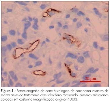
PURPOSE: to evaluate the effect of raloxifene on breast cancer angiogenesis of menopausal women. METHODS: sixteen menopausal women with stage II (>3 cm) estrogen receptor positive operable breast cancer were enrolled in this study. Following confirmation of the diagnosis by incisional biopsy, the patients received 60 mg raloxifene daily for 28 days prior to the definitive surgery. Immunohistochemical study was performed on the sample tumors obtained during the biopsy for the diagnosis and evaluation of the status of estrogen receptor and during the definitive surgery. The anti-CD34 monoclonal antibody was used as a marker for endothelial cells. The vascular unit was considered as any endothelial cell or group of cells of a brownish color, clearly separated from adjacent microvessels, tumor cells or other connective tissue, forming or not lumen. Microvessel count was performed in ten fields of each slide using a 40X objective lens (400X magnification). A microscope coupled to a system of capture and analysis of image was used (Imagelab®). Statistical analysis of data was carried out using the paired Student t-test and significance level was established at p<0.05. RESULTS: mean numbers of anti-CD34 antibody-stained microvessels before and after raloxifene treatment were 44.4±3.5 and 22.6±1.6, respectively. A significant reduction in the number of microvessels following raloxifene therapy was observed (p<0.001). CONCLUSIONS: when administered as primary therapy for menopausal women with breast carcinoma, raloxifene significantly reduced tumoral angiogenesis.
Summary
Revista Brasileira de Ginecologia e Obstetrícia. 2007;29(7):335-339
DOI 10.1590/S0100-72032007000700002
PURPOSE: to evaluate the effect of raloxifene on breast cancer angiogenesis of menopausal women. METHODS: sixteen menopausal women with stage II (>3 cm) estrogen receptor positive operable breast cancer were enrolled in this study. Following confirmation of the diagnosis by incisional biopsy, the patients received 60 mg raloxifene daily for 28 days prior to the definitive surgery. Immunohistochemical study was performed on the sample tumors obtained during the biopsy for the diagnosis and evaluation of the status of estrogen receptor and during the definitive surgery. The anti-CD34 monoclonal antibody was used as a marker for endothelial cells. The vascular unit was considered as any endothelial cell or group of cells of a brownish color, clearly separated from adjacent microvessels, tumor cells or other connective tissue, forming or not lumen. Microvessel count was performed in ten fields of each slide using a 40X objective lens (400X magnification). A microscope coupled to a system of capture and analysis of image was used (Imagelab®). Statistical analysis of data was carried out using the paired Student t-test and significance level was established at p<0.05. RESULTS: mean numbers of anti-CD34 antibody-stained microvessels before and after raloxifene treatment were 44.4±3.5 and 22.6±1.6, respectively. A significant reduction in the number of microvessels following raloxifene therapy was observed (p<0.001). CONCLUSIONS: when administered as primary therapy for menopausal women with breast carcinoma, raloxifene significantly reduced tumoral angiogenesis.
Summary
Revista Brasileira de Ginecologia e Obstetrícia. 2012;34(7):335-342
DOI 10.1590/S0100-72032012000700008
PURPOSE: To evaluate whether climacteric women undergoing liver transplantation had higher prevalence of decreased bone mass than those without any liver disease. METHODS: A cross-sectional study with 48 women receiving follow-up care at a university hospital in Southeastern Brazil, from February 4th 2009 to January 5th 2011, was conducted. Of these women, 24 were 35 years or older and had undergone liver transplantation at least one year before study entry. The remaining 24 women had no liver disease and their ages and menstrual patterns were similar to those of transplanted patients. Laboratorial tests (follicle-stimulating hormone and estradiol) and bone density measurements of the lumbar spine and femur (equipment Hologic, Discovery WI) were performed. Statistical analysis was carried out by Fisher's exact test, simple Odds Ratio (OR), and multiple logistic regression. RESULTS: Mean age of the women included in the study was 52.8 (±10.7) years-old, 27.1% were premenopausal and 72.9% were peri/postmenopausal. Approximately 14.6% of these women exhibited osteoporosis and 35.4% had low bone mass. The following items were associated with decreased bone mass: being postmenopausal (OR=71.4; 95%CI 3.8 - 1,339.7; p<0.0001), current age over 49 years-old (OR=11.4; 95%CI 2.9 - 44.0; p=0.0002), and serum estradiol levels lower than 44.5 pg/mL (OR=18.3; 95%CI 3.4 - 97.0; p<0.0001). Having a history of liver transplantation was not associated with decreased bone mass (OR=1.4; 95%CI 0.4 - 4.3; p=0.56). CONCLUSION: Liver transplantation was not associated with decreased bone mass in this group of climacteric women.
Summary
Revista Brasileira de Ginecologia e Obstetrícia. 1998;20(6):335-341
DOI 10.1590/S0100-72031998000600006
Objective: to analyze maternal and fetal folate status in cases of neural tube defects (NTD). Methods: a case-control study was designed with 14 cases of fetuses with neural tube defects (study group) and 14 cases of fetuses with other unrelated malformations (control group) gestational age matched, in low-risk pregnant women. Both total and methylated folic acid levels in fetal and maternal compartments using serum and tissular (red blood cells) concentrations and also average corpuscular volume, hematocrit and hemoglobin levels were determined. Fetal and maternal samples were obtained immediately before termination of pregnancy. Results in both groups were compared using a gestational age paired t-test. Results: there were no statistically significant differences in fetal folate levels and fetal hematologic parameters between both groups However, both total (239.9 ng/mL in NTD against 399.1 ng/mL in control group, p=0.01) and methylated (201.9 ng/mL in NTD against 314.0 ng/mL in control group, p=0.02) maternal red blood cells folate levels were significantly lower in the neural tube defect group. Maternal serum folate levels were similar in study and control groups. Conclusion: this study showed that maternal red blood cell folate but not serum folate was significantly reduced in mothers of fetuses with neural tube defects.
Summary
Revista Brasileira de Ginecologia e Obstetrícia. 2009;31(7):335-341
DOI 10.1590/S0100-72032009000700003
PURPOSE: to describe alcohol and tobacco use in adult pregnant women and determine its association with the obstetric outcome. METHODS: analytical transversal study, in which 433 adult pregnant women and their newborns have been included, attended at a public maternity hospital in Rio de Janeiro, from 1999 to 2006. Information on the mother and the newborn was collected at the moment of delivery and during puerperium through an interview and inspection of the medical records. "Use of alcohol during gestation" and "use of cigarette during gestation" have been considered when detected at any gestational age and written down on the medical record. RESULTS: it was observed that 5.5 and 7.7% of the pregnant women reported cigarette and alcohol use during gestation, respectively. Maternal features related to tobacco use during pregnancy were marital status (p=0.005), age (p=0.01) and pre-natal nutritional guidance (p=0.003). Tobacco use during pregnancy has been strongly associated with alcohol use, 31.3% of the women reporting concomitant use of both substances (p<0.05). No association between alcohol or tobacco use during gestation and obstetric outcome (gestational age, newborn weight at birth and newborn medical conditions; p>0.05) has been detected. CONCLUSIONS: these results suggest that tobacco and alcohol use should be investigated during pre-natal care among all women, particularly single women, over 35 years old, with history of miscarriage, and with unwanted pregnancy. Nutritional guidance had a protective effect against tobacco use during gestation, and thus pregnant women should be informed as to the harmful effects of substance use to ensure better obstetric outcome.
Summary
Revista Brasileira de Ginecologia e Obstetrícia. 2014;36(8):335-339
Summary
Revista Brasileira de Ginecologia e Obstetrícia. 2014;36(8):335-339