You searched for:"Régis Resende Paulinelli"
We found (9) results for your search.Summary
Revista Brasileira de Ginecologia e Obstetrícia. 2007;29(12)
Summary
Revista Brasileira de Ginecologia e Obstetrícia. 2007;29(12)
Summary
Revista Brasileira de Ginecologia e Obstetrícia. 2024;46:e-rbgo15
To compare the medical image interpretation's time between the conventional and automated methods of breast ultrasound in patients with breast lesions. Secondarily, to evaluate the agreement between the two methods and interobservers.
This is a cross-sectional study with prospective data collection. The agreement's degrees were established in relation to the breast lesions's ultrasound descriptors. To determine the accuracy of each method, a biopsy of suspicious lesions was performed, considering the histopathological result as the diagnostic gold standard.
We evaluated 27 women. Conventional ultrasound used an average medical time of 10.77 minutes (± 2.55) greater than the average of 7.38 minutes (± 2.06) for automated ultrasound (p<0.001). The degrees of agreement between the methods ranged from 0.75 to 0.95 for researcher 1 and from 0.71 to 0.98 for researcher 2. Among the researchers, the degrees of agreement were between 0.63 and 1 for automated ultrasound and between 0.68 and 1 for conventional ultrasound. The area of the ROC curve for the conventional method was 0.67 (p=0.003) for researcher 1 and 0.72 (p<0.001) for researcher 2. The area of the ROC curve for the automated method was 0. 69 (p=0.001) for researcher 1 and 0.78 (p<0.001) for researcher 2.
We observed less time devoted by the physician to automated ultrasound compared to conventional ultrasound, maintaining accuracy. There was substantial or strong to perfect interobserver agreement and substantial or strong to almost perfect agreement between the methods.
Summary
Revista Brasileira de Ginecologia e Obstetrícia. 1999;21(3):133-137
DOI 10.1590/S0100-72031999000300003
Purpose: to evaluate how knowledgeable medical students at the Universidade Federal de Goiás were concerning the basic diagnostic principles breast cancer. The study also aimed at promoting a debate among the students and at assessing the understanding of the students in the fifth year of medical school, who had already attended the Gynecology course. Methods: Through questionnaires given to 348 individuals, from the first to the fifth year, out of a total population of 550 students, the authors searched for information with regard to basic knowledge on the diagnosis of breast cancer. Of the 348 questionnaires, 55 (16%) were given to fifth-year students, who had already attended the Gynecology course. Furthermore, 43% of the students were women, 62% had medical doctors in their immediate family, and 17% had a family history of breast cancer. Results: in regard to the knowledge of diagnostic methods, 84% of the students were aware of the most frequent sign of breast cancer, 34% knew which was the best screening method, 49% knew when to refer asymptomatic women to mammography, 37% knew the recommended interval between mammography for women above the age of 50, and 24% knew when to associate ultrasound to mammography for the detection of breast cancer. The fifth-year students provided correct answers at a significantly higher rate, when compared to the others. Concerning gender, the only difference regarded the fact that women showed a better knowledge as to the best time for self-examination and when to recommend ultrasound associated with mammography. The presence of medical doctors in the family and a history of family members with breast cancer did not have any influence on the answers. Conclusion: The lack of information in regard to the diagnosis of breast cancer is very high, even among medical students. Nevertheless, the rate of information increases significantly after students are taught Gynecology, which is only offered during the fifth year of the medical school.
Summary
Revista Brasileira de Ginecologia e Obstetrícia. 2002;24(3):195-199
DOI 10.1590/S0100-72032002000300008
Purpose: to evaluate, in a prospective way, the importance of ultrasound features of solid breast lesions in the differentiation between benign and malignant lumps. Methods: one hundred and forty-two patients with solid breast lesions, from the Department of Gynecology and Obstetrics of the Federal University of Goias (Brazil), were included in the trial. All ultrasound examinations were performed by a training doctor, always supervised by an experienced professional. The characteristics of the lesions studied were: shape, retrotumoral echoes, internal echoes, oriented diameter, halo of bright echoes and Cooper ligaments. Each of the ultrasound features was compared to the results of the histological examination. Results: among the 142 patients included in the trial, 90 (63%) had their lesions excised, and 77 (86%) had pathologic diagnoses of benign tumors and 13 (14%) of malignant tumors. The following characteristics were statistically significant in the diagnosis of the breast cancer (c²): masses with retrotumoral shadowing (p=0.0001), irregular shape (p=0.0007), heterogeneous internal echoes (p=0.0015) and vertically oriented - taller than wide (p<0.0001). The presence of halo of bright echoes anterior to the lump and the presence of wider Cooper ligaments were not related to the correct diagnosis of malignancy in this trial. Conclusion: ultrasound is a diagnostic method that can help physicians between the differentiation of benign and malignant breast lumps. The presence of retrotumoral shadowing, irregular shape, heterogeneous internal echoes and vertical orientation - lesions taller than wide - were related to the pathologic diagnosis of breast malignancies.
Summary
Revista Brasileira de Ginecologia e Obstetrícia. 1999;21(5):287-290
DOI 10.1590/S0100-72031999000500007
Purpose: to evaluate the knowledge and practice of breast self-examination among medical students and to determine possible factors associated with this practice. Method: the authors used a questionnaire to gather information about the students and their knowledge of this self-examination. This questionnaire also allowed the authors to verify the frequency with which the female students performed breast self-examination. The chi² test and Student's "t" test were used, when applicable, to check the association of certain factors. Results: of the 348 questionnaires which were answered, 16% (55) were submitted by 5th year medical students, who had already attended the Gynecology course; 43% were answered by females, 62% of the students had medical doctors among their relatives, and 17% had a family history of breast cancer. In terms of breast self-examination, 95% knew about the method. Of the 149 females who answered the questionnaire, only 64% checked their breasts regularly. The reasons given for not performing self-examination varied: 24% considered themselves to be too young, 4% thought they would not have cancer, 9% listed fear as the reason, 19% reported they were too lazy, and 44% of the female students had no clear reason for not performing breast self-examination. Neither the knowledge nor the practice of the breast self-examination were associated with the subjects the students had or had not yet taken in medical school, with a family history of breast cancer or with the fact that one or more relatives were medical doctors. Conclusion: breast self-examination is known by practically all the medical students; nevertheless, only one third of the female students performed it regularly. This fact highlights the importance of emphasizing breast self-examination among medical students, so that they can help to disseminate this practice among the general population, rather than delegating this responsibility to the midia.
Summary
Revista Brasileira de Ginecologia e Obstetrícia. 2005;27(5):294-295
Summary
Revista Brasileira de Ginecologia e Obstetrícia. 2005;27(5):294-295
Summary
Revista Brasileira de Ginecologia e Obstetrícia. 2000;22(5):307-310
DOI 10.1590/S0100-72032000000500009
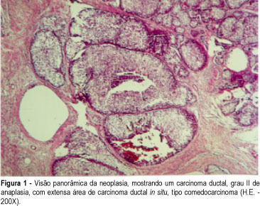
Polymastia is a usual problem in Mastology clinics and the possibility of cancer must be taken into consideration, as much as in any other mammary tissue. In the present study the case of a 48-year-old patient, submitted to the excision of the left axillary breast for cosmetic purposes is reported. The histological examination showed an invasive ductal carcinoma with an extensive in situ component. The patient was submitted to a wide excision plus axillary lymphadenectomy and radiation therapy. The frequency, diagnosis, prognosis and treatment of cancer in supernumerary breasts is also reviewed.
Summary
Revista Brasileira de Ginecologia e Obstetrícia. 2000;22(5):307-310
DOI 10.1590/S0100-72032000000500009
Polymastia is a usual problem in Mastology clinics and the possibility of cancer must be taken into consideration, as much as in any other mammary tissue. In the present study the case of a 48-year-old patient, submitted to the excision of the left axillary breast for cosmetic purposes is reported. The histological examination showed an invasive ductal carcinoma with an extensive in situ component. The patient was submitted to a wide excision plus axillary lymphadenectomy and radiation therapy. The frequency, diagnosis, prognosis and treatment of cancer in supernumerary breasts is also reviewed.
Summary
Revista Brasileira de Ginecologia e Obstetrícia. 2023;45(7):409-414
In this integrative review, we aimed to describe the records of time devoted by physicians to breast ultrasound in a review of articles in the literature, in order to observe whether the automation of the method enabled a reduction in these values. We selected articles from the Latin American and Caribbean Literature in Health Sciences (LILACS) and MEDLINE databases, through Virtual Health Library (BVS), SciELO (Scientific Electronic Library Online), PubMed, and Scopus. We obtained 561 articles, and, after excluding duplicates and screening procedures, 9 were selected, whose main information related to the guiding question of the research was synthesized and analyzed. It was concluded that the automation of breast ultrasound represents a possible strategy for optimization of the medical time dedicated to the method, but this needs to be better evaluated in comparative studies between both methods (traditional and automated), with methodology directed to the specific investigation of this potentiality.