Summary
Rev Bras Ginecol Obstet. 2003;25(4):283-288
DOI 10.1590/S0100-72032003000400010
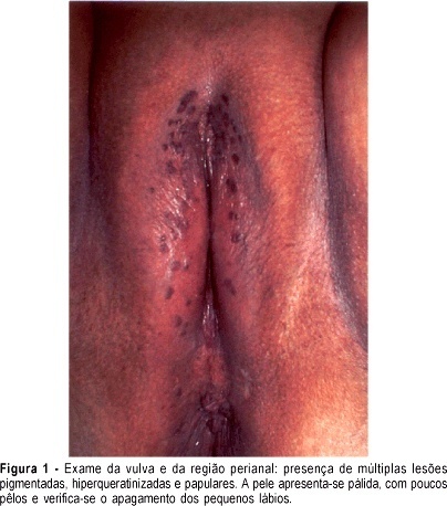
High-grade vulvar intraepithelial neoplasia (VIN III) is a visible lesion; therefore, it is accessible to biopsy and thus, to a histological diagnosis. There are two forms of vulvar cancer precursors: VIN caused by human papillomavirus (HPV) and VIN associated with untreated lichen simplex chronicus, squamous cell hyperplasia, and lichen sclerosus. There may be overlap of the two forms. The term bowenoid papulosis, although discouraged, identifics a clinical form of VIN III. Such lesion appears as pigmented, wart-like growths or papules. VIN III is associated with HPV in more than 80% of the cases, and there is perianal involvement in 40% of the times. Vulvar intraepithelial neoplasia is difficult to cure and relapses can occur at any time for many years. Although there is no defined standard treatment, studies point to surgery, respecting a free margin, as the most adequate one.
Summary
Rev Bras Ginecol Obstet. 2003;25(4):283-288
DOI 10.1590/S0100-72032003000400010
High-grade vulvar intraepithelial neoplasia (VIN III) is a visible lesion; therefore, it is accessible to biopsy and thus, to a histological diagnosis. There are two forms of vulvar cancer precursors: VIN caused by human papillomavirus (HPV) and VIN associated with untreated lichen simplex chronicus, squamous cell hyperplasia, and lichen sclerosus. There may be overlap of the two forms. The term bowenoid papulosis, although discouraged, identifics a clinical form of VIN III. Such lesion appears as pigmented, wart-like growths or papules. VIN III is associated with HPV in more than 80% of the cases, and there is perianal involvement in 40% of the times. Vulvar intraepithelial neoplasia is difficult to cure and relapses can occur at any time for many years. Although there is no defined standard treatment, studies point to surgery, respecting a free margin, as the most adequate one.
Summary
Rev Bras Ginecol Obstet. 2003;25(4):291-295
DOI 10.1590/S0100-72032003000400011
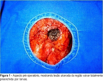
Myiasis located in the vulva is a rarely described disease. The objective of the present report is to describe a case of vulvar myiasis due to larvae of Cochliomyia hominivorax. A 77-year-old woman with precarious hygienic habits presented pain, pruritus and secretions with a fetid smell in the genital region for 10 days. Gynecological examination demonstrated an ulcerated lesion in the labium majus of the vulva measuring six centimeters that extended to the mons pubis and was found to be filled with larvae. The patient progressed favorably after removal of the larvae, surgical debridement and daily dressings. Fourteen days after the debridement, she was submitted to skin flap rotation, with good local scar formation. Two months after the intervention, she remained asymptomatic. Vulvar myiasis must be considered in the differential diagnosis of infectious diseases of the vulva in patients with precarious hygienic habits.
Summary
Rev Bras Ginecol Obstet. 2003;25(4):291-295
DOI 10.1590/S0100-72032003000400011
Myiasis located in the vulva is a rarely described disease. The objective of the present report is to describe a case of vulvar myiasis due to larvae of Cochliomyia hominivorax. A 77-year-old woman with precarious hygienic habits presented pain, pruritus and secretions with a fetid smell in the genital region for 10 days. Gynecological examination demonstrated an ulcerated lesion in the labium majus of the vulva measuring six centimeters that extended to the mons pubis and was found to be filled with larvae. The patient progressed favorably after removal of the larvae, surgical debridement and daily dressings. Fourteen days after the debridement, she was submitted to skin flap rotation, with good local scar formation. Two months after the intervention, she remained asymptomatic. Vulvar myiasis must be considered in the differential diagnosis of infectious diseases of the vulva in patients with precarious hygienic habits.