You searched for:"Sergio Britto Garcia"
We found (3) results for your search.Summary
Revista Brasileira de Ginecologia e Obstetrícia. 2019;41(11):668-672
To analyze the effect of thalidomide on the progression of endometriotic lesions experimentally induced in rats and to characterize the pattern of cell proliferation by immunohistochemical Proliferating Cell Nuclear Antigen (PCNA) labeling of eutopic and ectopic endometrium.
Fifteen female Wistar rats underwent laparotomy for endometriosis induction by resection of one uterine horn, isolation of the endometrium and fixation of a tissue segment to the pelvic peritoneum. Four weeks after, the animals were divided into 3 groups: control (I), 10mg/kg/day (II) and 1mg/kg/day (III) intraperitoneal thalidomide for 10 days. The lesion was excised together with the opposite uterine horn for endometrial gland and stroma analysis. Eutopic and ectopic endometrial tissue was submitted to immunohistochemistry for analysis of cell proliferation by PCNA labeling and the cell proliferation index (CPI) was calculated as the number of labeled cells per 1,000 cells.
Group I showed a mean CPI of 0.248 ± 0.0513 in the gland and of 0.178 ± 0.046 in the stroma. In contrast, Groups II and III showed a significantly lower CPI, that is, 0.088 ± 0.009 and 0.080 ± 0.021 for the gland (p < 0.001) and 0.0945 ± 0.0066 and 0.075 ± 0.018 for the stroma (p < 0.001), respectively. Also, the mean lesion area of Group I was 69.2mm2, a significantly higher value compared with Group II (49.4mm2, p = 0.023) and Group III (48.6mm2, p = 0.006). No significant difference was observed between Groups II and III.
Thalidomide proved to be effective in reducing the lesion area and CPI of the experimental endometriosis implants both at the dose of 1mg/kg/day and at the dose of 10 mg/kg/day.
Summary
Revista Brasileira de Ginecologia e Obstetrícia. 2018;40(11):705-712
To characterize the patterns of cell differentiation, proliferation, and tissue invasion in eutopic and ectopic endometrium of rabbits with induced endometriotic lesions via a well- known experimental model, 4 and 8 weeks after the endometrial implantation procedure.
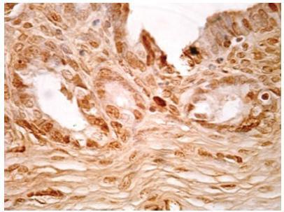
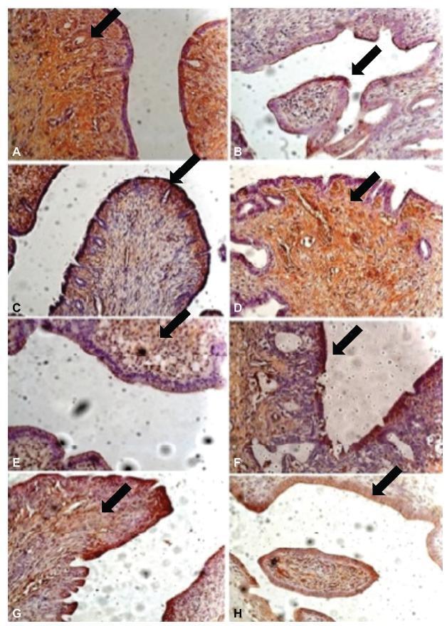
Twenty-nine female New Zealand rabbits underwent laparotomy for endometriosis induction through the resection of one uterine horn, isolation of the endometrium, and fixation of tissue segment to the pelvic peritoneum. Two groups of animals (one with 14 animals, and the other with15) were sacrificed 4 and 8 weeks after endometriosis induction. The lesion was excised along with the opposite uterine horn for endometrial gland and stroma determination. Immunohistochemical reactions were performed in eutopic and ectopic endometrial tissues for analysis of the following markers: metalloprotease (MMP-9) and tissue inhibitor of metalloprotease (TIMP-2), which are involved in the invasive capacity of the endometrial tissue; and metallothionein (MT) and p63, which are involved in cell differentiation and proliferation.
The intensity of the immunostaining for MMP9, TIMP-2, MT, and p63 was higher in ectopic endometria than in eutopic endometria. However, when the ectopic lesions were compared at 4 and 8 weeks, no significant difference was observed, with the exception of the marker p63, which was more evident after 8 weeks of evolution of the ectopic endometrial tissue.
Ectopic endometrial lesions seem to express greater power for cell differentiation and tissue invasion, compared with eutopic endometria, demonstrating a potentially invasive, progressive, and heterogeneous presentation of endometriosis.
Summary
Revista Brasileira de Ginecologia e Obstetrícia. 2018;40(11):705-712
To characterize the patterns of cell differentiation, proliferation, and tissue invasion in eutopic and ectopic endometrium of rabbits with induced endometriotic lesions via a well- known experimental model, 4 and 8 weeks after the endometrial implantation procedure.
Twenty-nine female New Zealand rabbits underwent laparotomy for endometriosis induction through the resection of one uterine horn, isolation of the endometrium, and fixation of tissue segment to the pelvic peritoneum. Two groups of animals (one with 14 animals, and the other with15) were sacrificed 4 and 8 weeks after endometriosis induction. The lesion was excised along with the opposite uterine horn for endometrial gland and stroma determination. Immunohistochemical reactions were performed in eutopic and ectopic endometrial tissues for analysis of the following markers: metalloprotease (MMP-9) and tissue inhibitor of metalloprotease (TIMP-2), which are involved in the invasive capacity of the endometrial tissue; and metallothionein (MT) and p63, which are involved in cell differentiation and proliferation.
The intensity of the immunostaining for MMP9, TIMP-2, MT, and p63 was higher in ectopic endometria than in eutopic endometria. However, when the ectopic lesions were compared at 4 and 8 weeks, no significant difference was observed, with the exception of the marker p63, which was more evident after 8 weeks of evolution of the ectopic endometrial tissue.
Ectopic endometrial lesions seem to express greater power for cell differentiation and tissue invasion, compared with eutopic endometria, demonstrating a potentially invasive, progressive, and heterogeneous presentation of endometriosis.
Summary
Revista Brasileira de Ginecologia e Obstetrícia. 2004;26(9):715-719
DOI 10.1590/S0100-72032004000900007
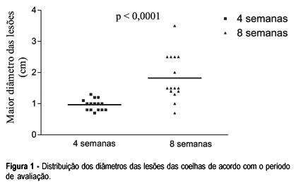
PURPOSE: development of a new experimental model of endometriosis induction in rabbits evaluating its temporal evolution both macro-and microscopically. METHODS: thirty female rabbits were submitted to endometriosis induction through the fixation of a piece of the left uterine horn to the abdominal peritoneum. After four or eight weeks the viability of the lesions was verified by laparoscopy. The lesions were observed endoscopically. The implants were measured and histological analyses were made. The groups were compared for the presence of endometriotic lesion on laparoscopy, presence of adhesions, implant size and histological aspects. For statistical analyses we utilized Student's t and Mann-Whitney's tests, with a statistical significance of 5%. RESULTS: endometriotic lesions were identified in all cases submitted to laparoscopy after 4 weeks of induction, 64% of them cystic, and in 80% of the rabbits after eight weeks, 66% of which cystic. The adhesions were present in 71% of the rabbits after 4 weeks (none in the implants) and in 80% of the rabbits after 8 weeks (13% in the implants). The lesions were significantly larger after 8 weeks (p<0,0001). The histological analyses showed 100% of endometrial tissue in both groups. CONCLUSION: this experimental model showed that it is possible to simulate endometriosis in rabbits with a viable and simple technique, also allowing to record the characteristics and development of the implants macro-and microscopically. Although the histological aspects were similar, the lesions after eight weeks were larger than after four, making their manipulation easier.
Summary
Revista Brasileira de Ginecologia e Obstetrícia. 2004;26(9):715-719
DOI 10.1590/S0100-72032004000900007
PURPOSE: development of a new experimental model of endometriosis induction in rabbits evaluating its temporal evolution both macro-and microscopically. METHODS: thirty female rabbits were submitted to endometriosis induction through the fixation of a piece of the left uterine horn to the abdominal peritoneum. After four or eight weeks the viability of the lesions was verified by laparoscopy. The lesions were observed endoscopically. The implants were measured and histological analyses were made. The groups were compared for the presence of endometriotic lesion on laparoscopy, presence of adhesions, implant size and histological aspects. For statistical analyses we utilized Student's t and Mann-Whitney's tests, with a statistical significance of 5%. RESULTS: endometriotic lesions were identified in all cases submitted to laparoscopy after 4 weeks of induction, 64% of them cystic, and in 80% of the rabbits after eight weeks, 66% of which cystic. The adhesions were present in 71% of the rabbits after 4 weeks (none in the implants) and in 80% of the rabbits after 8 weeks (13% in the implants). The lesions were significantly larger after 8 weeks (p<0,0001). The histological analyses showed 100% of endometrial tissue in both groups. CONCLUSION: this experimental model showed that it is possible to simulate endometriosis in rabbits with a viable and simple technique, also allowing to record the characteristics and development of the implants macro-and microscopically. Although the histological aspects were similar, the lesions after eight weeks were larger than after four, making their manipulation easier.