You searched for:"Carlos Eduardo Bacchi"
We found (2) results for your search.Summary
Revista Brasileira de Ginecologia e Obstetrícia. 2016;38(10):512-517
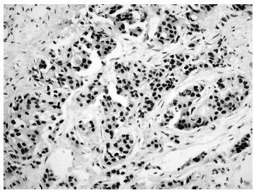
Triple-negative breast carcinomas (TNBCs) represent a heterogeneous group of neoplasias, even though they generally exhibit a clinically more aggressive phenotype, and are more prevalent in young women. To date, targeted therapies for this group of tumors have not been defined. The aim of this study was to evaluate the frequency of the apocrine subtype in TBNCs from premenopausal patients as defined by the immunohistochemical expression of the androgen receptor (AR) and its association with: histological type; tumor grade; proliferative activity; epidermal growth factor receptor (EGFR) expression; and a basal-like phenotype.
A total of 118 tumor samples from patients aged 45 years or younger were selected and reviewed according to histological type and grade. Ki-67 expression was also evaluated. Immunohistochemical expression of the AR, basal cytokeratin ⅚, and EGFR expression were analyzed in tissue microarrays. The apocrine subset was defined by AR-positive expression. The basal-like phenotype was characterized by cytokeratin ⅚ and/or EGFR expression.
An apocrine profile was identified in 6/118 (5.1%) cases. This subset of cases also exhibited a lower rate of Ki-67 expression (17.5% versus 70.0%, p= 0.02), and a trend toward a lower histological grade (66.7% versus 27.9%, p= 0.06).
The apocrine subtype of TNBCs is rare among premenopausal women, and it tends to present as carcinomas of lower grade and lower proliferative activity, suggesting a less aggressive biological phenotype.

Summary
Revista Brasileira de Ginecologia e Obstetrícia. 2014;36(8):340-346
DOI 10.1509/SO100-720320140005034
To examine the expression of AKT and PTEN in a series of HER2-positive primary invasive breast tumors using immunohistochemistry, and to associate these expression profiles with classic pathologic features such as tumor grade, hormone receptor expression, lymphatic vascular invasion, and proliferation.
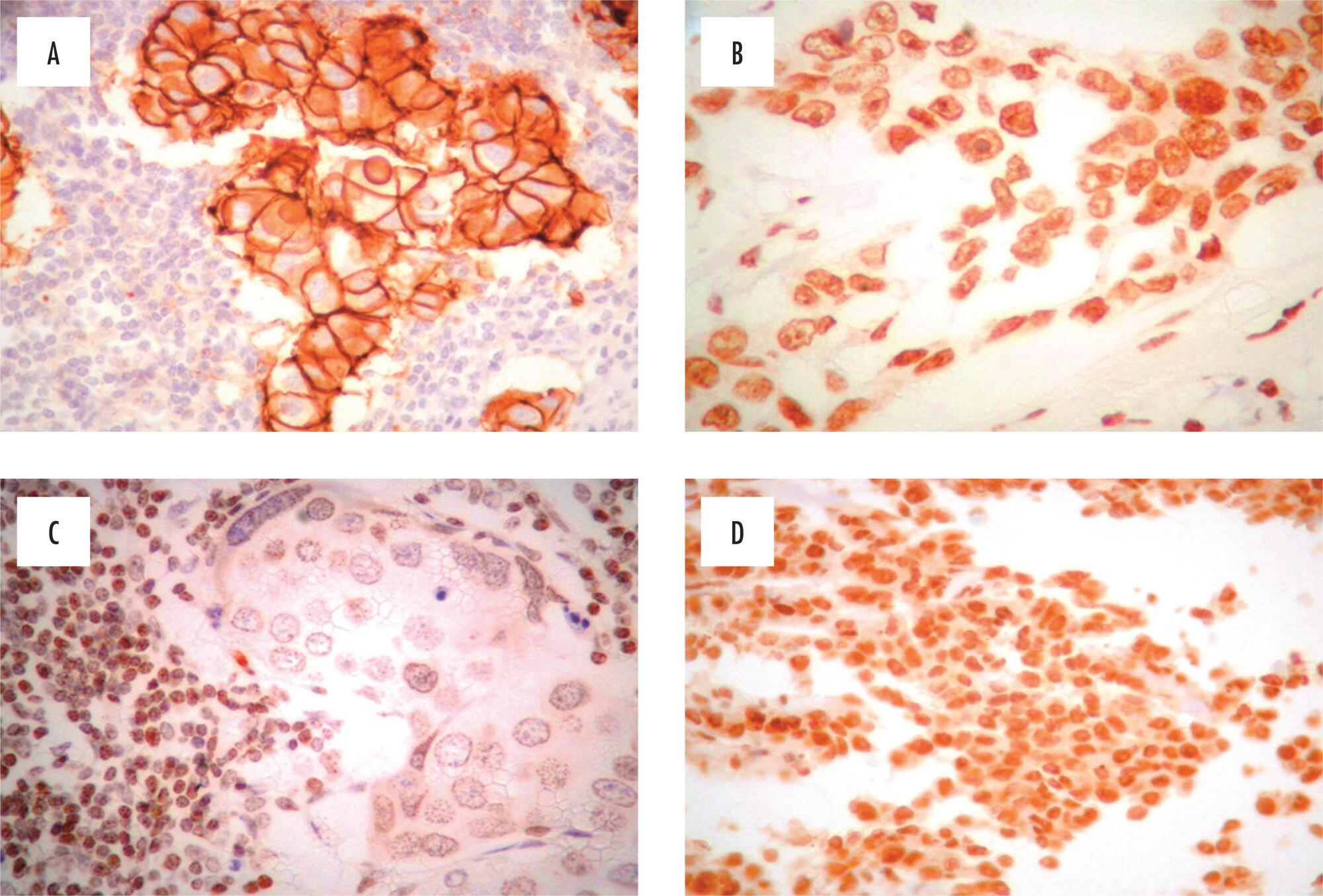
A total of 104 HER2-positive breast carcinoma specimens were prepared in tissue microarrays blocks for immunohistochemical detection of PTEN and phosphorylated AKT (pAKT). Original histologic sections were reviewed to assess pathological features, including HER2 status and Ki-67 index values. The associations between categorical and numeric variables were identified using Pearson's chi-square test and the Mann-Whitney, respectively.
Co-expression of pAKT and PTEN was presented in 59 (56.7%) cases. Reduced levels of PTEN expression were detected in 20 (19.2%) cases, and these 20 tumors had a lower Ki-67 index value. In contrast, tumors positive for pAKT expression [71 (68.3%)] were associated with a higher Ki-67 index value.
A role for AKT in the proliferation of HER2-positive breast cancers was confirmed. However, immunohistochemical detection of PTEN expression did not correlate with an inhibition of cellular proliferation or control of AKT phosphorylation, suggesting other pathways in these mechanisms of control.
